. 2016 Aug 9;24(2):324-31.
doi: 10.1016/j.cmet.2016.07.001. Epub 2016 Jul 28.

Circadian Rhythm Disruption Promotes Lung Tumorigenesis

Affiliations

Circadian Rhythm Disruption Promotes Lung Tumorigenesis

Thales Papagiannakopoulos et al. Cell Metab. .

Abstract

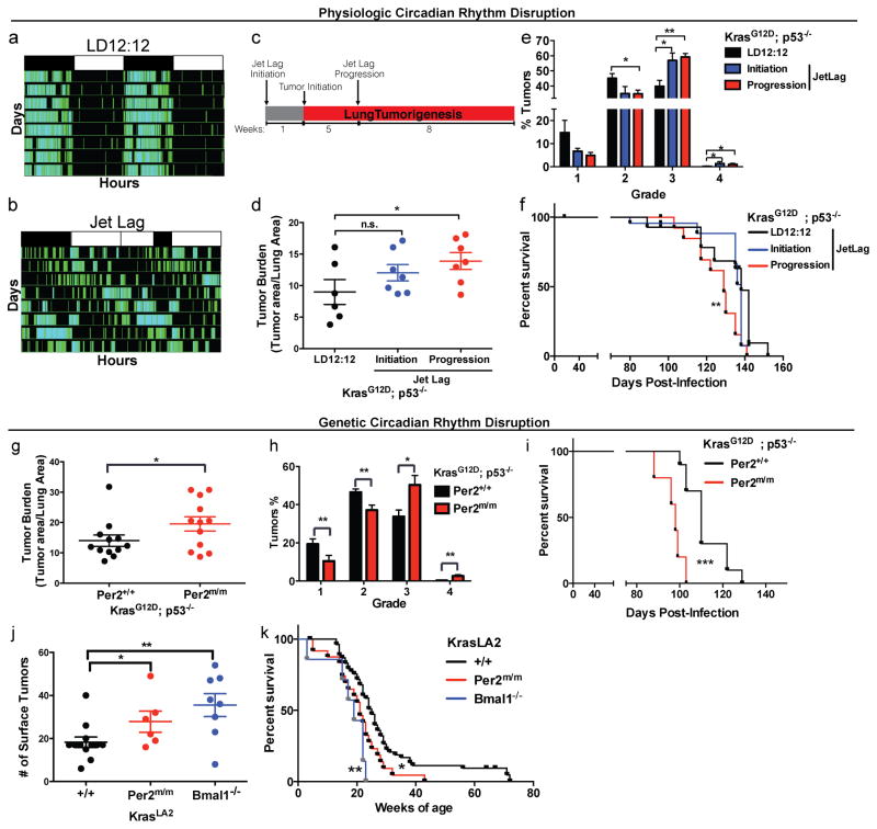
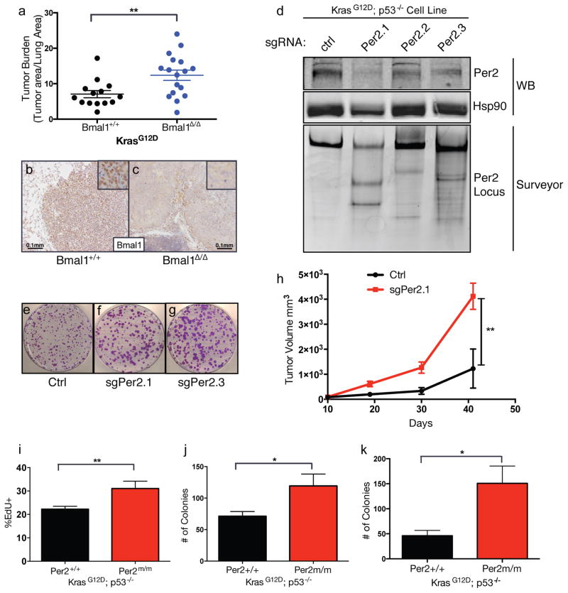
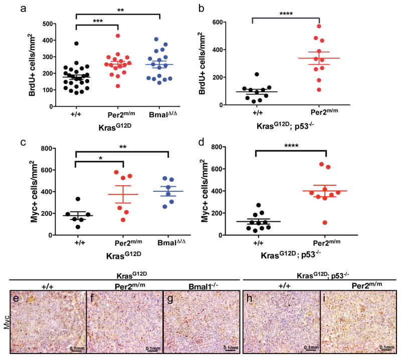
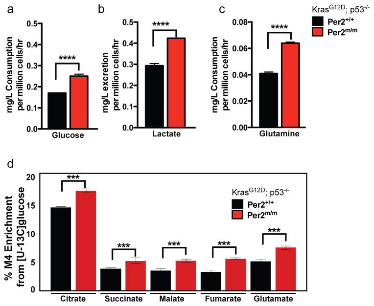
Circadian rhythms are 24-hr oscillations that control a variety of biological processes in living systems, including two hallmarks of cancer, cell division and metabolism. Circadian rhythm disruption by shift work is associated with greater risk for cancer development and poor prognosis, suggesting a putative tumor-suppressive role for circadian rhythm homeostasis. Using a genetically engineered mouse model of lung adenocarcinoma, we have characterized the effects of circadian rhythm disruption on lung tumorigenesis. We demonstrate that both physiologic perturbation (jet lag) and genetic mutation of the central circadian clock components decreased survival and promoted lung tumor growth and progression. The core circadian genes Per2 and Bmal1 were shown to have cell-autonomous tumor-suppressive roles in transformation and lung tumor progression. Loss of the central clock components led to increased c-Myc expression, enhanced proliferation, and metabolic dysregulation. Our findings demonstrate that both systemic and somatic disruption of circadian rhythms contribute to cancer progression.

Keywords: CRISPR; circadian rhythms; jet lag; lung cancer; physiology.


Figures

Figure 1
Figure 1. Physiologic and genetic disruption of circadian rhythms accelerates lung tumorigenesis
Representative double-plotted actogram of KP mice subjected to (A) regular LD12:12 (left) and (B) an 8 hour phase advance by Jet lag (right). (C) Schematic of the timeline of experiment to assess the effects of jet lag on lung tumorigenesis. Histological assessment of lung tumor burden (D) and grade (E) in KP mice placed in normal LD12:12 (n=6) or jet lag conditions at initiation (n=7) or tumor progression (n=7). (F) Kaplan-Meier survival analysis for KP animals placed in LD12:12 (n=15) or Jet lag initiation (n=13) and progression (n=13) conditions. Histological analysis of lung tumor burden (G) and grade (H) in KP mice with WT (Per2+/+) (n=12) or germline mutant (Per2m/m) Per2 (n=12). (I) Kaplan-Meier survival analysis for KP animals with WT (Per2+/+) (n=10) or mutant (Per2m/m) Per2 (n=5). (J) Surface tumor number in KrasLA2/+ WT (+/+) animals (n=12), systemic loss of Per2 (Per2m/m) (n=6) and Bmal1 loss (Bmal1−/−) (n=8). (K) Kaplan-Meier survival analysis for KrasLA2/+ animals with WT (+/+) (n=50), Per2m/m (n=31) and Bmal1−/− (n=7). Note: n.s. = not significant, * = p<0.05, ** = p<0.01, *** = p<0.001, obtained from two-sided Student’s t-test. All error bars denote s.e.m.
Figure 2
Figure 2. Cell autonomous loss of circadian genes enhances transformation and tumorigenesis
(A) Graph of histological assessment of lung tumor burden tumor burden in K animals with tumor-specific loss of Bmal1 (Bmal1Δ/Δ) (n=17) compared to WT controls (Bmal1+/+) (n=14). Immunohistochemical staining for Bmal1 in Bmal1+/+ (B) and Bmal1Δ/Δ (C) K tumors, insets represent high magnification images, scale bar=0.1mm. (D) Western blot (WB) and Surveyor analysis of Per2 protein and the Per2 locus, respectively, in KP cell lines where Per2 was target by three different short-guide RNAs (sgRNAs) using the CRISPR/Cas9 system. Representative of three independent replicate low-density clonogenicity assays in Ctrl (E), sgPer2.1 (F) and sgPer2.3 (G) targeted KP cells. (H) Tumor volume measurements of subcutaneous transplanted CRISPR/Cas9-targeted KP murine lung cancer cells. (I) Flow cytometry measurements of EdU incorporation in KP MEFs with WT (+/+) or mutant (Per2m/m) Per2. (J) Colony numbers from low-density clonogenicity assays in WT (+/+) or mutant (Per2m/m) Per2 KP cells. (K) Colony numbers from 3D agar assays of WT (+/+) or mutant (Per2m/m) Per2 KP cells. All experiments in (E–K) represent data from three independent experimental replicates from 3 independent MEF lines. Note: * = p<0.05, ** = p<0.01, obtained from two-sided Student’s t-test. All error bars denote s.e.m.
Figure 3
Figure 3. Enhanced proliferation and increased levels of c-Myc in circadian mutant tumors
Immunohistochemical analysis of BrdU incorporation in tumors from (A) K animals with WT (+/+) (n=24), mutant Per2 (Per2m/m) (n=16) and Bmal1 mutant (Bmal1Δ/Δ) (n=16) tumors; (B) KP animals with WT (+/+) (n=10), mutant Per2 (Per2m/m) (n=10). Immunohistochemical staining of c-Myc (C) K animals with WT (+/+) (n=6), mutant Per2 (Per2m/m) (n=6) and Bmal1 mutant (Bmal1Δ/Δ) (n=6) tumors; (D) KP animals with WT (+/+) (n=10), mutant Per2 (Per2m/m) (n=10). Representative c-Myc immunohistochemical images of: K, (E) WT (+/+), (F) Per2 mutant (Per2m/m), (G) Bmal1 mutant (Bmal1Δ/Δ) and KP (H) WT (+/+), (I) Per2 mutant (Per2m/m). Scale bars 0.1mm. n= individual tumors. Note: * = p<0.05, ** = p<0.01, *** = p<0.001, **** = p<0.0001 obtained from two-sided Student’s t-test. All error bars denote s.e.m.
Figure 4
Figure 4. Altered glucose and glutamine metabolism in circadian mutant cells
Measurements (mg/L per million cells/hour) of (A) glucose consumption and (B) lactate excretion, (C) glutamine consumption in KP WT (Per2+/+) or mutant (Per2m/m) cells. (D) Percent M4 carbon labeling enrichment in TCA cycle intermediates in in KP WT (Per2+/+) or mutant (Per2m/m) cells. Error bars denote standard deviation. Note: * = p<0.05, ** = p<0.01, **** = p<0.0001 obtained from two-sided Student’s t-test.

References

    1. Alhopuro P, Bjorklund M, Sammalkorpi H, Turunen M, Tuupanen S, Bistrom M, Niittymaki I, Lehtonen HJ, Kivioja T, Launonen V, et al. Mutations in the circadian gene CLOCK in colorectal cancer. Mol Cancer Res. 2010;8:952–960. - PubMed
    1. Altman BJ, Hsieh AL, Sengupta A, Krishnanaiah SY, Stine ZE, Walton ZE, Gouw AM, Venkataraman A, Li B, Goraksha-Hicks P, et al. MYC Disrupts the Circadian Clock and Metabolism in Cancer Cells. Cell metabolism. 2015;22:1009–1019. - PMC - PubMed
    1. Bass J, Takahashi JS. Circadian integration of metabolism and energetics. Science. 2010;330:1349–1354. - PMC - PubMed
    1. Bunger MK, Wilsbacher LD, Moran SM, Clendenin C, Radcliffe LA, Hogenesch JB, Simon MC, Takahashi JS, Bradfield CA. Mop3 is an essential component of the master circadian pacemaker in mammals. Cell. 2000;103:1009–1017. - PMC - PubMed
    1. Cancer Genome Atlas Research, N. Comprehensive molecular profiling of lung adenocarcinoma. Nature. 2014;511:543–550. - PMC - PubMed

MeSH terms