. 2016 Nov;110(Pt A):419-430.
doi: 10.1016/j.neuropharm.2016.07.028. Epub 2016 Jul 29.

Inhibition of the prostaglandin E2 receptor EP2 prevents status epilepticus-induced deficits in the novel object recognition task in rats

Affiliations

Inhibition of the prostaglandin E2 receptor EP2 prevents status epilepticus-induced deficits in the novel object recognition task in rats

Asheebo Rojas et al. Neuropharmacology. 2016 Nov.

Abstract

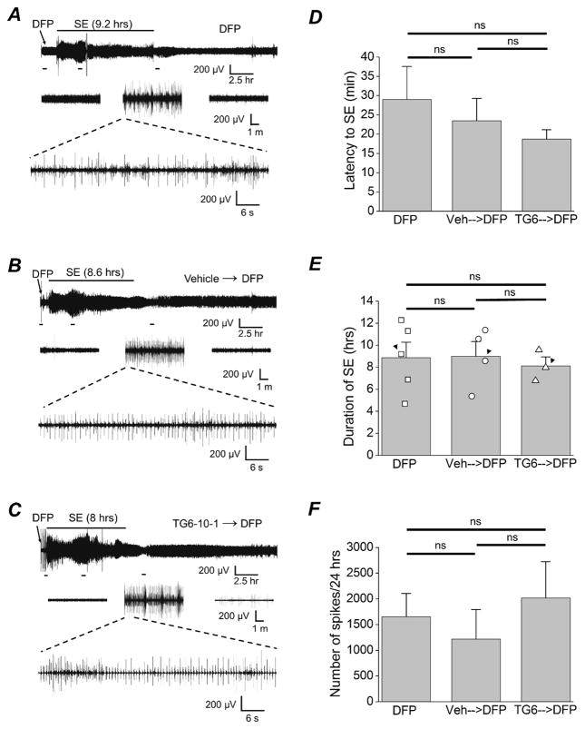
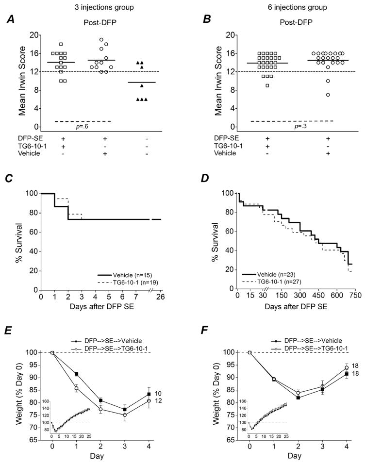
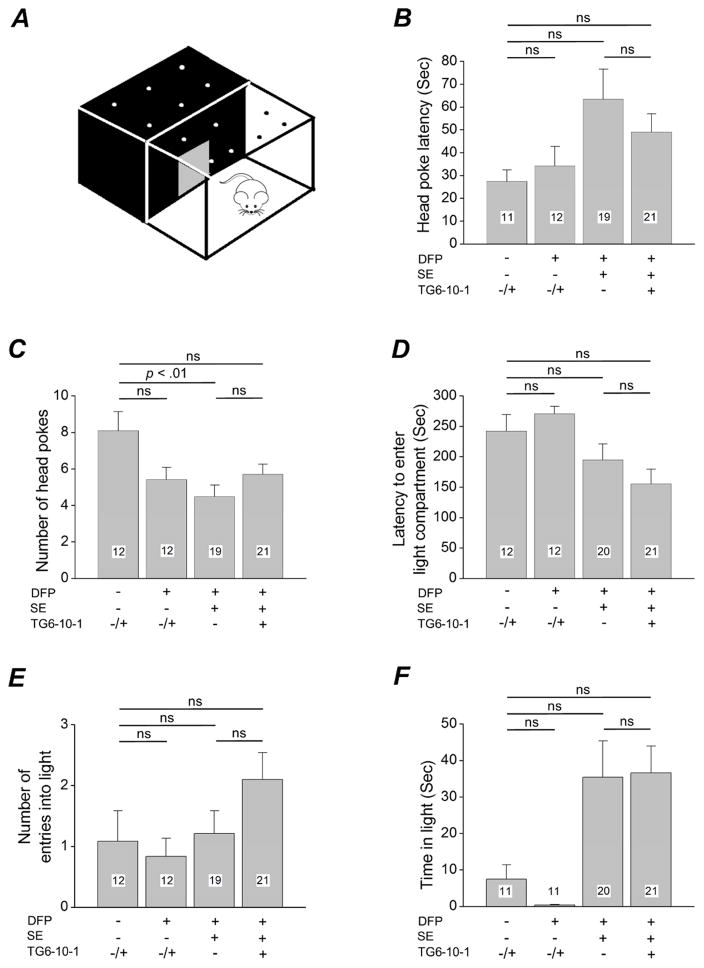
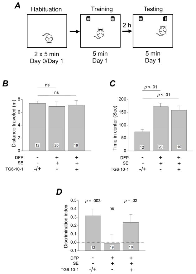
Survivors of exposure to an organophosphorus nerve agent may develop a number of complications including long-term cognitive deficits (Miyaki et al., 2005; Nishiwaki et al., 2001). We recently demonstrated that inhibition of the prostaglandin E2 receptor, EP2, attenuates neuroinflammation and neurodegeneration caused by status epilepticus (SE) induced by the soman analog, diisopropylfluorophosphate (DFP), which manifest within hours to days of the initial insult. Here, we tested the hypothesis that DFP exposure leads to a loss of cognitive function in rats that is blocked by early, transient EP2 inhibition. Adult male Sprague-Dawley rats were administered vehicle or the competitive EP2 antagonist, TG6-10-1, (ip) at various times relative to DFP-induced SE. DFP administration resulted in prolonged seizure activity as demonstrated by cortical electroencephalography (EEG). A single intraperitoneal injection of TG6-10-1 or vehicle 1 h prior to DFP did not alter the development of seizures, the latency to SE or the duration of SE. Rats administered six injections of TG6-10-1 starting 90 min after the onset of DFP-induced SE could discriminate between a novel and familiar object 6-12 weeks after SE, unlike vehicle treated rats which showed no preference for the novel object. By contrast, behavioral changes in the light-dark box and open field assays were not affected by TG6-10-1. Delayed mortality after DFP was also unaffected by TG6-10-1. Thus, selective inhibition of the EP2 receptor may prevent SE-induced memory impairment in rats caused by exposure to a high dose of DFP.

Keywords: Anxiety; DFP; EP2 receptor; Electroencephalography; Epilepsy; Hippocampus; Light-dark box preference test; Novel object recognition; Open field; Organophosphate; Seizure; Status epilepticus.


Figures

Figure 1
Figure 1. Pharmacokinetics of TG6-10-1 and temporal evolution of SE
A, Experimental paradigm of chemical administration in a rat model of DFP-induced status epilepticus. Following DFP, rats were injected with vehicle (40% water, 50% PEG400, 10% DMSO) or TG6-10-1 (5 mg/kg) dissolved in the vehicle multiple times beginning 90 min from the onset of SE (3 injections or 6 injections at times shown on the right after SE onset). B, plasma concentration of normal adult rats that received TG6-10-1 (10 mg/kg, ip) (n = 3 rats). C, the brain to plasma ratio in rats administered TG6-10-1 over the same time span (n = 3 rats for each time point). Open circles represents individual rats. D, the mean behavioral seizure scores of rats that received DFP followed by nothing (n = 10 rats), DFP followed by the vehicle (n = 45 rats) and DFP followed by TG6-10-1 (n = 53 rats) are plotted as a function of time (data from that received 3 injections and 6 injections were combined for this analysis as there was no difference in the groups). Also shown is the behavioral seizure activity of rats that did not enter status epilepticus (n = 11 rats). The dashed line indicates the behavioral seizure activity score at the onset of status epilepticus.
Figure 2
Figure 2. Characteristics of DFP induced SE
A, Cortical EEG activity was recorded prior, during and after SE induced by exposure to DFP for 24 hours. Shown is a representative EEG trace from the cortical recording of an adult male rat with large spikes that appear within 30 minutes after exposure to DFP. The duration of SE is denoted by the horizontal bar above the trace. B, representative EEG trace from the cortical recording of an adult male rat that was administered the vehicle (ip) 1 hour prior to DFP. C, representative EEG trace from the cortical recording of an adult male rat that was injected with a single dose of TG6-10-1 (5 mg/kg, ip) 1 hour prior to DFP. D, plot of the latency to the onset of status epilepticus comparing the three groups of rats [DFP-SE (n = 4), vehicle followed by DFP-SE (n = 4), TG6-10-1 followed by DFP-SE (n = 4)]. E, bar graph of the mean duration of SE for each group of rats tested [DFP-SE (n = 5), vehicle followed by DFP-SE (n = 4), TG6-10-1 followed by DFP-SE (n = 3)]. The open symbols represents each individual rat. The filled triangles denotes the traces shown in A, B and C. F, plot of the total number of spikes identified by dCLAMP during the 24 hour recording for the three groups of rats [DFP-SE (n = 5), vehicle followed by DFP-SE (n = 4), TG6-10-1 followed by DFP-SE (n = 3)]. Error bars represent the standard error of the mean. ns = p > .05, One-way ANOVA with Bonferroni posthoc.
Figure 3
Figure 3. Analysis of functional recovery and delayed mortality
Plot of the mean Irwin score for rats that received 3 injections (A) or 6 injections (B) of TG6-10-1 or vehicle following DFP (p = .6 for 3 injections; p = .3 for 6 injections, t test). Each open square represents an individual rat that received TG6-10-1 after DFP-SE [3 injections, n = 14; 6 injections, n = 25]. Open circles represent individual rats that received vehicle after DFP-SE [3 injections, n = 11, 6 injections n = 22]. The up facing closed triangles in A represent individual rats that did not enter status epilepticus (DFP-no-SE, n = 7 rats) following DFP administration. The short horizontal bold lines denotes the average of the individual animals within the group. The long horizontal dashed line represents the cutoff for determining whether an animal was healthy or impaired. C, D survival plots of rats that experienced DFP-induced SE and were administered TG6-10-1 or vehicle. Rats that received 3 injections are shown in panel C measured to day 26 after DFP-induced SE (p = 1, Fisher's exact test comparing treatment groups) and rats that received 6 injections measured to day 742 after DFP-induced SE are shown in panel D (p = .7, Fisher's exact test comparing treatment groups). The number in parentheses represents the total number of rats in each group. E, graph of the weight change for rats that received 3 injections of TG6-10-1 or vehicle (p = .5, t test, day 4). F, shown is the weight change for rats that received 6 injections of TG6-10-1 or vehicle (p = .3, t test, day 4). The small inserts show the weight change over 26 days. Rats that lost 30% or more of the original body weight on any day over the 4 day period was not included in the analysis.
Figure 4
Figure 4. Exploration and anxiety behavior 4 weeks following SE
A, schematic of light-dark exploration apparatus. Latency to the first head poke (B), the number of head pokes (C), latency to enter the light compartment (D), number of entries into the light compartment (E) and the time spent in the light compartment (F) are shown for the four groups of rats (sham treated controls, DFP-no-SE, DFP-SE followed by vehicle, DFP-SE followed by TG6-10-1). The bars show the mean of the group and the number in the white box within the bar represent the total number of rats in each group. The error bars represent the standard error of the mean. The “+/−“ symbol next to TG6-10-1 denotes sham treated control rats and DFP-no-SE rats that received TG6-10-1 (n=6) or vehicle (n=6). These rats were combined into one group as they were not different in any measure. p < .01, one-way ANOVA with Bonferroni posthoc; ns = p > .05 by One-way ANOVA with Bonferroni posthoc. Grubb’s test identified one sham treated control rat as an outlier. This rat was removed during analysis in panels B and F reducing the total number of sham treated control rats from 12 to 11. One DFP-SE rat that was injected with vehicle was also identified as an outlier by the Grubb’s test and removed from the analysis in panels B, C and E reducing the total number of rats in the DFP-SE followed by vehicle group from 20 to 19. Time in the light was not obtained for one DFP-no-SE rat reducing the number from 12 to 11.
Figure 5
Figure 5. Novel object recognition memory in rats that experienced SE. A
, schematic of novel object recognition testing consisting of three epochs carried out over two days. B, a graph of the mean total distanced traveled for each group (sham treated controls, DFP-SE followed by vehicle, DFP-SE followed by TG6-10-1) is shown. ns = p > .05, One-way ANOVA with Dunnett’s posthoc. C, time spent in the center is shown for the three groups tested. p < .01, One-way ANOVA, Dunnett’s posthoc. D, a discrimination index was used as a measure of memory retention. The individual groups were compared to zero by a 1-sample t test. The number in the white box within the bar represents the total number of rats in each group. The “+/−“ symbol next to TG6-10-1 denotes sham treated control rats that received TG6-10-1 (n=6) or vehicle (n=6). These rats were combined into one group as they were not different in any measure. The horizontal dashed line at 0 indicates the point at which there is no discrimination between the novel and familiar objects. ns = p > .05. One DFP-SE rat that received vehicle experienced a spontaneous seizure in the arena during NOR testing and was not included in that analysis of the discrimination index reducing the total number of rats from 20 to 19. One DFP-SE rat that received TG6-10-1 was deemed not fit to perform NOR testing and was not included in the analysis of the discrimination index reducing the total number in the group from 19 to 18.

References

    1. Bevins RA, Besheer J. Object recognition in rats and mice: a one-trial non-matching-to-sample learning task to study 'recognition memory'. Nat Protoc. 2006;1:1306–1311. - PubMed
    1. Binukumar BK, Bal A, Gill KD. Chronic dichlorvos exposure: microglial activation, proinflammatory cytokines and damage to nigrostriatal dopaminergic system. Neuromolecular Med. 2011;13:251–265. - PubMed
    1. Chen Y. Organophosphate-induced brain damage: mechanisms, neuropsychiatric and neurological consequences, and potential therapeutic strategies. Neurotoxicology. 2012;33:391–400. - PubMed
    1. Colovic MB, Krstic DZ, Lazarevic-Pasti TD, Bondzic AM, Vasic VM. Acetylcholinesterase inhibitors: pharmacology and toxicology. Curr Neuropharmacol. 2013;11:315–335. - PMC - PubMed
    1. Costa LG. Current issues in organophosphate toxicology. Clin Chim Acta. 2006;366:1–13. - PubMed

MeSH terms