. 2016 Aug 1:6:30637.
doi: 10.1038/srep30637.

Dose-dependent and cell type-specific cell death and proliferation following in vitro exposure to radial extracorporeal shock waves

Affiliations

Dose-dependent and cell type-specific cell death and proliferation following in vitro exposure to radial extracorporeal shock waves

Tanja Hochstrasser et al. Sci Rep. .

Abstract

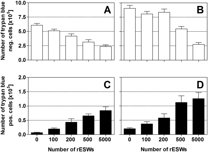
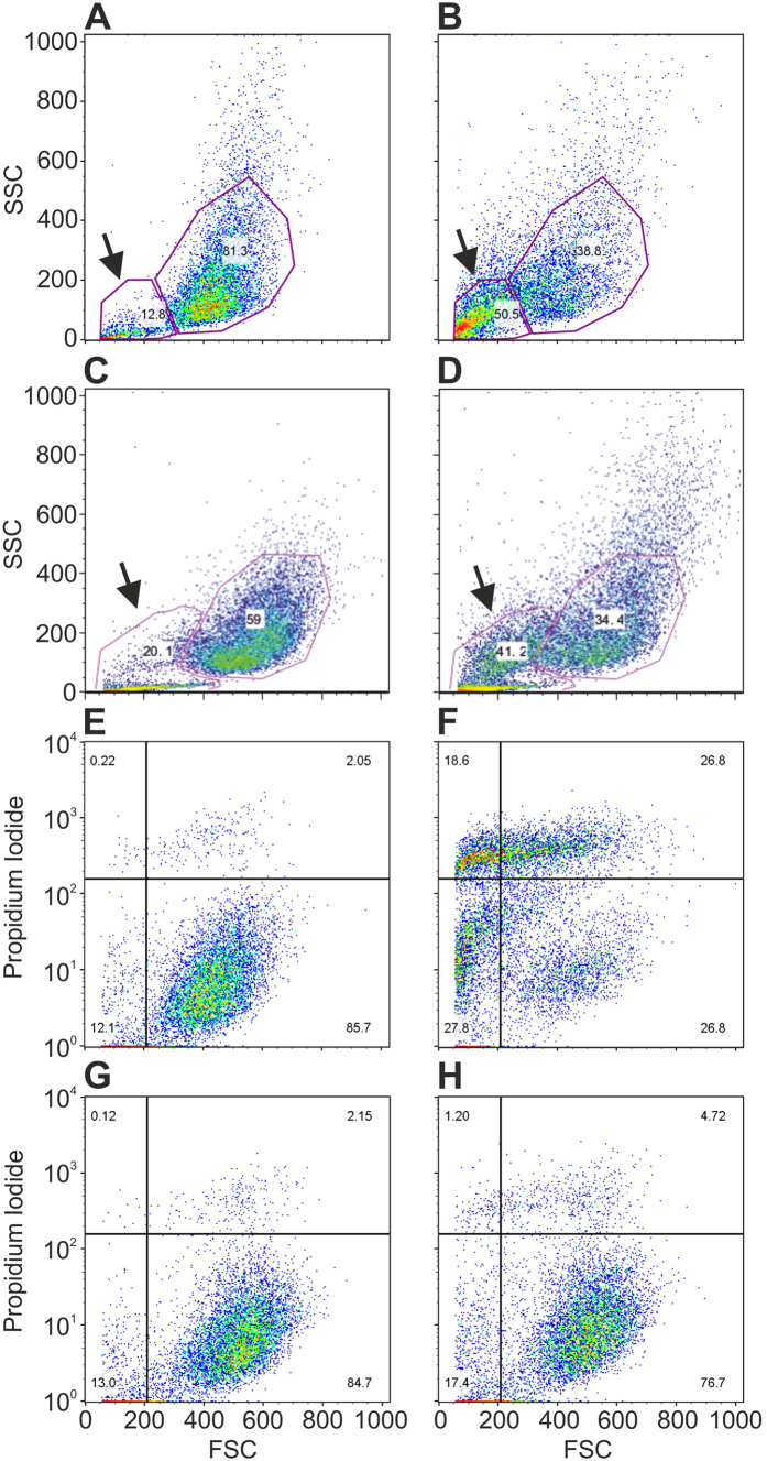
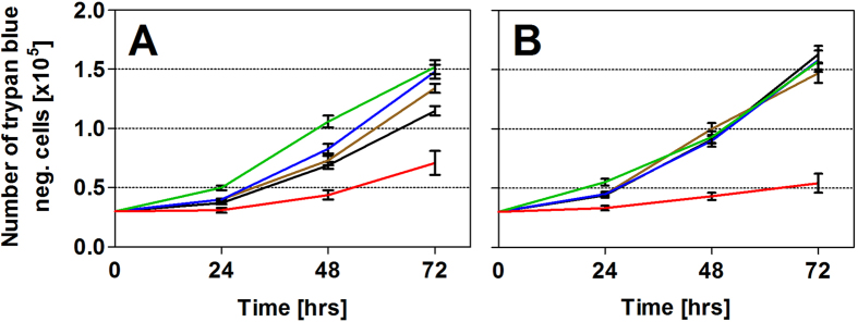
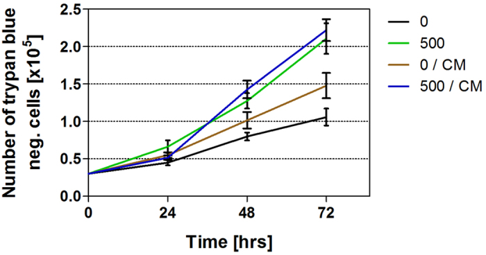
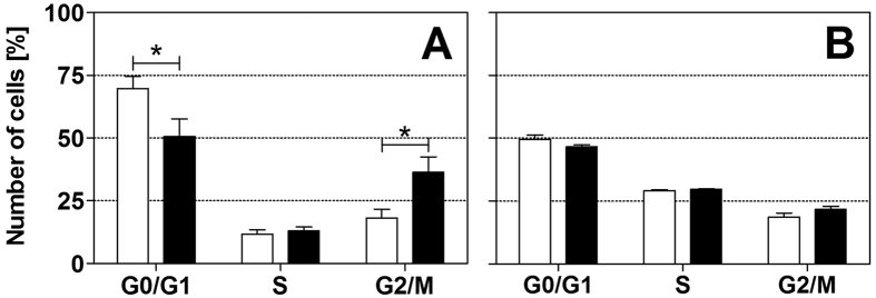
Radial extracorporeal shock wave (rESW) therapy is widely used in musculoskeletal disorders and wound repair. However, the mechanisms of action are still largely unknown. The current study compared the effects of rESWs on two cell types. Human fetal foreskin fibroblasts (HFFF2) and human placental choriocarcinoma cell line JEG-3 were exposed to 0, 100, 200, 500 or 5000 rESWs generated with a Swiss DolorClast device (2.5 bar, 1 Hz). FACS analysis immediately after rESW exposure showed that initially, rESWs rather induced mechanical cell destruction than regulated or programmed cell death. Cell damage was nearly negated by reducing cavitation. Furthermore, cell viability decreased progressively with higher numbers of rESWs. Exposure to rESWs had no impact on growth potential of JEG-3 cells, but dose-dependently increased growth potential of HFFF2 cells. Cultivation of cells that were initially exposed to sham-rESWs in conditioned media increased the growth potential of HFFF2 cells, nevertheless, an even stronger effect was achieved by direct exposure to rESWs. Additionally, cell cycle distribution analysis demonstrated a shift in proportion from G0/G1 to G2/M phase in HFFF2 cells, but not in JEG-3 cells. These data demonstrate that rESWs leads to initial and subsequent dose-dependent and cell type-specific effects in vitro.


Conflict of interest statement

C.S. serves as paid consultant for and receives benefits from Electro Medical Systems, the manufacturer and distributor of the radial extracorporeal shock wave device, Swiss DolorClast. However, Electro Medical Systems had no any role in study design, data collection and analysis, decision to publish, or preparation of this manuscript, and C.S. has not received any honoraria or consultancy fee in writing this manuscript. No other potential conflicts of interest relevant to this article were reported.

Figures

Figure 1
Figure 1. Morphological appearance of adherent cells before and after exposure to radial extracorporeal shock waves.
The morphological appearance of HFFF2 (A,C) and JEG-3 (B,D) monolayers exposed to sham-rESWs (A,B) and rESWs (C,D) was assessed by immunofluorescence staining with phalloidin (green) and DAPI (4,6-diamidino-2-phenyl-indole (blue). Cells in (C,D) were exposed to 100 rESWs as explained in detail in the main text. Cells exposed to sham-rESWs showed a homogeneous cell distribution (A,B). Exposure to rESWs caused cellular detachment and, thus, holes in the monolayers (asterisks in C,D) as well as disruption of actin filaments in cells located next to the holes (arrows in the upper insets in C,D). Cells distant to the holes in the monolayers appeared normal (arrowheads in the lower insets in C,D). The scale bar represents 100 μm in the low-power photomicrographs in (A–D) and 14 μm in the high-power insets in (A–D).
Figure 2
Figure 2. Cell viability after exposure to radial extracorporeal shock waves.
Data show absolute numbers (mean ± SEM) of trypan blue negative HFFF2 (A) and JEG-3 (B) cells as well as of trypan blue positive HFFF2 (C) and JEG-3 (D) cells as a function of the number of applied rESWs. Results of statistical analysis are summarized in Table 2.
Figure 3
Figure 3. Viable cells and debris/dead cells after exposure to radial extracorporeal shock waves.
(AD) Original dot-plots of side light scatter (SSC) vs. forward light scatter (FSC) obtained by flow cytometry (FACS Calibur flow cytometer, BD Biosciences, Heidelberg, Germany) of HFFF2 (A,B) and JEG-3 (C,D) cells after exposure to sham-rESWs (A,C) or 500 rESWs (B,D). The arrows indicate the fraction of debris/dead cells that was increased between two-fold (JEG-3 cells; C,D) and four-fold (HFFF2 cells; A,B) immediately after exposure to 500 rESWs compared to exposure to sham-rESWs. (E–H), original dot-plots of propidium iodide vs. FSC of HFFF2 cells undergoing cell death after exposure to sham-rESWs (E,G) or 500 rESWs (F,H) in culture medium (E,F) or 10% polyvinyl alcohol solution (G,H).
Figure 4
Figure 4. Cell count after exposure to radial extracorporeal shock waves.
Data show absolute numbers (mean ± SEM) of HFFF2 (A) and JEG-3 (B) cells that were trypan blue negative after exposure to 0 (sham-rESWs, black), 100 (brown), 200 (blue), 500 (green) and 5000 (red) rESWs as a function of time after exposure (0, 24, 48 and 72 hours). Results of statistical analysis are summarized in Table 3.
Figure 5
Figure 5. Effect of conditioned medium on cell count.
Data show absolute numbers (mean ± SEM) of the following groups of HFFF2 cells that were trypan blue negative as a function of time after exposure (0, 24, 48 and 72 hours): (i) cells exposed to sham-ESW and cultured in fresh culture medium (black); (ii) cells exposed to 500 rESW impulses and cultured in fresh culture medium (green); (iii) cells exposed to sham-ESW and cultured in conditioned medium (brown); and (iv) cells exposed to 500 rESW impulses and cultured in conditioned medium (blue). Results of statistical analysis are summarized in Table 4.
Figure 6
Figure 6. Cell cycle phase distribution after exposure to radial extracorporeal shock waves.
Data show relative numbers (mean ± SEM) of HFFF2 (A) and JEG-3 (B) cells in the G0/G1, S and G2/M phases 24 h after exposure to sham-rESWs (open bars) or to 500 rESWs (closed bars). *p < 0.05.

References

    1. Schmitz C., Csaszar N. B., Rompe J. D., Chaves H. & Furia J. P. Treatment of chronic plantar fasciopathy with extracorporeal shock waves (review). J Orthop Surg Res 8, 31 (2013). - PMC - PubMed
    1. Speed C. A systematic review of shockwave therapies in soft tissue conditions: focusing on the evidence. Br J Sports Med 48, 1538–1542 (2014). - PubMed
    1. Butterworth P. A. et al.. The effectiveness of extracorporeal shock wave therapy for the treatment of lower limb ulceration: a systematic review. J Foot Ankle Res 8, 3 (2015). - PMC - PubMed
    1. Schmitz C. et al.. Efficacy and safety of extracorporeal shock wave therapy for orthopedic conditions: a systematic review on studies listed in the PEDro database. Br Med Bull 116, 115–138 (2015). - PMC - PubMed
    1. Frairia R. & Berta L. Biological effects of extracorporeal shock waves on fibroblasts. A review. Muscles Ligaments Tendons J 1, 138–147 (2011). - PMC - PubMed