

Review
doi: 10.1055/s-0036-1584823.
Common Adult Skin and Soft Tissue Lesions
Affiliations
- PMID: 27478418
- PMCID: PMC4961504
- DOI: 10.1055/s-0036-1584823
Item in Clipboard
Review
Common Adult Skin and Soft Tissue Lesions
Semin Plast Surg.
2016 Aug.
Abstract
A strong foundational knowledge of dermatologic disease is crucial for a successful practice in plastic surgery. A plastic surgeon should be able to identify and appreciate common dermatologic diseases that may require medical and/or surgical evaluation and management. In this article, the authors describe epidermal/dermal, infectious, pigmented, and malignant cutaneous lesions that are commonly encountered in practice. Descriptions include the epidemiology, pathogenesis, clinical course, and management options for each type of lesion.
Keywords: benign; common; cutaneous; dermatology; malignant.
Figures
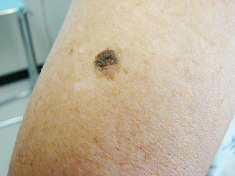
Seborrheic keratosis. (Photograph courtesy of Dr. Theodore Rosen.)
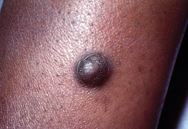
Dermatofibroma. (Photograph courtesy of Dr. Theodore Rosen.)
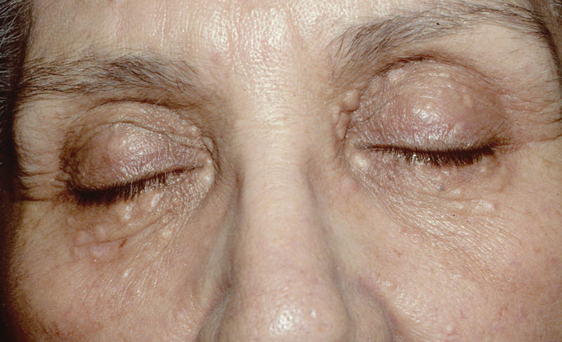
Syringoma. (Photograph courtesy of Dr. Theodore Rosen.)
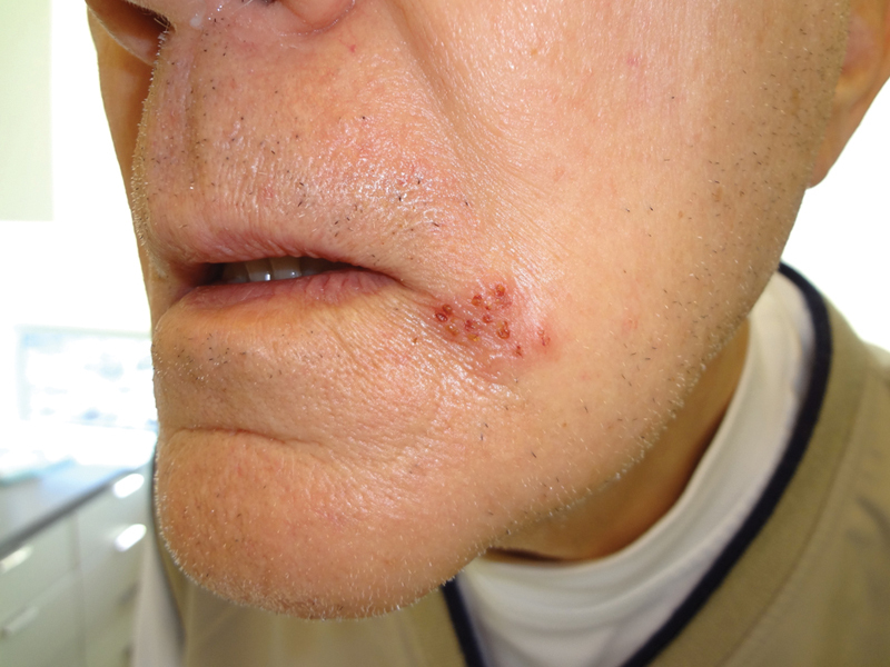
Herpes simplex virus. (Photograph courtesy of Dr. Theodore Rosen.)
Eczema herpeticum in a transplant patient.

Varicella zoster virus (shingles).

Multiple melanocytic nevi.
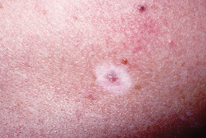
Halo nevus. (Photograph courtesy of Dr. Theodore Rosen.)
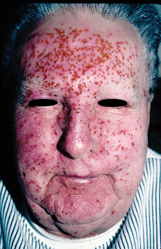
Cutaneous horn.
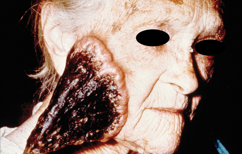
Ulcerated basal cell carcinoma.
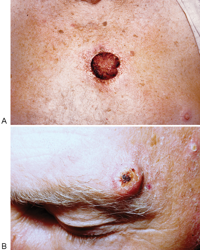
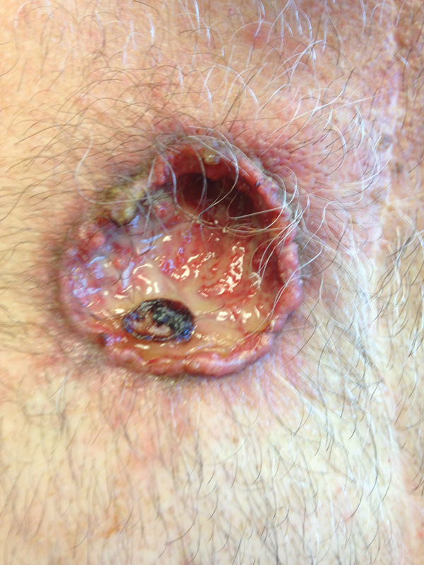
(A) Squamous cell carcinoma. (B) Squamous cell carcinoma, keratoacanthoma type.
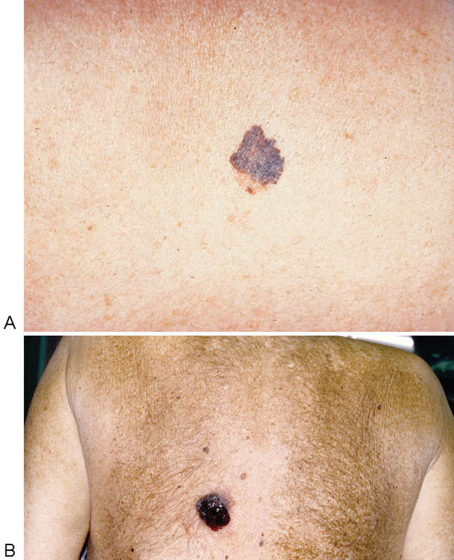
(A) Superficial spreading malignant melanoma. (B) Nodular malignant melanoma.
References
-
- Bolognia J L, Jorizzo J L, Schaffer J V. Philadelphia: Elsevier Saunders; 2012. Dermatology. 3rd ed.
-
- Hafner C, Vogt T. Seborrheic keratosis. J Dtsch Dermatol Ges. 2008;6(8):664–677. - PubMed
-
- Sari R, Akman A, Alpsoy E, Balci M K. The metabolic profile in patients with skin tags. Clin Exp Med. 2010;10(3):193–197. - PubMed
-
- Fitzpatrick T B, Wolff K, Johnson R A, Suurmond D. New York: McGraw-Hill; 2005. Fitzpatrick's Color Atlas and Synopsis of Clinical Dermatology. 5th ed.
-
- Thomas V D, Snavely N R, Lee K K, Swanson N A. New York: McGraw-Hill; 2012. Benign epithelial tumors, hamartomas, and hyperplasias; p. 1334.
Publication types
LinkOut - more resources
Full Text Sources
Other Literature Sources
