. 2016 Jul 28;8(21):902-14.
doi: 10.4254/wjh.v8.i21.902.

Anti-hepatitis C virus potency of a new autophagy inhibitor using human liver slices model

Affiliations

Anti-hepatitis C virus potency of a new autophagy inhibitor using human liver slices model

Sylvie Lagaye et al. World J Hepatol. .

Abstract

Aim: To evaluate the antiviral potency of a new anti-hepatitis C virus (HCV) antiviral agent targeting the cellular autophagy machinery.

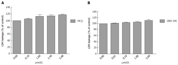
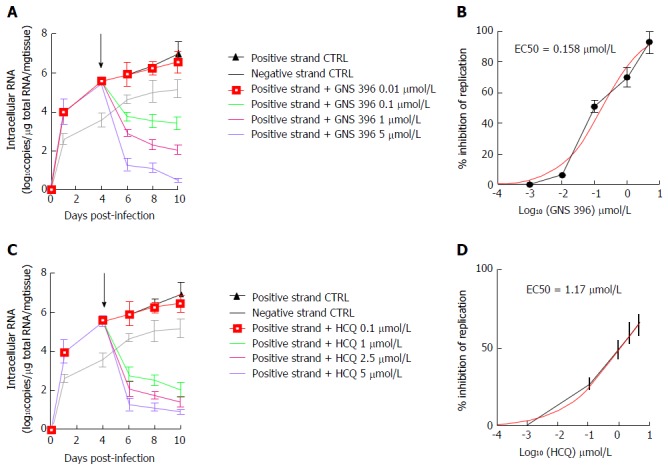
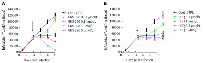
Methods: Non-infected liver slices, obtained from human liver resection and cut in 350 μm-thick slices (2.7 × 10(6) cells per slice) were infected with cell culture-grown HCV Con1b/C3 supernatant (multiplicity of infection = 0.1) cultivated for up to ten days. HCV infected slices were treated at day 4 post-infection with GNS-396 for 6 d at different concentrations. HCV replication was evaluated by strand-specific real-time quantitative reverse transcription - polymerase chain reaction. The infectivity titers of supernatants were evaluated by foci formation upon inoculation into naive Huh-7.5.1 cells. The cytotoxic effect of the drugs was evaluated by lactate dehydrogenase leakage assays.

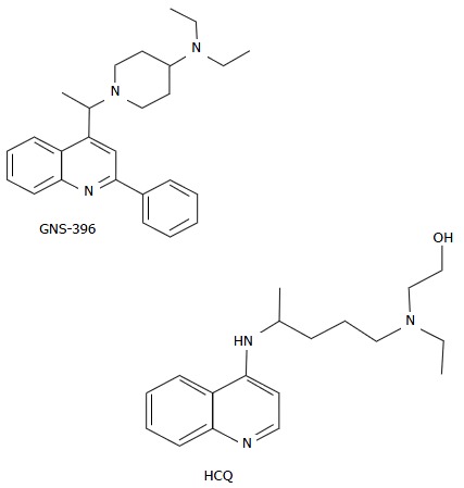
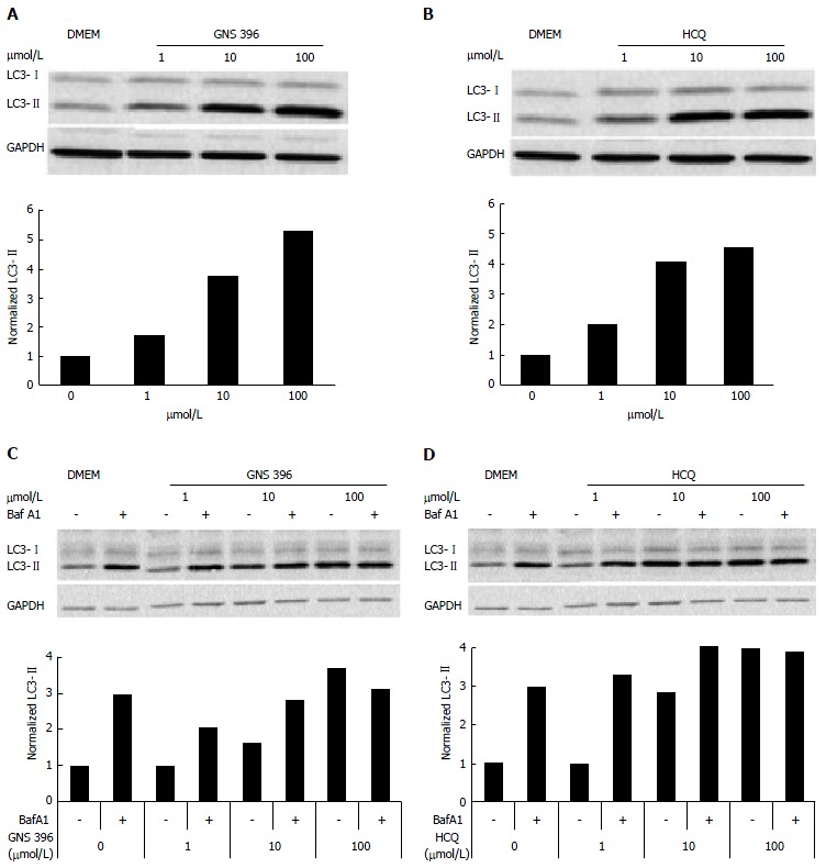
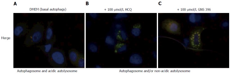
Results: The antiviral efficacy of a new antiviral drug, GNS-396, an autophagy inhibitor, on HCV infection of adult human liver slices was evidenced in a dose-dependent manner. At day 6 post-treatment, GNS-396 EC50 was 158 nmol/L without cytotoxic effect (compared to hydroxychloroquine EC50 = 1.17 μmol/L).

Conclusion: Our results demonstrated that our ex vivo model is efficient for evaluation the potency of autophagy inhibitors, in particular a new quinoline derivative GNS-396 as antiviral could inhibit HCV infection in a dose-dependent manner without cytotoxic effect.

Keywords: Autophagy; Hepatitis C virus; Host antiviral therapy; Quinoline derivative; Tissue culture.


Figures

Figure 1
Figure 1
GNS-396 and hydroxychloroquine structures. HCQ: Hydroxychloroquine.
Figure 2
Figure 2
Cytotoxicity assays of the drugs used hydroxychloroquine (A) and GNS-396 (B). Percentages of lactate deshydrogenase (LDH) leakage are relative to DMSO control-treated liver slices. Drugs cytotoxicity is significant at % of control > 120. All experiments were performed on triplicate. Values are expressed as means ± SE, comparisons were performed using the Mann-Whitney rank-sum test (P < 0.001). HCQ: Hydroxychloroquine; DMSO: Dimethylsufoxide.
Figure 3
Figure 3
Modulation and inhibition of autophagy by treatment with GNS-396 in HeLa cells. A and B: Autophagy modulation was evaluated using HeLa cells treated either with (A) GNS-396 or (B) HCQ at different concentrations (1, 10 and 100 μmol/L) during 6 h. HCQ was used as a positive control of autophagy modulation. Intracellular expression of proteins LC3 was evaluated by Western-blot assay and normalized for LC3-II; C and D: Autophagy inhibition by treatment with GNS-396 in HeLa cells. Autophagy inhibition was evaluated using HeLa cells treated either with (C) GNS-396 or (D) HCQ at different concentrations (1, 10 and 100 μmol/L) during 4 h in the presence or absence of 100 nmol/L bafilomycine A1. HCQ was used as a positive control of autophagy inhibition. LC3-II intracellular expression was evaluated by Western-blot assay and normalized. HCQ: Hydroxychloroquine; LC3: Microtubule-associated protein 1A/1B-light chain; DMEM: Dulbecco’s modified eagles’s medium; GAPDH: Glyceraldehyde-3-phosphate dehydrogenase; BafA1: Bafilomycin A1.
Figure 4
Figure 4
Inhibition of autophagic flux by treatment with GNS-396 in SkBr3 mRFP-EGFP-LC3 stable cell line. Autophagic flux was monitored using the mRFP-EGFP-LC3 tandem-tagged fluorescent protein in SkBr3 mRFP-EGFP-LC3 stable cell line. A: SkBr3 mRFP-EGFP-LC3 stable cell line without any treatment is representative of basal autophagy; SkBr3 mRFP1-EGFP-LC3 stable cell line was treated either with (B) 100 μmol/L HCQ or (C) 100 μmol/L GNS-396 during 6 h. In green/red merged images, yellow punctua (i.e., mRFP+EGFP+) indicate autophagosomes or non-acidic autolysosomes, while red punctua (i.e., mRFP+EGFP) indicate autolysosomes. HCQ is used as a positive control of autophagy inhibition. HCQ: Hydroxychloroquine; DMEM: Dulbecco’s modified eagles’s medium; LC3: Microtubule-associated protein 1A/1B-light chain.
Figure 5
Figure 5
Inhibition of autophagy by treatment with GNS-396 (1 μmol/L) in primary adult human liver slices infected with cell culture-grown hepatitis C virus Con1 (multiplicity of infection = 1). A: Intracellular expression of LC3-I/LC3-II proteins in non-infected liver slices without treatment; B: Intracellular expression of LC3-I/LC3-II proteins and the normalization of intracellular protein LC3-II expression, for 10 d in non-infected liver slices with treatment either by GNS-396 (1 μmol/L) (G) or HCQ (1 μmol/L) (H) or without treatment (D: day); C: Expression of LC3-I/LC3-II proteins and HCV core and NS5A proteins in HCVcc Con1 infected liver slices either without treatment or (D) either with treatment by GNS-396 (1 μmol/L) or HCQ (1 μmol/L) and the normalization of Intracellular protein LC3-II expression for 10 d in HCVcc Con1 infected liver slices with treatment either by GNS-396 (1 μmol/L) (G) or HCQ (1 μmol/L) (H) or without treatment (D: day). LC3: Microtubule-associated protein 1A/1B-light chain; HCVcc: Cell culture-grown hepatitis C virus; NS5A: HCV nonstructural protein 5A.
Figure 6
Figure 6
Inhibition of hepatitis C virus RNA replication by treatment either with GNS-396 or hydroxychloroquine in a dose-dependent manner in primary adult human cell culture-grown hepatitis C virus Con1 infected liver slices. Human liver slices were infected overnight with HCVcc Con1 (MOI = 0.1). The supernatant is then removed, the human liver slices washed and cultured. The liver slices and culture supernatants were collected different times post-infection. At day 4 post-infection, the liver slices were treated with increasing concentrations either of GNS-396 (0.01, 0.1, 1, 5 μmol/L) (A, B) or HCQ (C, D) for 6 d (black arrow: Start of the treatment either with GNS-396 or HCQ). Human HCVcc Con1 infected liver slices were lysed to evaluate intracellular levels of positive- and negative-strand HCV RNA by specific strand RT-qPCR at 1, 4, 6, 8, 10 d post-infection. The results were compared using the two-paired Student’s test. Values are expressed as means ± standard errors: (A) HCV RNA replication by treatment with GNS-396: Positive strand (black line), P < 0.03; negative strand (grey line), P < 0.013, GNS-396 0.01 μmol/L (red line), P < 0.04; GNS-396 0.1 μmol/L (green line), P < 0.05; GNS-396 1 μmol/L (pink line), P < 0.05; GNS-396 5 μmol/L (blue line), P < 0.05; (C) HCV RNA replication by treatment with HCQ: Positive strand (black line), P < 0.03; negative strand (grey line), P < 0.015; HCQ 0.1 µmol/L (red line), P < 0.0001; HCQ 1 μmol/L (green line), P < 0.0001; HCQ 2.5 μmol/L (pink line), P < 0.01; HCQ 5 μmol/L (blue line), P < 0.03. The detection of negative strand of HCV RNA evidences active replication as well as the increase overtime of both positive and negative strands of HCV RNA; B: Inhibition of HCV replication (%) with GNS-396 treatment P < 0.0038; D: Inhibition of HCV replication (%) with HCQ treatment P < 0.0013. The replication was significantly inhibited in a dose-dependent manner in presence of increasing concentrations either of GNS-396 (B) or HCQ (D) for 6 d. HCVcc: Cell culture-grown hepatitis C virus; HCQ: Hydroxychloroquine; qRT-PCR: Quantitative technique consisting of reverse transcription followed by real-time polymerase chain reaction; MOI: Multiplicity of infection; CTRL: Control-treated liver slices.
Figure 7
Figure 7
Dose-dependent inhibition of primary-culture-derived virus infectivity in primary adult human cell culture–grown hepatitis C virus Con1 infected liver slices by treatment either with GNS-396 (A) or hydroxychloroquine (B). Kinetics of infectivity of culture supernatants from human liver slices infected by HCV Con1 (MOI = 0.1) and treated either GNS-396 (A) or with HCQ (B) or at day 4 post-infection for 6 d. A: Con1 (black line), P < 0.0001; GNS-396 0.01 μmol/L (green line), P < 0.0003; GNS-396 0.1 μmol/L (blue line), P < 0.019; GNS-396 1 μmol/L (red line), P < 0.05; GNS-396 5 μmol/L (purple line), P < 0.05; B: Con1 (black line), P < 0.0001; HCQ 0.1 μmol/L (red line), P < 0.0001; HCQ 1 μmol/L (green line), P < 0.0001; HCQ 2.5 μmol/L (red line), P < 0.0001; HCQ 5 μmol/L (purple line), P < 0.0003. Each curve represented the average of 2 independent infections performed in triplicate from 2 different donors. Values are expressed as means ± SE. The results were compared using the two-paired student’s test. HCV: Hepatitis C virus; HCQ: Hydroxychloroquine; MOI: Multiplicity of infection; CTRL: Control-treated liver slices.

References

    1. Szabó E, Lotz G, Páska C, Kiss A, Schaff Z. Viral hepatitis: new data on hepatitis C infection. Pathol Oncol Res. 2003;9:215–221. - PubMed
    1. Corouge M, Pol S. New treatments for chronic hepatitis C virus infection. Med Mal Infect. 2011;41:579–587. - PubMed
    1. Mallet V, Gilgenkrantz H, Serpaggi J, Verkarre V, Vallet-Pichard A, Fontaine H, Pol S. Brief communication: the relationship of regression of cirrhosis to outcome in chronic hepatitis C. Ann Intern Med. 2008;149:399–403. - PubMed
    1. Ashfaq UA, Javed T, Rehman S, Nawaz Z, Riazuddin S. An overview of HCV molecular biology, replication and immune responses. Virol J. 2011;8:161. - PMC - PubMed
    1. López-Labrador FX. Hepatitis C Virus NS3/4A Protease Inhibitors. Recent Pat Antiinfect Drug Discov. 2008;3:157–167. - PubMed