Review
. 2016:2016:6285620.
doi: 10.1155/2016/6285620. Epub 2016 Jul 11.

Impact of Dental Implant Surface Modifications on Osseointegration

Affiliations
Review

Impact of Dental Implant Surface Modifications on Osseointegration

Ralf Smeets et al. Biomed Res Int. 2016.

Abstract

Objective. The aim of this paper is to review different surface modifications of dental implants and their effect on osseointegration. Common marketed as well as experimental surface modifications are discussed. Discussion. The major challenge for contemporary dental implantologists is to provide oral rehabilitation to patients with healthy bone conditions asking for rapid loading protocols or to patients with quantitatively or qualitatively compromised bone. These charging conditions require advances in implant surface design. The elucidation of bone healing physiology has driven investigators to engineer implant surfaces that closely mimic natural bone characteristics. This paper provides a comprehensive overview of surface modifications that beneficially alter the topography, hydrophilicity, and outer coating of dental implants in order to enhance osseointegration in healthy as well as in compromised bone. In the first part, this paper discusses dental implants that have been successfully used for a number of years focusing on sandblasting, acid-etching, and hydrophilic surface textures. Hereafter, new techniques like Discrete Crystalline Deposition, laser ablation, and surface coatings with proteins, drugs, or growth factors are presented. Conclusion. Major advancements have been made in developing novel surfaces of dental implants. These innovations set the stage for rehabilitating patients with high success and predictable survival rates even in challenging conditions.


Figures
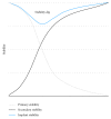
Figure 1
Figure 1
Mechanical stability of a dental implant after insertion. Primary stability decreases subsequently to implant insertion while secondary stability increases. After 2-3 weeks, the implant stability is the lowest in a phase called implant stability dip.
Figure 2
Figure 2
Roxolid implant with SLA surface (Straumann Holding AG, Basel, Switzerland). Roxolid dental implants (a) are made of titanium zirconium alloy. Large grit-blasting generates the macrolevel aspects of the surface (b), while the microtopographic features (c) are induced by acid-etching with HCl/H2SO4. Courtesy of Straumann Holding AG.
Figure 3
Figure 3
FRIADENT plus surface (DENTSPLY Implants, Mannheim, Germany). The surface of the FRIADENT plus surface (a) is created by large grit-blasting, etching, and a proprietary neutralizing technique. The hydrophilic surface features grooves that are interspersed with micropores (b). Courtesy of DENTSPLY Implants.
Figure 4
Figure 4
3i T3 (BIOMET 3i, Palm Beach Gardens, FL, USA). Small calcium phosphate particles are deposited on a double acid-etched surface in 3i dental implants (a). These particles are 20–100 nm (c) in size and form about half of the implant's total surface (b). Courtesy of BIOMET 3i.
Figure 5
Figure 5
Laser-Lok implant (BioHorizons, Birmingham, AL, USA). A pattern of microchannels around the implant collar (b) is created by laser ablation. These cell-sized microchannels have been shown to act as a biological seal around the implant by fostering the attachment of connective tissue (c). Courtesy of BioHorizons IPH Inc.
Figure 6
Figure 6
TiUnite surface (Nobel Biocare Holding AG, Zürich, Switzerland). The NobelReplace dental implant (a) is equipped with the TiUnite surface. The porous microstructure of the surface (b) has been suggested to promote osseointegration by providing additional retention in bone formation (c). Courtesy of Nobel Biocare.
Figure 7
Figure 7
OsseoSpeed implant (DENTSPLY Implants, Mannheim, Germany). The nanolevel aspect (c) of the OsseoSpeed dental implant (a, b) is the result of titanium oxide blasting followed by etching with hydrofluoric acid. Accumulation of fluoride on the surface is a beneficial side effect of the manufacturing process. Courtesy of DENTSPLY Implants.
Figure 8
Figure 8
Concept of hydrophilicity. The hydrophilic surface on the left exhibits a water contact angle α < 90°, whereas the hydrophobic surface on the right shows a contact angle of β > 90°.
Figure 9
Figure 9
SLActive (Straumann Holding AG, Basel, Switzerland). In SLActive dental implants, the degree of hydrophilicity has been enhanced by rinsing under nitrogen protection and storage in saline solution. The SLActive surface (a, b) possesses elements of nanotopography. Courtesy of Straumann Holding AG.
Figure 10
Figure 10
Hydrophilic effects of UV treatment. Pure titanium discs were subjected to photofunctionalization using UV light. Droplets of water (20 μL) were placed on untreated (a) and photofunctionalized discs (b). The water contact angle is drastically decreased by UV treatment (b), illustrating the hydrophilic effects of photofunctionalization. Courtesy of Henningsen.

Similar articles

Cited by

References

    1. Brånemark P.-I., Adell R., Breine U., Hansson B. O., Lindström J., Ohlsson A. Intra-osseous anchorage of dental prostheses: I. Experimental studies . Scandinavian Journal of Plastic and Reconstructive Surgery. 1969;3(2):81–100. doi: 10.3109/02844316909036699. - DOI - PubMed
    1. Brånemark P. I., Adell R., Albrektsson T., Lekholm U., Lundkvist S., Rockler B. Osseointegrated titanium fixtures in the treatment of edentulousness. Biomaterials. 1983;4(1):25–28. doi: 10.1016/0142-9612(83)90065-0. - DOI - PubMed
    1. Junker R., Dimakis A., Thoneick M., Jansen J. A. Effects of implant surface coatings and composition on bone integration: a systematic review. Clinical Oral Implants Research. 2009;20(supplement 4):185–206. doi: 10.1111/j.1600-0501.2009.01777.x. - DOI - PubMed
    1. Esposito M., Ardebili Y., Worthington H. V. Interventions for replacing missing teeth: different types of dental implants. The Cochrane Database of Systematic Reviews. 2014;7 doi: 10.1002/14651858.cd003815.pub4.CD003815 - DOI - PubMed
    1. Dohan Ehrenfest D. M., Coelho P. G., Kang B.-S., Sul Y.-T., Albrektsson T. Classification of osseointegrated implant surfaces: materials, chemistry and topography. Trends in Biotechnology. 2010;28(4):198–206. doi: 10.1016/j.tibtech.2009.12.003. - DOI - PubMed

Substances

LinkOut - more resources