. 2016:2016:9345495.
doi: 10.1155/2016/9345495. Epub 2016 Jul 13.

Thrombin Cleavage of Osteopontin Modulates Its Activities in Human Cells In Vitro and Mouse Experimental Autoimmune Encephalomyelitis In Vivo

Affiliations

Thrombin Cleavage of Osteopontin Modulates Its Activities in Human Cells In Vitro and Mouse Experimental Autoimmune Encephalomyelitis In Vivo

Elena Boggio et al. J Immunol Res. 2016.

Abstract

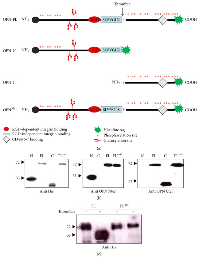
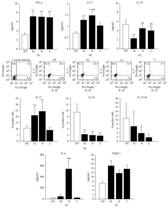
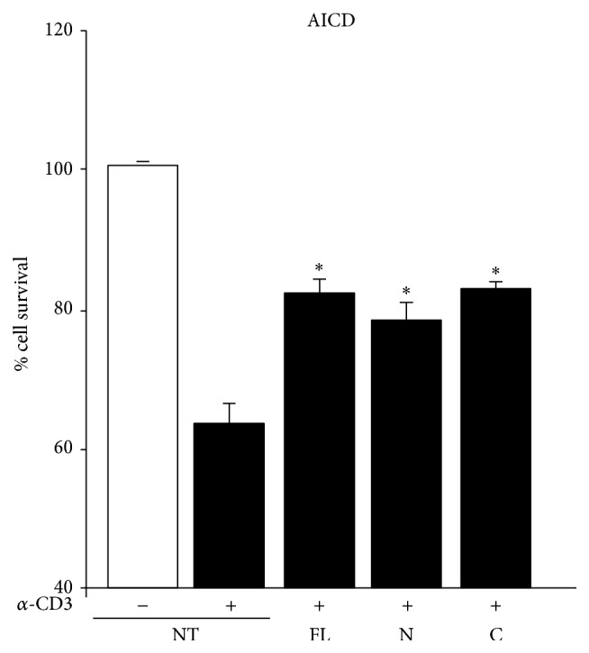
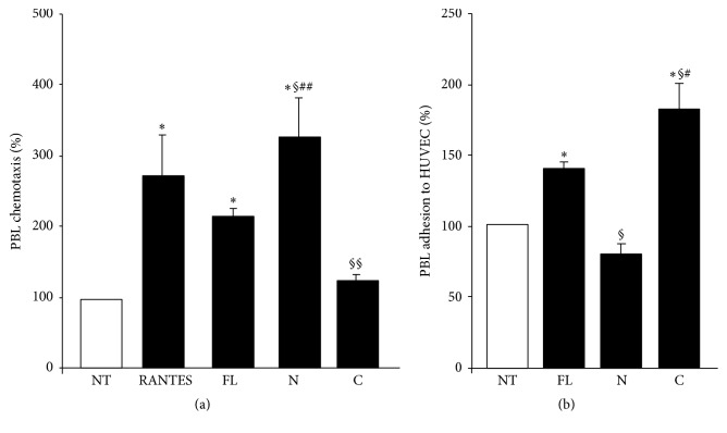
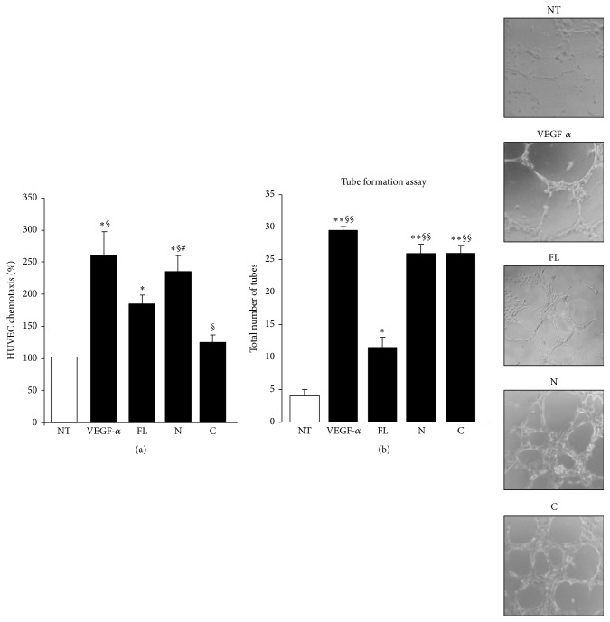
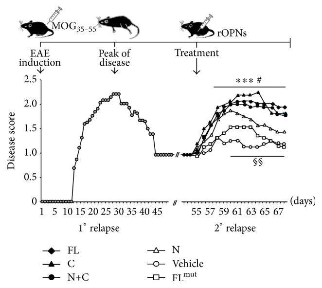
Osteopontin is a proinflammatory cytokine and plays a pathogenetic role in multiple sclerosis and its animal model, experimental autoimmune encephalomyelitis (EAE), by recruiting autoreactive T cells into the central nervous system. Osteopontin functions are modulated by thrombin cleavage generating N- and C-terminal fragment, whose individual roles are only partly known. Published data are difficult to compare since they have been obtained with heterogeneous approaches. Interestingly, thrombin cleavage of osteopontin unmasks a cryptic domain of interaction with α 4 β 1 integrin that is the main adhesion molecule involved in lymphocyte transmigration to the brain and is the target for natalizumab, the most potent drug preventing relapses. We produced recombinant osteopontin and its N- and C-terminal fragments in an eukaryotic system in order to allow their posttranslational modifications. We investigated, in vitro, their effect on human cells and in vivo in EAE. We found that the osteopontin cleavage plays a key role in the function of this cytokine and that the two fragments exert distinct effects both in vitro and in vivo. These findings suggest that drugs targeting each fragment may be used to fine-tune the pathological effects of osteopontin in several diseases.


Figures

Figure 1
Figure 1
Recombinant OPN variants. (a) The figure depicts the recombinant OPN variants: OPN-FL (aa 17–314 human and aa 17–294 mouse), OPN-N including aa 17–168 (human) or 17–153 (mouse) of OPN; OPN-C including aa 169–314 (human) or 154–294 of OPN; mouse OPN-FLmut carrying a mutated thrombin cleavage site (from R153-S154 to S153-F154). (b) Western blotting showing the recombinant proteins after purification probed with the anti-His-tag (left panel) or antibodies specific for the N (middle panel) or C-terminal portion (right panel). (c) OPN-FL but not OPN-FLmut is cleaved by thrombin.
Figure 2
Figure 2
Effect of OPN fragments on cytokine secretion. (a) IFN-γ, (b) IL-17A, and (c) IL-10 protein evaluated in the culture supernatants from CD4+ T cells by ELISA or (d) by intracellular staining with anti-IL-17A and anti-IL-10 after 5 days of treatment with OPN variants. (e) IL-6 and (f) TIMP-1 protein secreted by monocytes after 2 days of treatment with OPN variants. Data are expressed as the mean ± SE from 6 independent experiments ( p < 0.05; ∗∗ p ≤ 0.01 versus the control; § p < 0.05; §§ p ≤ 0.01 versus OPN-FL; # p < 0.05; ## p ≤ 0.01 versus OPN-C; Wilcoxon's signed rank test).
Figure 3
Figure 3
Effect of OPN variants on AICD of T cells. AICD was induced in PHA-derived T cell lines from healthy controls in the presence/absence of anti-CD3 and OPN variants. Results are expressed as relative cell survival % and are the mean ± SEM from 6 experiments. ( p < 0.05 versus the control; Wilcoxon's signed rank test).
Figure 4
Figure 4
Effect of OPN variants on PBL migration and adhesion. (a) PBL were plated onto the apical side of matrigel-coated filters in 50 μL of medium in the presence or absence of either 10 μg/mL OPN-FL, 5 μg/mL OPN-N, or OPN-C; RANTES (10 ng/mL) was loaded in the basolateral chamber as a positive control for migration. The cells that migrated to the bottom of the filters were stained using crystal violet and counted (5 fields for each triplicate filter) using an inverted microscope. (b) PBL were pretreated or not with OPN-FL, OPN-N, or OPN-C (10 μg/mL) for 30 min, washed, and then incubated together for 1 h in the adhesion assay. Data are expressed as the mean ± SEM of the percentage of migration or adhesion versus the control obtained from untreated cells set at 100% from 5 independent experiments. ( p < 0.05; versus the control; § p < 0.05; §§ p ≤ 0.01 versus OPN-FL; # p < 0.05; ## p ≤ 0.01 versus OPN-C or OPN-N; Wilcoxon's signed rank test).
Figure 5
Figure 5
Effect of OPN variants on angiogenesis. (a) HUVECs were plated onto the apical side of matrigel-coated filters in 50 μL of medium in the presence or absence of either 10 μg/mL OPN-FL, 5 μg/mL OPN-N, or OPN-C; VEGF-α (10 ng/mL) was loaded in the basolateral chamber as a positive control for migration. The cells that migrated to the bottom of the filters were stained using crystal violet and counted (5 fields for each triplicate filter) using an inverted microscope. Results are expressed as in Figure 4. (b) In the tube formation assay, HUVECs were plated in the presence and absence of OPN-FL (10 μg/mL), OPN-N (5 μg/mL), OPN-C (5 μg/mL), or VEGF-α (10 ng/mL), as a control. The morphology of capillary-like structures formed by HUVECs was analyzed 6 h after culturing. Results are expressed as means ± SEM from 3 experiments ( p < 0.05; ∗∗ p ≤ 0.01 versus the control; § p < 0.05; §§ p ≤ 0.01 versus OPN-FL; # p < 0.05; versus OPN-C; Wilcoxon's signed rank test). Right panels show a representative tubulogenesis experiment.
Figure 6
Figure 6
Effect of different forms of OPN on the EAE remission phase. The upper schema depicts the timing of the experiment: n = 48 mice were immunized at day 0 with MOG35–55 peptide to induce EAE. The mice were examined daily for clinical signs of EAE and scored as reported in the lower panel (grey circles). Twenty days after the remission, at day 55 after EAE induction, mice were randomized into different experimental groups and received daily injection of either OPN-FL (black diamonds), OPN-FLmut (white squares), OPN-N (white triangles), OPN-C (black triangles), OPN-C + OPN-N (black circles), or vehicle (white circles). A nonparametric ANOVA test was used for clinical score comparisons (∗∗∗ p < 0.001 PBS versus OPN-FL, OPN-C, and OPN-N + OPN-C; # p < 0.05 PBS versus OPN-N; §§ p < 0.01 OPN-C versus OPN-N and versus OPN-FLmut).

References

    1. Cho H.-J., Cho H.-J., Kim H.-S. Osteopontin: a multifunctional protein at the crossroads of inflammation, atherosclerosis, and vascular calcification. Current Atherosclerosis Reports. 2009;11(3):206–213. doi: 10.1007/s11883-009-0032-8. - DOI - PubMed
    1. Liu J., Liu Q., Wan Y., et al. Osteopontin promotes the progression of gastric cancer through the NF-κB pathway regulated by the MAPK and PI3K. International Journal of Oncology. 2014;45(1):282–290. doi: 10.3892/ijo.2014.2393. - DOI - PubMed
    1. Brown A. Osteopontin: a key link between immunity, inflammation and the central nervous system. Translational Neuroscience. 2012;3(3):288–293. doi: 10.2478/s13380-012-0028-7. - DOI - PMC - PubMed
    1. Chen G., Zhang X., Li R., et al. Role of osteopontin in synovial Th17 differentiation in rheumatoid arthritis. Arthritis and Rheumatism. 2010;62(10):2900–2908. doi: 10.1002/art.27603. - DOI - PubMed
    1. Santamaría M. H., Corral R. S. Osteopontin-dependent regulation of Th1 and Th17 cytokine responses in Trypanosoma cruzi-infected C57BL/6 mice. Cytokine. 2013;61(2):491–498. doi: 10.1016/j.cyto.2012.10.027. - DOI - PubMed

MeSH terms