. 2016 Aug 1:3:160064.
doi: 10.1038/sdata.2016.64.

Coherent diffraction of single Rice Dwarf virus particles using hard X-rays at the Linac Coherent Light Source

Affiliations

Coherent diffraction of single Rice Dwarf virus particles using hard X-rays at the Linac Coherent Light Source

Anna Munke et al. Sci Data. .

Abstract

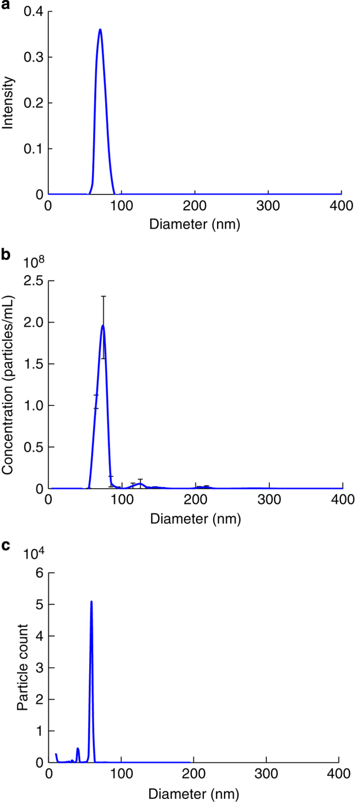
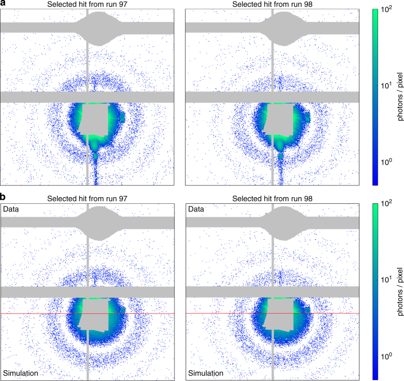
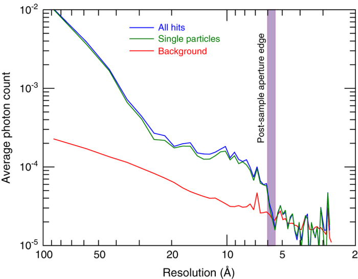
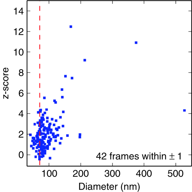
Single particle diffractive imaging data from Rice Dwarf Virus (RDV) were recorded using the Coherent X-ray Imaging (CXI) instrument at the Linac Coherent Light Source (LCLS). RDV was chosen as it is a well-characterized model system, useful for proof-of-principle experiments, system optimization and algorithm development. RDV, an icosahedral virus of about 70 nm in diameter, was aerosolized and injected into the approximately 0.1 μm diameter focused hard X-ray beam at the CXI instrument of LCLS. Diffraction patterns from RDV with signal to 5.9 Ångström were recorded. The diffraction data are available through the Coherent X-ray Imaging Data Bank (CXIDB) as a resource for algorithm development, the contents of which are described here.


Conflict of interest statement

The authors declare no competing financial interests.

Figures

Figure 1
Figure 1. Experimental design.
(1) As the first step in the experiment, an analysis of candidate samples was carried out and a primary target (Rice Dwarf Virus, RDV) was selected. RDV was purified from grasshopper nymphs, which were fed infected rice plants as described in the text. (2) Purified virus particles were then injected into the X-ray beam of the LCLS and diffraction patterns were recorded on the front and back detectors of the CXI instrument. (3) The raw data were pre-processed using psana and converted to XTC files. 175 frames of strong hits were selected and converted into the CXI file form.
Figure 2
Figure 2. The purity of RDV was analyzed with DLS, NTA and DMA.
(a), Size distribution determined with DLS. The average size of the RDV particles was approximately 76 nm. (b), Size distribution determined with NTA. The mean and mode of the RDV particles were approximately 76 and 73 nm, respectively. (c), DMA measurement of RDV gave an apparent particle diameter of approximately 60 nm. Here the actual gas flow was lower than the set value, which resulted in an under estimation of the particle size by about 10%. The main purpose of the DMA measurement was to assess sample purity and the results show that.
Figure 3
Figure 3. Background image of the back detector, averaged from 1000 non-hits and non-dark frames.
The grey areas are masked out. They correspond to the beamstop, the gap between sensors, and a shadow mask of an additional beamstop from the beamstop holder. Unbonded pixels that do not read out signal, are likewise masked out.
Figure 4
Figure 4. Manifold of raw back detector data in two dimensions.
Each point represents a diffraction pattern. The axes are orthogonal coordinates provided by the manifold embedding algorithm.
Figure 5
Figure 5. Measured and simulated diffraction patterns of single RDV particles.
(a), Two hits on the back detector, selected on the expected size for RDV and high diffraction intensity. (b), Measured and simulated data combined. The top half of each of the two patterns shows the measured signal and is identical to the top halves of the patterns in (a). The bottom half of each pattern shows simulated diffraction data from a homogenous sphere of size 71 nm and with a mass density of 1.381 gcm−3. The simulation assumes a photon energy of 7 keV, a detector distance of 2.4 m, a pixel size of 110 microns and a conversion of 33 ADUs per photon. Regions of beam-stops and gaps between the detector panels are masked in grey.
Figure 6
Figure 6. Radial average of signal on the front detector from blank frames compared to radial average from frames determined to be hits.
Elevated photon counts from the sample are visible up to an angle commensurate with 5.9 Å resolution, this being the resolution limit set by the angular acceptance of the post-sample aperture. ‘All hits’ is the average of all hits and includes all particles independent of size (including clusters of particles), while ‘single particle’ is the average of hits that are of the appropriate size to be isolated single particles.
Figure 7
Figure 7. Front detector normalized surprise (z-score) versus back detector particle size fits.
The dashed red line indicates the diameter (70.8 nm) of the RDV model. The normalized surprise function, or its z-score, measures the agreement of the data with a known model: The data are inconsistent with the model when the absolute value of the z-score is much greater than unity: a z-score much greater than unity is consistent with the data being ‘surprising’ given the assumed model.

Comment in

References

Data Citations

    1. Munke A. 2016. Figshare. http://dx.doi.org/10.6084/m9.figshare.c.2342581 - DOI
    1. Munke A. 2016. Coherent X-ray Imaging Data Bank. http://dx.doi.org/10.11577/1252456 - DOI

References

    1. Henderson R. The potential and limitations of neutrons, electrons and X-rays for atomic resolution microscopy of unstained biological molecules. Q. Rev. Biophys. 28, 171–193 (1995). - PubMed
    1. Neutze R., Wouts R., van der Spoel D., Weckert E. & Hajdu J. Potential for biomolecular imaging with femtosecond X-ray pulses. Nature 406, 752–757 (2000). - PubMed
    1. Howells M. R. et al. An assessment of the resolution limitation due to radiation-damage in X-ray diffraction microscopy. J. Electron Spectros. Relat. Phenomena 170, 4–12 (2009). - PMC - PubMed
    1. Chapman H. N. et al. Femtosecond diffractive imaging with a soft-X-ray free-electron laser. Nature Phys 2, 839–843 (2006).
    1. Seibert M. M. et al. Single mimivirus particles intercepted and imaged with an X-ray laser. Nature 470 78–81 (2011). - PMC - PubMed

Publication types

LinkOut - more resources