Comparative Study
. 2016 Aug 1;11(8):e0160208.
doi: 10.1371/journal.pone.0160208. eCollection 2016.

Pathological Investigation of Congenital Bicuspid Aortic Valve Stenosis, Compared with Atherosclerotic Tricuspid Aortic Valve Stenosis and Congenital Bicuspid Aortic Valve Regurgitation

Affiliations
Comparative Study

Pathological Investigation of Congenital Bicuspid Aortic Valve Stenosis, Compared with Atherosclerotic Tricuspid Aortic Valve Stenosis and Congenital Bicuspid Aortic Valve Regurgitation

Yasuhiro Hamatani et al. PLoS One. .

Abstract

Background: Congenital bicuspid aortic valve (CBAV) is the main cause of aortic stenosis (AS) in young adults. However, the histopathological features of AS in patients with CBAV have not been fully investigated.

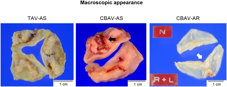
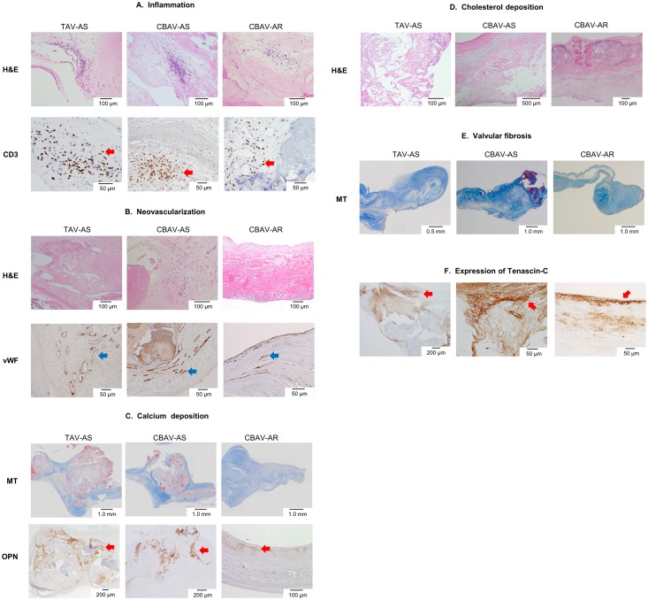
Methods and results: We examined specimens of aortic valve leaflets obtained from patients who had undergone aortic valve re/placement at our institution for severe AS with CBAV (n = 24, CBAV-AS group), severe AS with tricuspid aortic valve (n = 24, TAV-AS group), and severe aortic regurgitation (AR) with CBAV (n = 24, CBAV-AR group). We compared the histopathological features among the three groups. Pathological features were classified using semi-quantitative methods (graded on a scale 0 to 3) by experienced pathologists without knowledge of the patients' backgrounds. The severity of inflammation, neovascularization, and calcium and cholesterol deposition did not differ between the CBAV-AS and TAV-AS groups, and these four parameters were less marked in the CBAV-AR group than in the CBAV-AS (all p<0.01). Meanwhile, the grade of valvular fibrosis was greater in the CBAV-AS group, compared with the TAV-AS and CBAV-AR groups (both p<0.01). In AS patients, thickness of fibrotic lesions was greater on the aortic side than on the ventricular side (both p<0.01). Meanwhile, thickness of fibrotic lesions was comparable between the aortic and ventricular sides in CBAV-AR patients (p = 0.35).

Conclusions: Valvular fibrosis, especially on the aortic side, was greater in patients with CBAV-AS than in those without, suggesting a difference in the pathogenesis of AS between CBAV and TAV.


Conflict of interest statement

Competing Interests: The authors have declared that no competing interests exist.

Figures

Fig 1
Fig 1. Macroscopic appearances of representative excised aortic valves for aortic stenosis and regurgitation.
Tricuspid aortic valve stenosis (TAV-AS); Severe calcified sclerotic tricuspid valve with fused commissures is seen. Congenital bicuspid aortic valve stenosis (CBAV-AS); Two calcified cusps with raphe (black arrow) and severe fibrous thickening are seen. Congenital bicuspid aortic valve regurgitation (CBAV-AR); R (right coronary cusp) and L (left coronary cusp) are fused. Coaptation sites are rather thicker than the other portions (white arrow).
Fig 2
Fig 2. Histological and immunohistochemical findings of aortic valve in TAV-AS, CBAV-AS, and CBAV-AR.
A: Inflammation; Inflammation with CD3 is seen in each group. More inflammatory cell infiltration is noted in TAV-AS and CBAV-AS compared with CBAV-AR. Red arrows indicate T cells infiltrations in valves. Upper photomicrographs are hematoxylin-eosin (H&E) staining, and lower photomicrographs are immunohistochemical staining for CD3. B: Neovascularization; Neovascularizations in TAV-AS and CBAV-AS are more intensive than those in CBAV-AR. Upper photomicrographs are H&E staining, and lower photomicrographs are immunohistochemical staining for von Willebrand factor (vWF). Blue arrows indicate the expression of vWF. C: Calcium deposition; The degrees of calcium deposition are higher in TAV-AS and CBAV-AS compared with CBAV-AR. Upper photomicrographs are Masson’s trichrome (MT) staining, and lower photomicrographs are immunohistochemical staining for osteopontin (OPN). Red arrows indicate the expression of osteopontin. D: Cholesterol deposition; The degrees of cholesterol deposition are higher in TAV-AS and CBAV-AS compared with CBAV-AR. Photomicrographs are H&E staining. E: Fibrosis; Valvular fibrosis is more severely noted in CBAV-AS compared with TAV-AS and CBAV-AR. Photomicrographs are MT staining. F: Immunohistochemical staining for tenascin-C. Red arrows indicate the expression of tenascin-C. Tenascin-C deposit is more diffuse and intensive in CBAV-AS valve.

References

    1. Furukawa H, Tanemoto K. Current Topics on Bicuspid Aortic Valve: Clinical Aspects and Surgical Management. Ann Thorac Cardiovasc Surg. 2015. August 20;21(4):314–21. 10.5761/atcs.ra.15-00130 - DOI - PMC - PubMed
    1. Ward C. Clinical significance of the bicuspid aortic valve. Heart. 2000. January;83(1):81–5. - PMC - PubMed
    1. Siu SC, Silversides CK. Bicuspid aortic valve disease. J Am Coll Cardiol. 2010. June 22;55(25):2789–800. 10.1016/j.jacc.2009.12.068 - DOI - PubMed
    1. Goldbarg SH, Elmariah S, Miller MA, Fuster V. Insights into degenerative aortic valve disease. J Am Coll Cardiol. 2007. September 25;50(13):1205–13. - PubMed
    1. Mohler ER 3rd, Gannon F, Reynolds C, Zimmerman R, Keane MG, Kaplan FS. Bone formation and inflammation in cardiac valves. Circulation. 2001. March 20;103(11):1522–8. - PubMed

Publication types