

Characterization of the Methylthioadenosine Phosphorylase Polymorphism rs7023954 - Incidence and Effects on Enzymatic Function in Malignant Melanoma
- PMID: 27479139
- PMCID: PMC4968798
- DOI: 10.1371/journal.pone.0160348
Characterization of the Methylthioadenosine Phosphorylase Polymorphism rs7023954 - Incidence and Effects on Enzymatic Function in Malignant Melanoma
Abstract
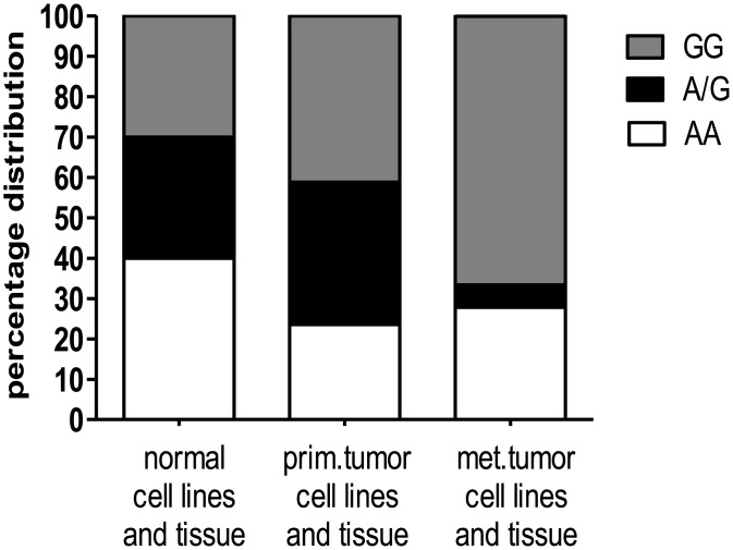
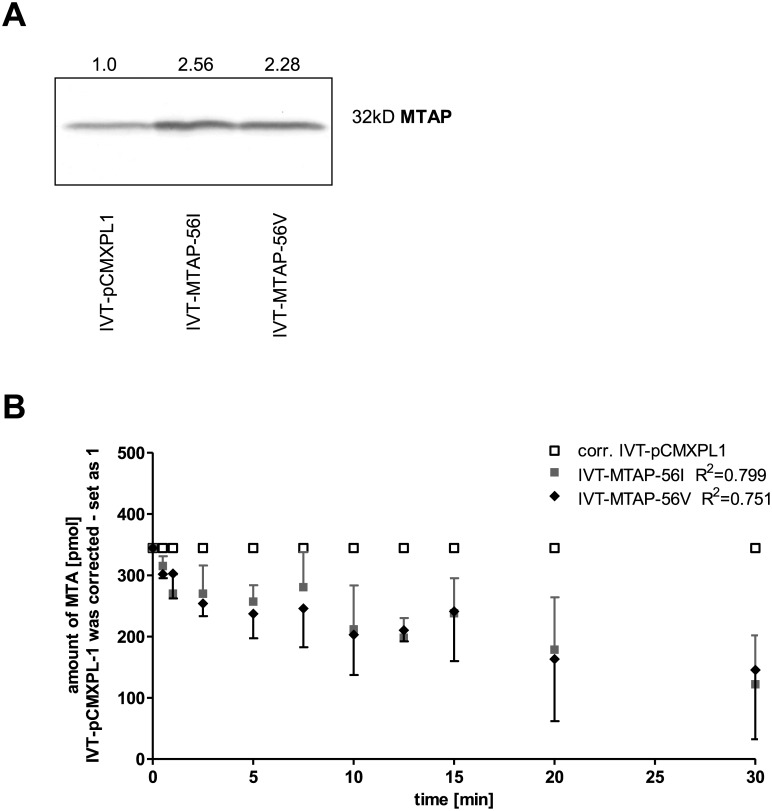
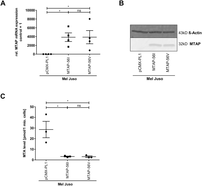
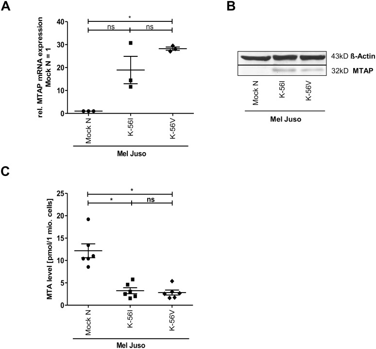
Deficiency of methylthioadenosine phosphorylase (MTAP) supports melanoma development and progression through accumulation of its substrate 5'-methylthioadenosine (MTA), which leads amongst others to a constitutive inhibition of protein arginine methyltransferases (PRMTs) and activation of the transcription factor AP-1 via the receptor ADORA2B. Genetic association studies have also suggested that genetic polymorphism in MTAP may modulate the risk of melanoma. Here, we investigated the only globally common non-synonymous single nucleotide polymorphism (SNP) reported to date for MTAP. The SNP rs7023954 is located in exon 3 (c.166G>A), and leads to the conservative substitution of one branched-chain amino acid residue (valine) for another (isoleucine) at position 56 (p.Val56Ile). Whereas genotype frequencies in normal and primary melanoma tissues or cell lines were in Hardy-Weinberg equilibrium based on cDNA amplicon sequencing, a marked (P = 0.00019) deviation was observed in metastatic melanoma tissues and cell lines due to a deficit of heterozygotes. Enzyme assays conducted on the co-dominantly expressed alleles revealed no difference in the conversion rate of MTA to adenine and 5-methylthioribose-1-phosphate, indicating that this known enzymatic activity does not modulate the tumor suppressive function of MTAP.
Conflict of interest statement
Figures
Similar articles
-
Characterization of methylthioadenosin phosphorylase (MTAP) expression in malignant melanoma.Am J Pathol. 2003 Aug;163(2):683-90. doi: 10.1016/S0002-9440(10)63695-4. Am J Pathol. 2003. PMID: 12875987 Free PMC article.
-
Gene deletion chemoselectivity: codeletion of the genes for p16(INK4), methylthioadenosine phosphorylase, and the alpha- and beta-interferons in human pancreatic cell carcinoma lines and its implications for chemotherapy.Cancer Res. 1996 Mar 1;56(5):1083-90. Cancer Res. 1996. PMID: 8640765
-
Deregulation of protein methylation in melanoma.Eur J Cancer. 2013 Apr;49(6):1305-13. doi: 10.1016/j.ejca.2012.11.026. Epub 2012 Dec 19. Eur J Cancer. 2013. PMID: 23265702
-
Targeting tumors that lack methylthioadenosine phosphorylase (MTAP) activity: current strategies.Cancer Biol Ther. 2011 Apr 1;11(7):627-32. doi: 10.4161/cbt.11.7.14948. Epub 2011 Apr 1. Cancer Biol Ther. 2011. PMID: 21301207 Free PMC article. Review.
-
A potential predictive marker for response to interferon in malignant melanoma.J Dtsch Dermatol Ges. 2007 Jun;5(6):456-9. doi: 10.1111/j.1610-0387.2007.06303.x. J Dtsch Dermatol Ges. 2007. PMID: 17537037 Review. English, German.
Cited by
-
Cold atmospheric plasma causes a calcium influx in melanoma cells triggering CAP-induced senescence.Sci Rep. 2018 Jul 3;8(1):10048. doi: 10.1038/s41598-018-28443-5. Sci Rep. 2018. PMID: 29968804 Free PMC article.
-
Molecular signatures that can be transferred across different omics platforms.Bioinformatics. 2017 Jul 15;33(14):i333-i340. doi: 10.1093/bioinformatics/btx241. Bioinformatics. 2017. PMID: 28881975 Free PMC article.
-
Acidification is an Essential Process of Cold Atmospheric Plasma and Promotes the Anti-Cancer Effect on Malignant Melanoma Cells.Cancers (Basel). 2019 May 14;11(5):671. doi: 10.3390/cancers11050671. Cancers (Basel). 2019. PMID: 31091795 Free PMC article.
References
-
- Kirovski G, Stevens AP, Czech B, Dettmer K, Weiss TS, Wild P, et al. Down-Regulation of Methylthioadenosine Phosphorylase (MTAP) Induces Progression of Hepatocellular Carcinoma via Accumulation of 5′-Deoxy-5′-Methylthioadenosine (MTA). Am J Pathol. 2011;178: 1145–1152. 10.1016/j.ajpath.2010.11.059 - DOI - PMC - PubMed
MeSH terms
Substances
LinkOut - more resources
Full Text Sources
Other Literature Sources
Medical
