. 2016 Sep;18(9):917-29.
doi: 10.1038/ncb3394. Epub 2016 Aug 1.

Snail/Slug binding interactions with YAP/TAZ control skeletal stem cell self-renewal and differentiation

Affiliations

Snail/Slug binding interactions with YAP/TAZ control skeletal stem cell self-renewal and differentiation

Yi Tang et al. Nat Cell Biol. 2016 Sep.

Abstract

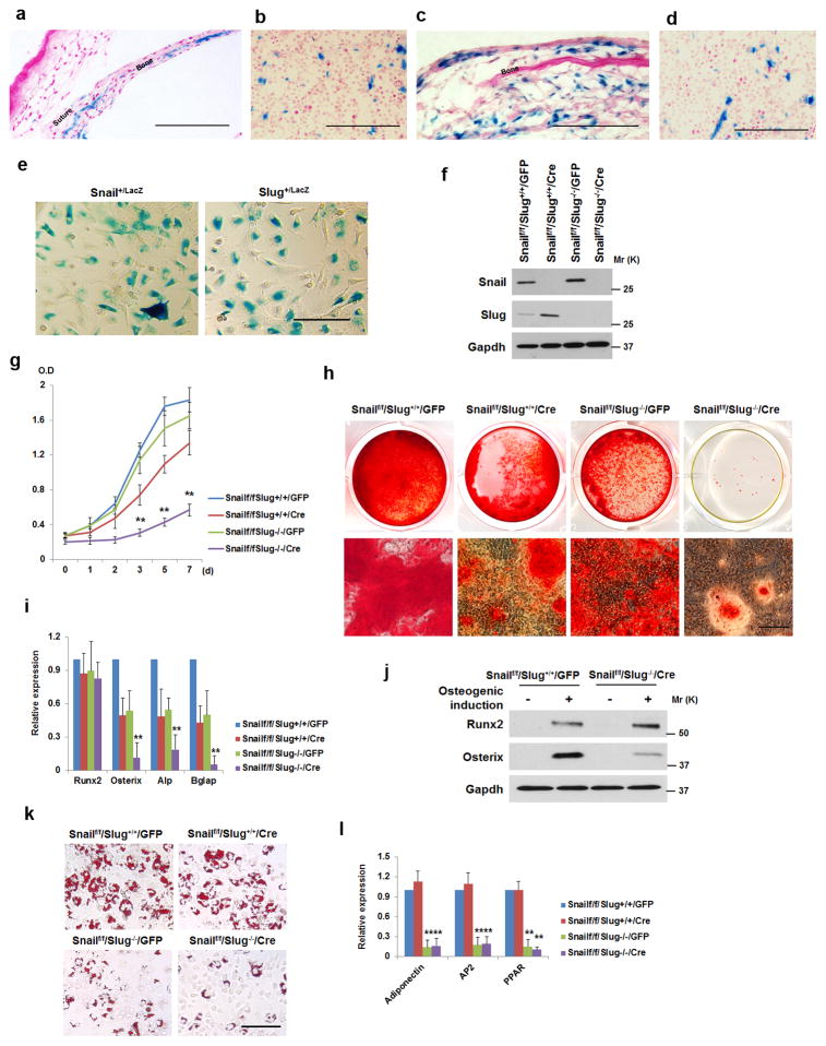
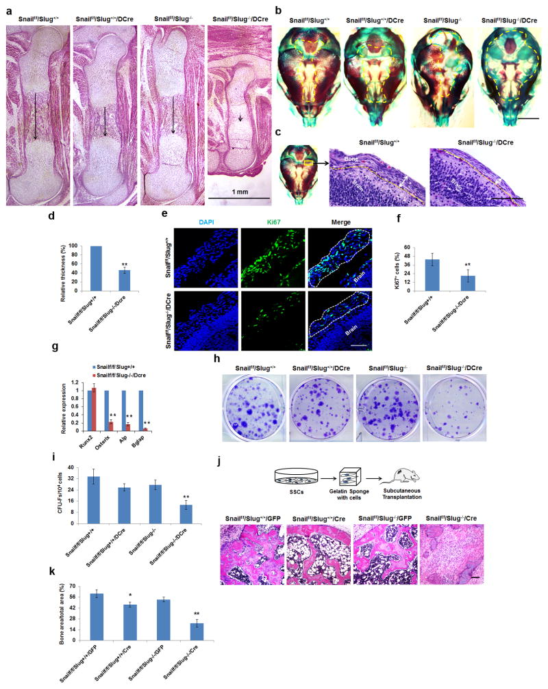
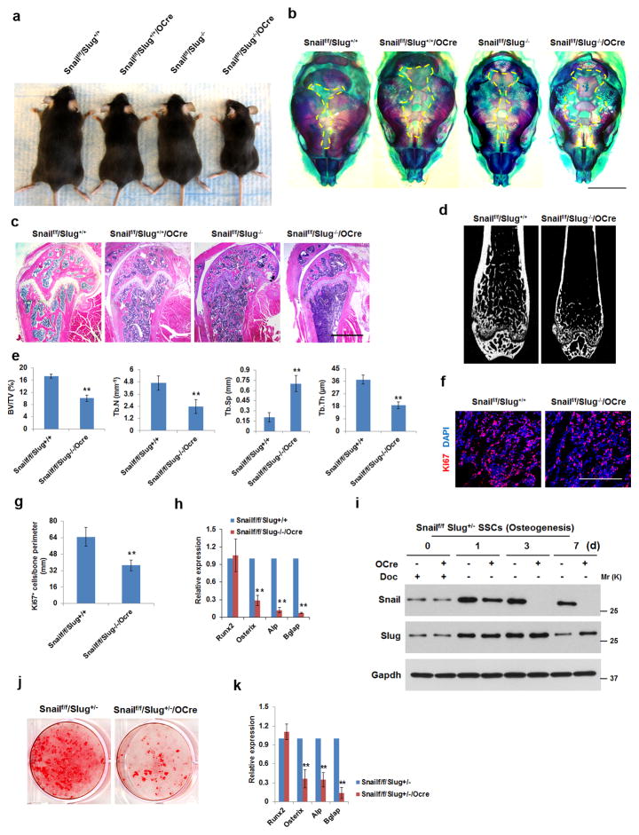
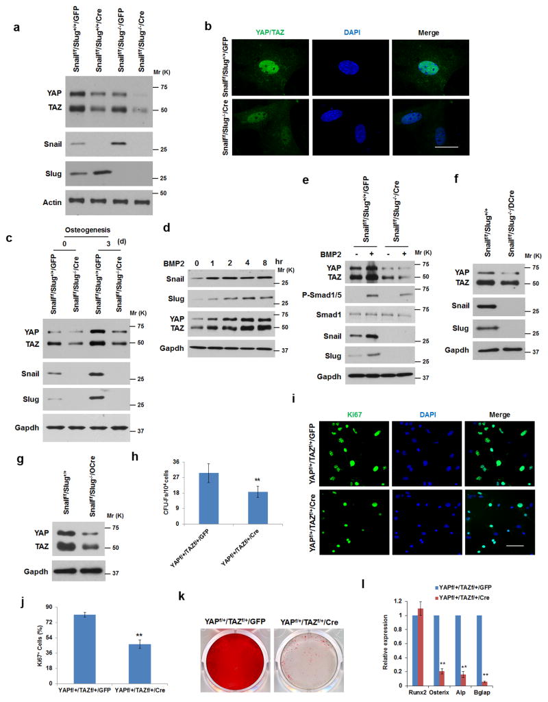
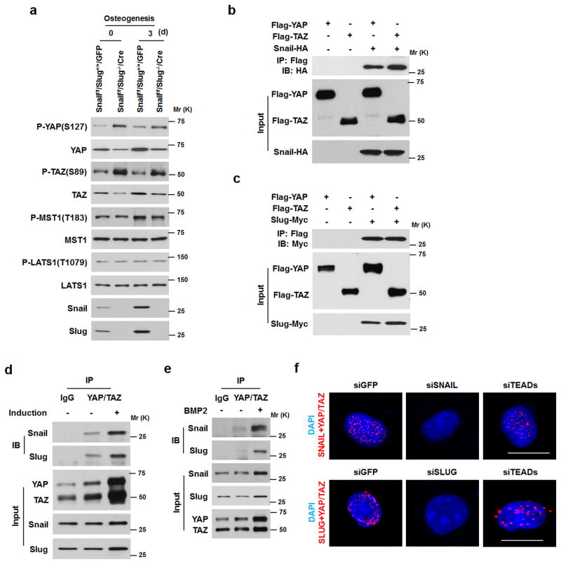
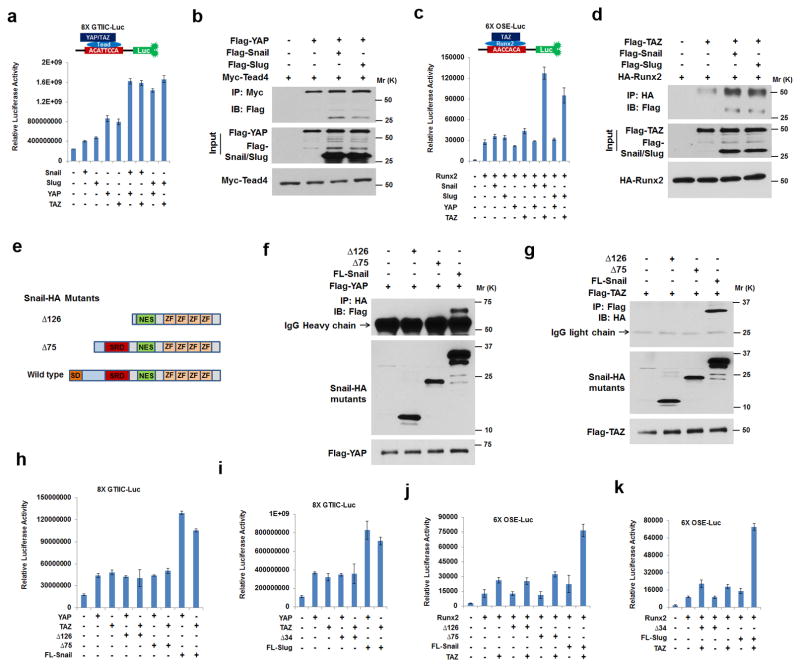
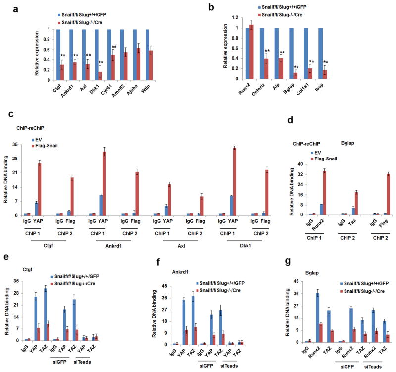
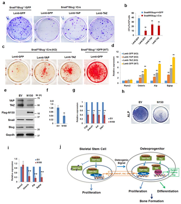
Bone-marrow-derived skeletal stem/stromal cell (SSC) self-renewal and function are critical to skeletal development, homeostasis and repair. Nevertheless, the mechanisms controlling SSC behaviour, particularly bone formation, remain ill-defined. Using knockout mouse models that target the zinc-finger transcription factors Snail or Slug, or Snail and Slug combined, a regulatory axis has been uncovered wherein Snail and Slug cooperatively control SSC self-renewal, osteoblastogenesis and bone formation. Mechanistically, Snail/Slug regulate SSC function by forming complexes with the transcriptional co-activators YAP and TAZ in tandem with the inhibition of the Hippo-pathway-dependent regulation of YAP/TAZ signalling cascades. In turn, the Snail/Slug-YAP/TAZ axis activates a series of YAP/TAZ/TEAD and Runx2 downstream targets that control SSC homeostasis and osteogenesis. Together, these results demonstrate that SSCs mobilize Snail/Slug-YAP/TAZ complexes to control stem cell function.


Figures

Figure 1
Figure 1. Snail/Slug Regulate SSC Proliferation and Differentiation
a) LacZ expression in E17.5 Snail+/LacZ neonatal sagittal suture and parietal bone. Scale bar: 100 μm. Results are representative of 3 experiments performed. b) LacZ expression in a sub-population of bone marrow cells in the femur of a 2-wk old Snail+/LacZ mouse. Scale bar: 100 μm. Results are representative of 3 experiments performed. c) LacZ expression in Slug+/LacZ 7-d old sagittal suture and parietal bone. Scale bar: 100 μm. Results are representative of 3 experiments performed. d) LacZ expression in a sub-population of bone marrow cells in the femur of a 2-wk old Slug+/LacZ mouse. Scale bar: 100 μm. Results are representative of 3 experiments performed. e) LacZ expression in bone marrow-derived SSCs isolated from 4-wk old Snail+/LacZ or Slug+/LacZ mice. Scale bar: 100 μm. Results are representative of 3 experiments performed. f) Western blot of Snail and Slug in SSCs isolated from Snailf/f/Slug+/+ and Snailf/f/Slug−/− mice transduced with adeno-GFP or Cre expression vectors. Results are representative of 3 experiments performed. g) Growth curve of SSCs isolated from Snailf/f/Slug+/+ and Snailf/f/Slug−/− mice, and transduced with adeno-GFP or Cre expression vectors (mean ± s.d. n=3 independent experiments). **p<0.01; one-way ANOVA. h) SSCs were isolated from Snailf/f/Slug+/+ and Snailf/f/Slug−/− mice and transduced with adeno-GFP or Cre. Cells were cultured under osteogenic conditions for 14 d, and stained with Alizarin Red S. Lower panels are magnified images of the upper panels. Scale bar: 100 μm. Results are representative of 3 experiments performed. i) Relative mRNA expression of osteogenic markers in cultures from (h) (mean ± s.d., n=3 independent experiments). **p<0.01; one-way ANOVA. j) SSCs were isolated from Snailf/f/Slug+/+ and Snailf/f/Slug−/− mice and transduced with adeno-GFP or Cre. After culture with or without osteogenic medium for 7 d, cell lysates were immunoblotted. Results are representative of 3 experiments performed. k) SSCs were isolated from Snailf/f/Slug+/+ and Snailf/f/Slug−/− mice and transduced with adeno-GFP or Cre. After culture under adipogenic conditions for 7 d, cultures were stained with Oil Red O. Scale bar: 100 μm. Results are representative of 3 experiments performed. l) Relative mRNA expression of adipogenic markers in cultures from (k) (mean ± s.d., n=3 independent experiments). **p<0.01; one-way ANOVA.
Figure 2
Figure 2. Skeletal and SSC-Associated Defects in Snail/Slug-Targeted Mice
(a) Femur histology of E17.5 Snailf/f/Slug+/+, Snailf/f/Slug+/+/Dermo1-Cre Snailf/f/Slug−/− and Snailf/f/Slug−/−/Dermo1-Cre embryos. Scale bar: 1 mm. Results are representative of 5 experiments performed. (b) Alizarin Red/Alcian Blue staining of skulls isolated from E17.5 Snailf/f/Slug+/+, Snailf/f/Slug+/+/Dermo1-Cre, Snailf/f/Slug−/− and Snailf/f/Slug−/−/Dermo1-Cre embryos. Scale bar: 500 μm. Results are representative of 5 experiments performed. (c) Histology of E15 Snailf/f/Slug+/+ and Snailf/f/Slug+/+/Dermo1-Cre skulls across the parietal area. Scale bar: 100 μm. Results are representative of 5 experiments performed. (d) Relative thickness of skull mesenchymal cell layers from (c) (mean ± s.d., n=5 mice). **p<0.01; unpaired t-test. (e) Ki67 Immunofluorescence of parietal suture mesenchymal cell layers from E15 Snailf/f/Slug+/+ and Snailf/f/Slug−/−/Dermo1-Cre skulls. Scale bar: 25 μm. Results are representative of 4 experiments performed. (f) Quantification of Ki67-positive cells from (e) (mean ± s.d., n=4 mice). **p<0.01; unpaired t-test. (g) Relative mRNA expression of osteogenic markers in E17.5 calvarial RNA extracts from Snailf/f/Slug+/+ versus Snailf/f/Slug−/−/Dermo1-Cre embryos (mean ± s.d., n=5 mice). **p<0.01; unpaired t-test. (h) CFU-F generation from bone marrow cells isolated from E18.5 Snailf/f/Slug+/+, Snailf/f/Slug+/+/Dermo1-Cre, Snailf/f/Slug−/− and Snailf/f/Slug−/−/Dermo1-Cre mice. Results are representative of 5 experiments performed. (i) CFU-F colony counts from (h) (mean ± s.d., n=5 mice). **p<0.01; one-way ANOVA. (j) Schematic of the overall experimental design for in vivo implantation (upper panels).The lower panels show histology of tissues isolated from nude mice transplanted with Snailf/f/Slug+/+/GFP or Snailf/f/Slug−/−/Cre SSCs. Results are representative of 3 performed. Scale bar: 100 μm. (k) Quantification bone formation in tissues from (j) (mean ± s.d., n=3 mice). **p<0.01, *p<0.05; one-way ANOVA.
Figure 3
Figure 3. Osteogenic Defects in Osterix-Cre-Targeted Conditional Knockouts of Snail/Slug
(a) Gross view of 3-month old Snailf/f/Slug+/+/Osterix-Cre, Snailf/f/Slug−/−, Snailf/f/Slug−/−/Osterix-Cre mice and a control littermate (Snailf/f/Slug+/+). Results are representative of 5 experiments performed. (b) Alizarin Red/Alcian Blue staining of skulls isolated from neonatal Snailf/f/Slug+/+, Snailf/f/Slug+/+/Osterix-Cre, Snailf/f/Slug−/− and Snailf/f/Slug−/−/Osterix-Cre mice. Scale bar: 2 mm. Results are representative of 3 experiments performed. (c) Femur histology of 3-month old Snailf/f/Slug+/+, Snailf/f/Slug+/+/Osterix-Cre, Snailf/f/Slug−/− and Snailf/f/Slug−/−/Osterix-Cre mice. Scale bar: 1 mm. Results are representative of 5 experiments performed. (d) Microcomputed tomography of the proximal femur of 12-wk old Snailf/f/Slug+/+ and Snailf/f/Slug−/−/Osterix-Cre mice. Results are representative of 10 experiments performed. (e) Bone Mineral Density (BMD), Bone Volume/Tissue Volume (BV/TV), Trabecular Thickness (Tb.Th), Trabecular Number (Tb.N) and Trabecular Separation (Tb.Sp) were measured by microcomputed tomography in 12-wk old Snailf/f/Slug+/+ and Snailf/f/Slug−/−/Osterix-Cre mice (mean ± s.d., n=10 mice). **p<0.01; unpaired t-test. (f) Ki67 expression in proximal femurs isolated from neonatal Snailf/f/Slug+/+ and Snailf/f/Slug−/−/Osterix-Cre mice. Scale bar: 100 μm. Results are representative of 5 experiments performed. (g) Quantification of Ki67-positive cells from (f) (mean ± s.d., n=5 mice). **p<0.01; unpaired t-test. (h) Relative mRNA expression of osteogenic markers in neonatal calvarial RNA extracts from Snailf/f/Slug+/+ and Snailf/f/Slug−/−/Osterix-Cre mice (mean ± s.d., n=5 mice). **p<0.01; unpaired t-test. (i) Western blot of Snail and Slug expression in SSCs isolated from Snailf/f/Slug+/− or Snailf/f/Slug+/−/Osterix-Cre mice cultured with doxycycline (Doc; 1.5 μg/ml) under control or osteogenic conditions for the indicated time periods. Results are representative of 3 experiments performed. (j) SSCs from (i) were cultured under osteogenic conditions for 14 d and stained with Alizarin Red S. Results are representative of 3 experiments performed. (k) Relative mRNA expression of osteogenic markers in cultures from (j) (mean ± s.d., n=3 independent experiments). **p<0.01; unpaired t-test.
Figure 4
Figure 4. Snail/Slug Regulate YAP/TAZ Levels in SSCs
(a) YAP and TAZ expression levels in SSCs isolated from Snailf/f/Slug+/+ and Snailf/f/Slug−/− mice and transduced with adeno-GFP or Cre. Results are representative of 3 experiments performed. (b) YAP and TAZ localization in Snailf/f/Slug+/+ and Snailf/f/Slug−/− SSCs following transduction with adeno-LacZ or Cre. Nuclei were counterstained with DAPI (blue). Scale bar: 10 μm. Results are representative of 3 experiments performed. (c) YAP and TAZ expression levels in Snailf/f/Slug+/+/GFP and Snailf/f/Slug−/−/Cre SSCs cultured under osteogenic conditions as indicated. Results are representative of 3 experiments performed. (d) BMP2 induces Snail/Slug and YAP/TAZ levels in osteoblast progenitors. Calvarial osteoblast progenitors were isolated from E18 mice embryos, treated with 100 ng/ml BMP2 and protein expression monitored by Western blot. Results are representative of 3 experiments performed. (e) Deletion of Snail/Slug in osteoblast progenitors blunts the BMP2-induced up-regulation of YAP/TAZ. Calvarial osteoblast progenitors were isolated from Snailf/f/Slug+/+ and Snailf/f/Slug−/− mice and transduced with adeno-GFP or Cre. Cells were then treated with 100 ng/ml BMP2 for 2 h, and protein expression monitored by Western blot. Results are representative of 3 experiments performed. (f) YAP and TAZ expression levels in E17.5 Snailf/f/Slug+/+ and Snailf/f/Slug−/−/Dermo1-Cre calvarial lysates. Results are representative of 5 experiments performed. (g) YAP and TAZ expression levels in E17.5 Snailf/f/Slug+/+ and Snailf/f/Slug−/−/Osterix-Cre femur lysates. Results are representative of 5 experiments performed. (h) CFU-F colony count from bone marrow cells isolated from YAPf/+/TAZf/+ mice following transduction with lentivirus GFP or Cre expression vectors (mean ± s.d., n=5 independent experiments). **p<0.01; unpaired t-test. (i) Ki67 expression in SSCs isolated from YAPf/+/TAZf/+ mice following transduction with adeno-GFP or Cre. Scale bar: 50 μm. Results are representative of 3 experiments performed. (j) Quantification of Ki67-positive cells from (i) (mean ± s.d., n=3 independent experiments). **p<0.01; unpaired t-test. (k) Alizarin Red S staining of SSCs isolated from YAPf/+/TAZf/+ mice following transduction with adeno-GFP or Cre and cultured under osteogenic conditions for 14 d. Results are representative of 3 experiments performed. (l) Relative mRNA expression of osteogenic markers in cultures from (k) (mean ± s.d., n=3 independent experiments). **p<0.01; unpaired t-test.
Figure 5
Figure 5. Snail/Slug-Dependent Regulation of YAP/TAZ Stability
(a) Western blot of p-YAP, p-TAZ, p-Lats1 and p-Mst1 in SSCs isolated from Snailf/f/Slug+/+ and Snailf/f/Slug−/− mice and transduced with adeno-GFP or Cre. Results are representative of 3 experiments performed. (b) Association of YAP/TAZ with Snail was detected by immunoprecipitation in Cos-1 cells transfected with Flag-YAP, Flag-TAZ and Snail-HA, respectively, as indicated. Results are representative of 3 experiments performed. (c) Association of YAP/TAZ with Slug was detected by immunoprecipitation in Cos-1 cells transfected with Flag-YAP, Flag-TAZ and Slug-Myc, respectively, as indicated. Results are representative of 3 experiments performed. (d) Associations between endogenous YAP/TAZ and Snail/Slug were detected in SSCs cultured in the absence or presence of osteogenic medium. Results are representative of 3 experiments performed. (e) BMP2 increases Snail/Slug-YAP/TAZ complex formation. Calvarial osteoblast progenitors isolated from E17.5 mice were treated with 100 ng/ml BMP2 for 2 h. Endogenous Snail/Slug-YAP/TAZ complexes were detected by immunoprecipitation. Results are representative of 3 experiments performed. (f) In situ proximity ligation assay detection of endogenous Snail/YAP/TAZ and Slug/YAP/TAZ interactions in human SSCs. siRNAs: siGFP, siSNAIL, siSLUG or siTEADs(1–4) were transfected, respectively. Nuclei were counterstained with DAPI (blue). The detected interaction sites are marked by fluorescent dots (red). Scale bar: 5μm. Results are representative of 3 experiments performed.
Figure 6
Figure 6. Mapping of Snail/Slug-YAP/TAZ Binding Interactions and Regulation of Transcriptional Activity
(a) 8XGTIIC luciferase activity was determined in Cos-1 cells co-transfected with YAP, TAZ, Snail or Slug (mean ± s.d., n=3 independent experiments). (b) Snail/Slug binding to YAP/Tead4 complexes. Epitope-tagged Snail, Slug, YAP and Tead4 were co-transfected into Cos-1 cells, lysates immunoprecipitated with anti-Myc antibody (Tead4 tagged with Myc) and immunoblotted. Results are representative of 3 experiments performed. (c) 6XOSE luciferase activity was monitored in Cos-1 cells co-transfected with Runx2, Snail, Slug, YAP or TAZ (mean ± s.d., n=3 independent experiments). (d) Snail/Slug incorporation into TAZ/Runx2 complexes. Epitope-tagged Snail, Slug, YAP or Runx2 were co-transfected into Cos-1 cells, lysates immunoprecipitated with anti-HA antibody (Runx2 tagged with HA) and samples immunoblotted. Results are representative of 3 experiments performed. (e) Schematic of HA-tagged Snail mutants. (f) YAP interacts with the SNAG domain of Snail. Cos-1 cells were co-transfected with the indicated Snail-HA or mutant constructs in tandem with Flag-YAP. Cell lysates were subjected to anti-HA immunoprecipitation, and YAP in the complexes determined by anti-Flag immunoblotting. Results are representative of 3 experiments performed. (g) TAZ interacts with the SNAG domain of Snail. Cos-1 cells were co-transfected with the indicated Snail-HA or mutant constructs in tandem with Flag-TAZ. Cell lysates were subjected to anti-Flag immunoprecipitation, and wild-type Snail or Snail mutants in the complexes assessed following anti-HA immunoblotting. Results are representative of 3 experiments performed. (h) 8XGTIIC luciferase activity was measured in Cos-1 cells following co-transfection with YAP, TAZ, Snail or Snail mutants (mean ± s.d., n=3 independent experiments). (i) 8XGTIIC luciferase activity was determined in Cos-1 cells co-transfected with YAP, TAZ, Slug or Slug mutants (mean ± s.d., n=3 independent experiments). (j) 6XOSE luciferase activity was determined in Cos-1 cells co-transfected with Runx2, TAZ, Snail or Snail mutants (mean ± s.d., n=3 independent experiments). (k) 6XOSE luciferase activity was monitored in Cos-1 cells co-transfected with Runx2, TAZ, Slug or Slug mutants (mean ± s.d., n=3 independent experiments).
Figure 7
Figure 7. Snail/Slug-YAP/TAZ Complexes Regulate Gene Expression
(a) Snail/Slug regulates YAP/TAZ/TEAD targeted genes. mRNA expression of YAP/TAZ/TEAD-direct targets in calvarial osteoblast progenitors isolated from Snail1f/f/Slug+/+ or Snail1f/f/Slug−/− mice and transduced with adeno-GFP or Cre expression vectors (mean ± s.d., n=3 independent experiments). **p<0.01, unpaired t-test. (b) Relative mRNA expression of Runx2 and its downstream genes were assessed in calvarial osteoblast progenitors isolated from Snail1f/f/Slug+/+ or Snail1f/f/Slug−/− mice and transduced with adeno-GFP or Cre expression vectors, cultured under osteogenic conditions (mean ± s.d., n=3 independent experiments). **p<0.01; unpaired t-test. (c) Co-occupation of YAP and Snail in the YAP/TAZ/TEAD targeted promoters. ChIP experiments using anti-YAP antibody were performed in Snail/Slug double null osteoblast progenitors re-expressed with Flag-Snail and then re-ChIPed with anti-Flag antibody. Results are shown as the fold-enrichment relative to IgG IP controls (mean ± s.d., n=3 independent experiments). (d) Co-occupation of Runx2, TAZ and Snail in the Bglap2 promoter. ChIP experiments using anti-Runx2 antibody were performed in Snail/Slug double null osteoblast progenitors re-expressed with Flag-Snail under osteogenic culture condition and re-ChIPed with anti-TAZ or anti-Flag antibody. Results are shown as fold-enrichment relative to IgG IP controls (mean ± s.d., n=3 independent experiments). (e) Snail and Slug increase YAP/TAZ/TEADs binding to the Ctgf promoter. ChIP experiments using anti-YAP or anti-TAZ were performed in SSC lysates isolated from Snailf/f/Slug+/+ and Snailf/f/Slug−/− mice that were transduced with adeno- GFP or Cre, and then transfected with siGFP or siTEADs(1–4). Results are presented as the fold-enrichment relative to IgG IP controls (mean ± s.d., n=3 independent experiments). (f) Snail and Slug increases YAP/TAZ/TEADs binding to the Ankrd1 promoter. ChIP experiments using anti-YAP or anti-TAZ were performed in SSCs isolated from Snailf/f/Slug+/+ and Snailf/f/Slug−/− mice and transduced with adeno-GFP or Cre followed by transfection with siGFP or siTEADS(1–4). Results are presented as fold-enrichment relative to IgG IP controls (mean ± s.d., n=3 independent experiments). (g) Snail and Slug increase Runx2 binding to the Bglap2 promoter. ChIP experiments using anti-Runx2 or anti-TAZ were performed in lysates of calvarial osteoblast progenitors isolated from Snailf/f/Slug+/+ and Snailf/f/Slug−/− mice and transduced with adeno-GFP or Cre followed by transfection with siGFP or siTEADs(1–4). Results are presented as fold-enrichment relative to IgG IP controls (mean ± s.d., n=3 independent experiments).
Figure 8
Figure 8. A Snail/Slug-YAP/TAZ Axis Regulates SSC Function
(a) CFU-F generation from Snailf/f/Slug+/+/GFP or Snailf/f/Slug−/−/Cre SSCs that were transduced with lentiviral GFP, YAP or TAZ constructs. Results are representative of 5 experiments performed. (b) CFU-F colony counts from (a) (mean ± s.d., n=5 independent experiments). **p<0.01; one-way ANOVA. (c) Alizarin Red S staining of Snailf/f/Slug−/−/Cre or Snailf/f/Slug+/+/GFP SSCs transduced with lentiviral GFP, YAP or TAZ, and cultured under osteogenic conditions for 14 d. Results are representative of 3 experiments performed. (d) Relative mRNA expression of osteogenic markers (Runx2, Osterix, Alp or Bglap2) in cultures from (c) (mean ± s.d., n=3 independent experiments). **p<0.01; one-way ANOVA. (e) Immunoblotting of YAP/TAZ, Snail/Slug and mutant Snail (N150). C3H10T1/2 cells were transfected with mutant Snail (N150) or empty vector (EV). Results are representative of 3 experiments performed. (f) Mutant Snail (N150) inhibits proliferation of C3H10T1/2 cells. Proliferative response in cells from (e) were assayed by XTT assay (mean ± s.d., n=3 independent experiments). **p<0.01; unpaired t-test. (g) Relative mRNA expression of YAP/TAZ/TEAD targets was assessed in cells from (e) (mean ± s.d., n=3 independent experiments). **p<0.01; unpaired t-test. (h) Mutant Snail (N150) inhibits osteogenesis of C3H10T1/2 cells. Cells from (e) were cultured under osteogenic conditions for 7 d. Osteogenesis was monitored by ALP staining. (i) Relative mRNA expression of osteogenic markers (Runx2, Osterix, Alp and Bglap2) was assessed in cells from (h) (mean ± s.d., n=3 independent experiments). **p<0.01; unpaired t-test. (j) Schematic model of Snail/Slug in regulating SSC proliferation and osteogenic differentiation.

References

    1. Kfoury Y, Scadden DT. Mesenchymal cell contributions to the stem cell niche. Cell Stem Cell. 2015;16:239–253. - PubMed
    1. Bianco P, Robey PG. Skeletal stem cells. Development. 2015;142:1023–1027. - PMC - PubMed
    1. Batlle R, et al. Snail1 controls TGF-beta responsiveness and differentiation of mesenchymal stem cells. Oncogene. 2013;32:3381–3389. - PMC - PubMed
    1. Barrallo-Gimeno A, Nieto MA. Evolutionary history of the Snail/Scratch superfamily. Trends Genet. 2009;25:248–252. - PubMed
    1. Nieto MA. Epithelial plasticity: a common theme in embryonic and cancer cells. Science. 2013;342:1234850. - PubMed

MeSH terms