

Lipids, oxidized lipids, oxidation-specific epitopes, and Age-related Macular Degeneration
- PMID: 27480216
- PMCID: PMC5280582
- DOI: 10.1016/j.bbalip.2016.07.013
Lipids, oxidized lipids, oxidation-specific epitopes, and Age-related Macular Degeneration
Abstract
Age-related Macular Degeneration (AMD) is the leading cause of blindness among the elderly in western societies. While antioxidant micronutrient treatment is available for intermediate non-neovascular disease, and effective anti-vascular endothelial growth factor treatment is available for neovascular disease, treatment for early AMD is lacking due to an incomplete understanding of the early molecular events. The role of lipids, which accumulate in the macula, and their oxidation, has emerged as an important factor in disease development. These oxidized lipids can either directly contribute to tissue injury or react with amine on proteins to form oxidation-specific epitopes, which can induce an innate immune response. If inadequately neutralized, the inflammatory response from these epitopes can incite tissue injury during disease development. This review explores how the accumulation of lipids, their oxidation, and the ensuing inflammatory response might contribute to the pathogenesis of AMD. This article is part of a Special Issue entitled: Lipid modification and lipid peroxidation products in innate immunity and inflammation edited by Christoph J. Binder .
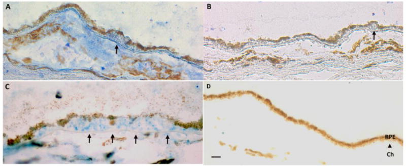
Keywords: Age-related Macular Degeneration; Basal deposits; Bruch's membrane; Drusen; Oxidation-specific epitopes; Retinal pigmented epithelium (RPE); Very low-density lipoprotein (LDL).
Copyright © 2016 Elsevier B.V. All rights reserved.
Conflict of interest statement
Drs. Handa and Cano receive grant funding from Bayer Pharmaceuticals, Inc.
Figures
References
-
- Congdon N, O’Colmain B, Klaver CC, Klein R, Munoz B, Friedman DS, Kempen J, Taylor HR, Mitchell P. Causes and prevalence of visual impairment among adults in the United States. Arch Ophthalmol. 2004;122:477–485. - PubMed
-
- Friedman DS, O’Colmain BJ, Munoz B, Tomany SC, McCarty C, de Jong PT, Nemesure B, Mitchell P, Kempen J. Prevalence of age-related macular degeneration in the United States. Arch Ophthalmol. 2004;122:564–572. - PubMed
-
- Mukesh BN, Dimitrov PN, Leikin S, Wang JJ, Mitchell P, McCarty CA, Taylor HR. Five-year incidence of age-related maculopathy: the Visual Impairment Project. Ophthalmology. 2004;111:1176–1182. - PubMed
Publication types
MeSH terms
Substances
Grants and funding
LinkOut - more resources
Full Text Sources
Other Literature Sources
Medical
