

Immunohistochemical techniques for the human inner ear
- PMID: 27480257
- PMCID: PMC5444466
- DOI: 10.1007/s00418-016-1471-2
Immunohistochemical techniques for the human inner ear
Erratum in
-
Erratum to: Immunohistochemical techniques for the human inner ear.Histochem Cell Biol. 2016 Oct;146(4):389. doi: 10.1007/s00418-016-1491-y. Histochem Cell Biol. 2016. PMID: 27639397 No abstract available.
Abstract
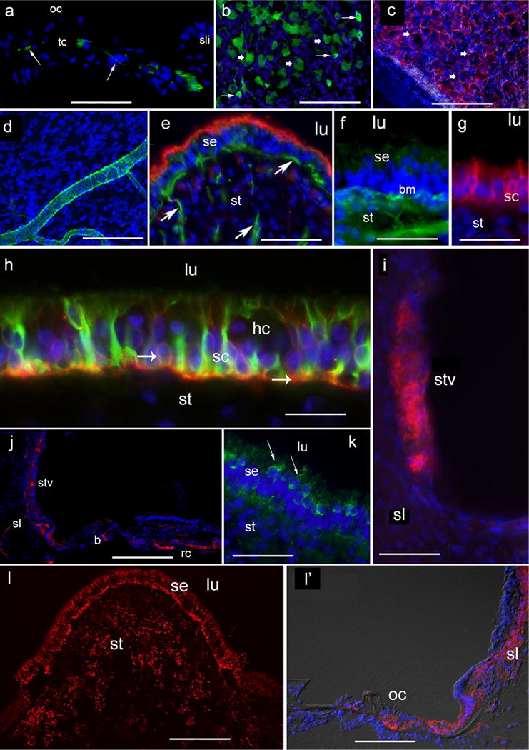
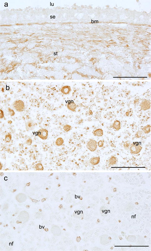
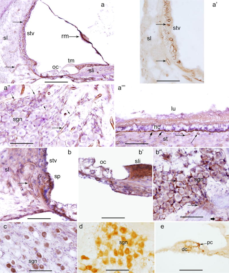
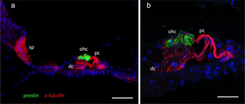
In this review, we provide a description of the recent methods used for immunohistochemical staining of the human inner ear using formalin-fixed frozen, paraffin and celloidin-embedded sections. We also show the application of these immunohistochemical methods in auditory and vestibular endorgans microdissected from the human temporal bone. We compare the advantages and disadvantages of immunohistochemistry (IHC) in the different types of embedding media. IHC in frozen and paraffin-embedded sections yields a robust immunoreactive signal. Both frozen and paraffin sections would be the best alternative in the case where celloidin-embedding technique is not available. IHC in whole endorgans yields excellent results and can be used when desiring to detect regional variations of protein expression in the sensory epithelia. One advantage of microdissection is that the tissue is processed immediately and IHC can be made within 1 week of temporal bone collection. A second advantage of microdissection is the excellent preservation of both morphology and antigenicity. Using celloidin-embedded inner ear sections, we were able to detect several antigens by IHC and immunofluorescence using antigen retrieval methods. These techniques, previously applied only in animal models, allow for the study of numerous important proteins expressed in the human temporal bone potentially opening up a new field for future human inner ear research.
Keywords: Antigen retrieval; Archival human temporal bone; Celloidin-embedded sections; Cochlea; Cryosections; Immunohistochemistry; Microdissection; Paraffin; Vestibule.
Conflict of interest statement
Figures

Similar articles
-
Antigen retrieval immunohistochemistry used for routinely processed celloidin-embedded human temporal bone sections: standardization and development.Auris Nasus Larynx. 1998 Dec;25(4):425-43. doi: 10.1016/s0385-8146(98)00042-x. Auris Nasus Larynx. 1998. PMID: 9853668 Review.
-
Immunohistochemical study of intermediate filament proteins on routinely processed, celloidin-embedded human temporal bone sections by using a new technique for antigen retrieval.Acta Otolaryngol. 1993 Jan;113(1):48-54. doi: 10.3109/00016489309135766. Acta Otolaryngol. 1993. PMID: 7680182
-
S-100 protein in human inner ear: use of a novel immunohistochemical technique on routinely processed, celloidin-embedded human temporal bone sections.Laryngoscope. 1992 Jul;102(7):734-8. doi: 10.1288/00005537-199207000-00002. Laryngoscope. 1992. PMID: 1614244
-
Gene expression analysis of distinct populations of cells isolated from mouse and human inner ear FFPE tissue using laser capture microdissection--a technical report based on preliminary findings.Brain Res. 2006 May 26;1091(1):289-99. doi: 10.1016/j.brainres.2006.01.057. Epub 2006 Mar 10. Brain Res. 2006. PMID: 16529721
-
Antigen retrieval techniques: current perspectives.J Histochem Cytochem. 2001 Aug;49(8):931-7. doi: 10.1177/002215540104900801. J Histochem Cytochem. 2001. PMID: 11457921 Review.
Cited by
-
Single-cell transcriptomic atlas reveals increased regeneration in diseased human inner ear balance organs.Nat Commun. 2024 Jun 6;15(1):4833. doi: 10.1038/s41467-024-48491-y. Nat Commun. 2024. PMID: 38844821 Free PMC article.
-
Mouse Models of Human Pathogenic Variants of TBC1D24 Associated with Non-Syndromic Deafness DFNB86 and DFNA65 and Syndromes Involving Deafness.Genes (Basel). 2020 Sep 24;11(10):1122. doi: 10.3390/genes11101122. Genes (Basel). 2020. PMID: 32987832 Free PMC article.
-
Histology of the Cochlear Outer Sulcus Cells in Normal Human Ears, Presbycusis, and Menière's Disease.Otol Neurotol. 2020 Apr;41(4):e507-e515. doi: 10.1097/MAO.0000000000002535. Otol Neurotol. 2020. PMID: 32176147 Free PMC article.
-
Expression of Brain-Derived Neurotrophic Factor in Human Spiral Ganglia Neurons after Cochlear Implantation.Otol Neurotol. 2024 Mar 1;45(3):326-333. doi: 10.1097/MAO.0000000000004104. Epub 2024 Jan 17. Otol Neurotol. 2024. PMID: 38238917 Free PMC article.
-
Emerging Mechanisms in the Pathogenesis of Menière's Disease: Evidence for the Involvement of Ion Homeostatic or Blood-Labyrinthine Barrier Dysfunction in Human Temporal Bones.Otol Neurotol. 2023 Dec 1;44(10):1057-1065. doi: 10.1097/MAO.0000000000004016. Epub 2023 Sep 20. Otol Neurotol. 2023. PMID: 37733989 Free PMC article.
References
-
- Anniko M, Arnold W, Thornell LE. Localization of the integral membrane glycoprotein synaptophysin and the surface glycoprotein Egp-34 in the embryonic and adult human inner ear ORL. J Otorhinolaryngol Relat Spec. 1989a;51(4):221–228. - PubMed
-
- Anniko M, Thornell LE, Ramaekers FC, Stigbrans T. Cytokeratin diversity in epithelia of the human inner ear. Acta Otolaryngol. 1989b;108(5–6):385–396. - PubMed
-
- Arnold W. Immunohistochemical investigation of the human inner ear. Limitations and prospects. Acta Otolaryngol. 1988;105(5–6):392–397. - PubMed
Publication types
MeSH terms
Grants and funding
LinkOut - more resources
Full Text Sources
Other Literature Sources
