

193 nm Ultraviolet Photodissociation Mass Spectrometry of Tetrameric Protein Complexes Provides Insight into Quaternary and Secondary Protein Topology
- PMID: 27480400
- PMCID: PMC5479422
- DOI: 10.1021/jacs.6b03905
193 nm Ultraviolet Photodissociation Mass Spectrometry of Tetrameric Protein Complexes Provides Insight into Quaternary and Secondary Protein Topology
Abstract
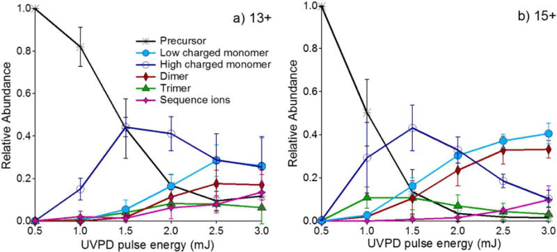
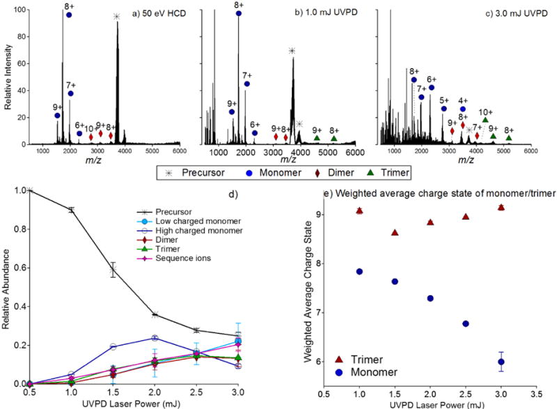
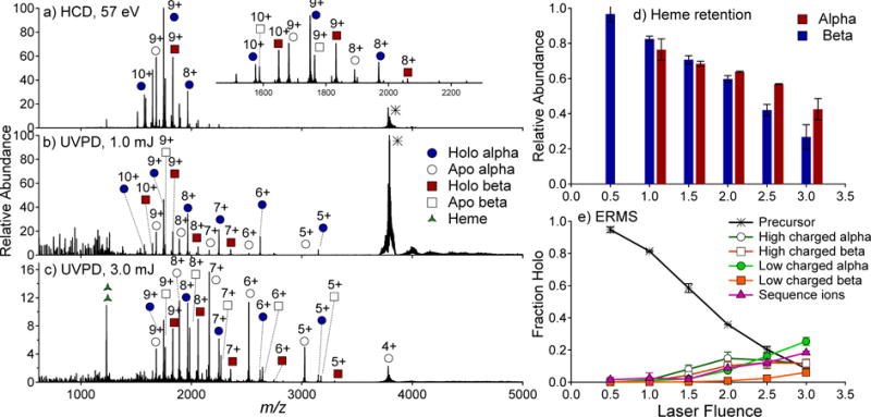
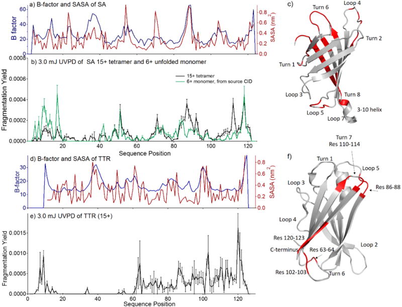
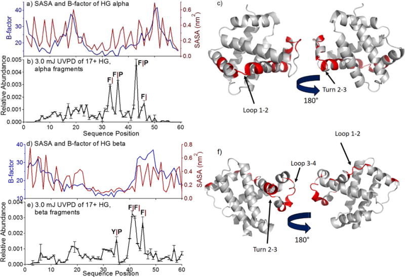
Protein-protein interfaces and architecture are critical to the function of multiprotein complexes. Mass spectrometry-based techniques have emerged as powerful strategies for characterization of protein complexes, particularly for heterogeneous mixtures of structures. In the present study, activation and dissociation of three tetrameric protein complexes (streptavidin, transthyretin, and hemoglobin) in the gas phase was undertaken by 193 nm ultraviolet photodissociation (UVPD) for the characterization of higher order structure. High pulse energy UVPD resulted in the production of dimers and low charged monomers exhibiting symmetrical charge partitioning among the subunits (the so-called symmetrical dissociation pathways), consistent with the subunit organization of the complexes. In addition, UVPD promoted backbone cleavages of the monomeric subunits, the abundances of which corresponded to the more flexible loop regions of the proteins.
Figures

Similar articles
-
Impact of charge state on 193 nm ultraviolet photodissociation of protein complexes.Phys Chem Chem Phys. 2019 May 8;21(18):9265-9276. doi: 10.1039/c9cp01144g. Phys Chem Chem Phys. 2019. PMID: 31016301 Free PMC article.
-
Symmetry of Charge Partitioning in Collisional and UV Photon-Induced Dissociation of Protein Assemblies.J Am Chem Soc. 2016 Aug 31;138(34):10860-8. doi: 10.1021/jacs.6b05147. Epub 2016 Aug 16. J Am Chem Soc. 2016. PMID: 27480281 Free PMC article.
-
Structural Characterization of Dihydrofolate Reductase Complexes by Top-Down Ultraviolet Photodissociation Mass Spectrometry.J Am Chem Soc. 2015 Jul 22;137(28):9128-35. doi: 10.1021/jacs.5b04628. Epub 2015 Jul 9. J Am Chem Soc. 2015. PMID: 26125523
-
[Applications of native mass spectrometry and ultraviolet photodissociation in protein structure and interaction analysis].Se Pu. 2024 Jul;42(7):681-692. doi: 10.3724/SP.J.1123.2024.01021. Se Pu. 2024. PMID: 38966976 Free PMC article. Review. Chinese.
-
Leveraging ultraviolet photodissociation and spectroscopy to investigate peptide and protein three-dimensional structure with mass spectrometry.Analyst. 2016 Aug 7;141(15):4534-40. doi: 10.1039/c6an01020b. Epub 2016 Jun 8. Analyst. 2016. PMID: 27270260 Review.
Cited by
-
Surface-Induced Dissociation of Noncovalent Protein Complexes in an Extended Mass Range Orbitrap Mass Spectrometer.Anal Chem. 2019 Mar 5;91(5):3611-3618. doi: 10.1021/acs.analchem.8b05605. Epub 2019 Feb 12. Anal Chem. 2019. PMID: 30688442 Free PMC article.
-
Influence of Primary Structure on Fragmentation of Native-Like Proteins by Ultraviolet Photodissociation.J Am Soc Mass Spectrom. 2021 Dec 1;32(12):2860-2873. doi: 10.1021/jasms.1c00269. Epub 2021 Oct 29. J Am Soc Mass Spectrom. 2021. PMID: 34714071 Free PMC article.
-
High-Resolution Native Mass Spectrometry.Chem Rev. 2022 Apr 27;122(8):7269-7326. doi: 10.1021/acs.chemrev.1c00212. Epub 2021 Aug 20. Chem Rev. 2022. PMID: 34415162 Free PMC article. Review.
-
Multistage Ultraviolet Photodissociation Mass Spectrometry To Characterize Single Amino Acid Variants of Human Mitochondrial BCAT2.Anal Chem. 2018 Aug 21;90(16):9904-9911. doi: 10.1021/acs.analchem.8b02099. Epub 2018 Aug 1. Anal Chem. 2018. PMID: 30016590 Free PMC article.
-
Surface-Induced Dissociation: An Effective Method for Characterization of Protein Quaternary Structure.Anal Chem. 2019 Jan 2;91(1):190-209. doi: 10.1021/acs.analchem.8b05071. Epub 2018 Dec 18. Anal Chem. 2019. PMID: 30412666 Free PMC article. Review.
References
-
- Marsh JA, Teichmann SA. Annu Rev Biochem. 2015;84:551. - PubMed
-
- Shen H-B, Chou K-C. J Proteome Res. 2009;8:1577. - PubMed
-
- Konijnenberg A, van Dyck JF, Kailing LL, Sobott F. Biol Chem. 2015;396:991. - PubMed
-
- Mehmood S, Allison TM, Robinson CV. Annu Rev Phys Chem. 2015;66:453. - PubMed
-
- Snijder J, Heck AJR. Annu Rev Anal Chem. 2014;7:43. - PubMed
Publication types
MeSH terms
Substances
Grants and funding
LinkOut - more resources
Full Text Sources
Other Literature Sources
Research Materials
