. 2016 Aug 2:6:30843.
doi: 10.1038/srep30843.

Small Molecular-Sized Artesunate Attenuates Ocular Neovascularization via VEGFR2, PKCα, and PDGFR Targets

Affiliations

Small Molecular-Sized Artesunate Attenuates Ocular Neovascularization via VEGFR2, PKCα, and PDGFR Targets

Yao Zong et al. Sci Rep. .

Abstract

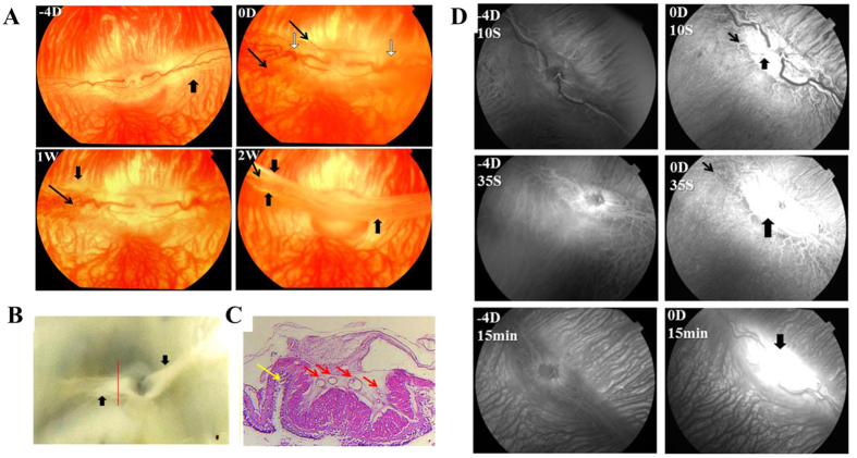
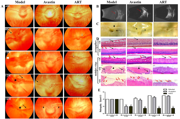
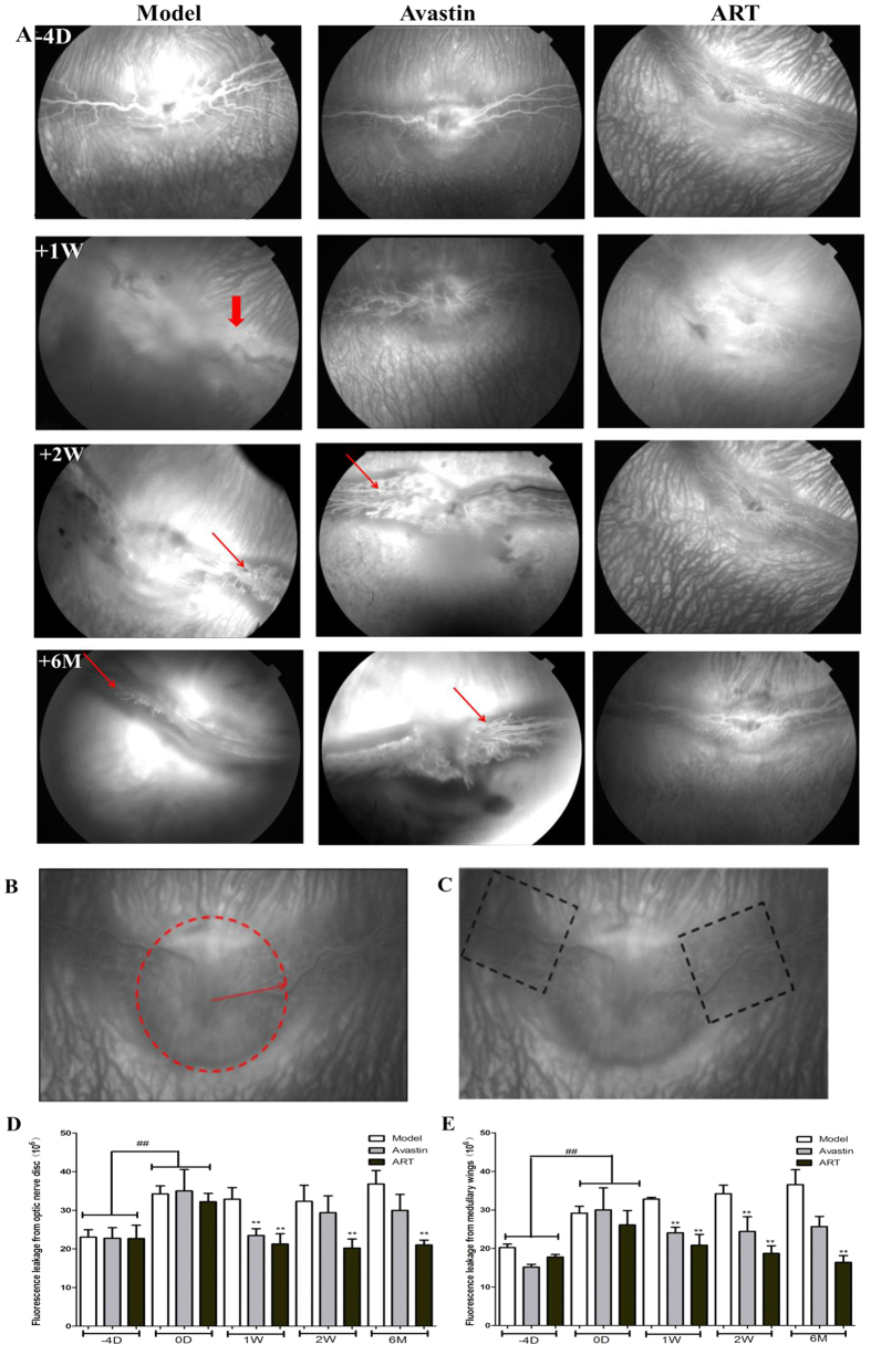
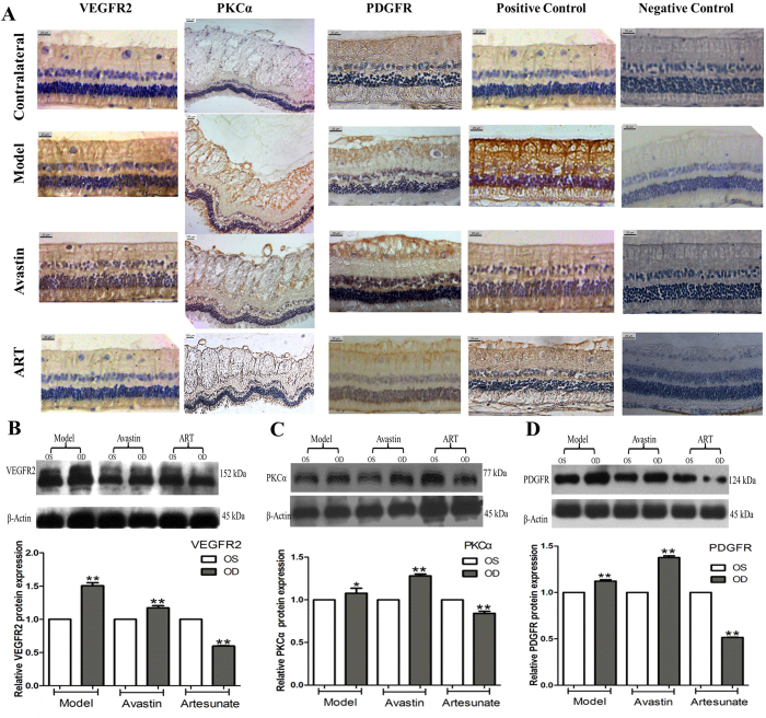
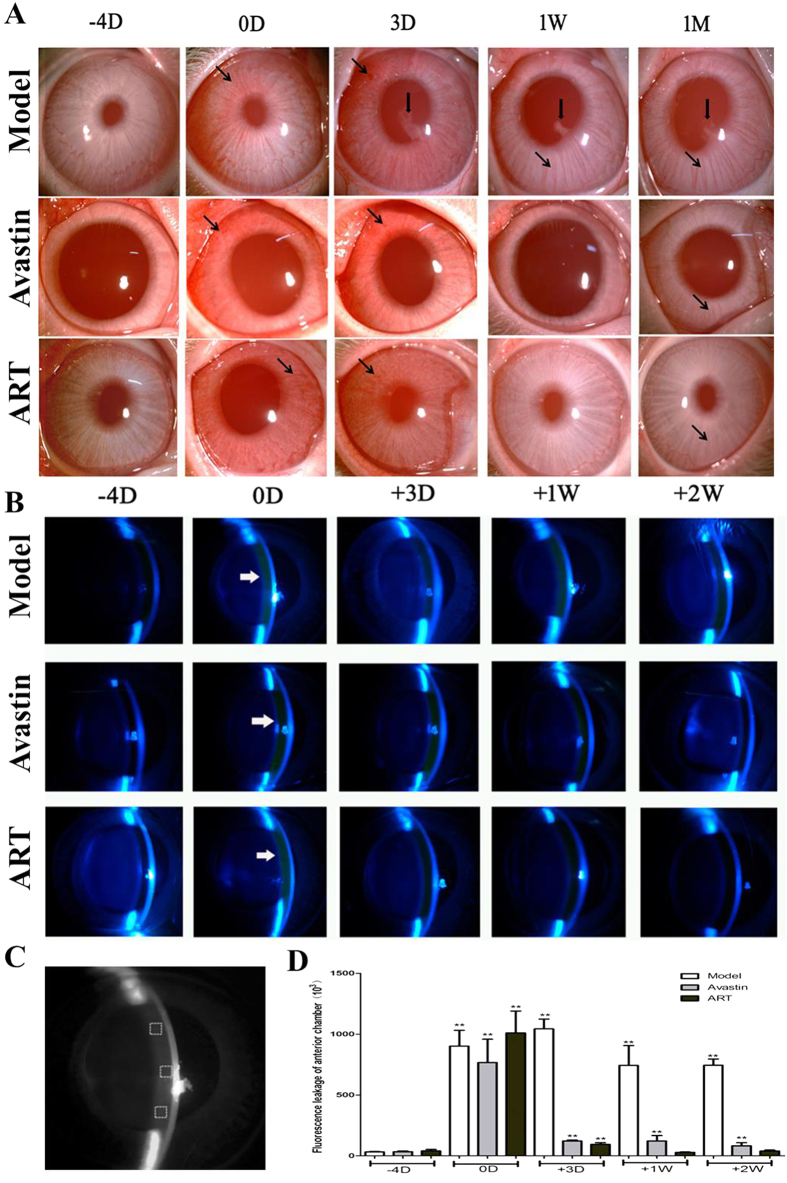
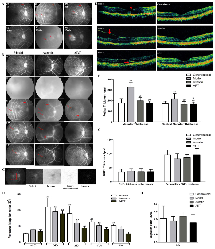
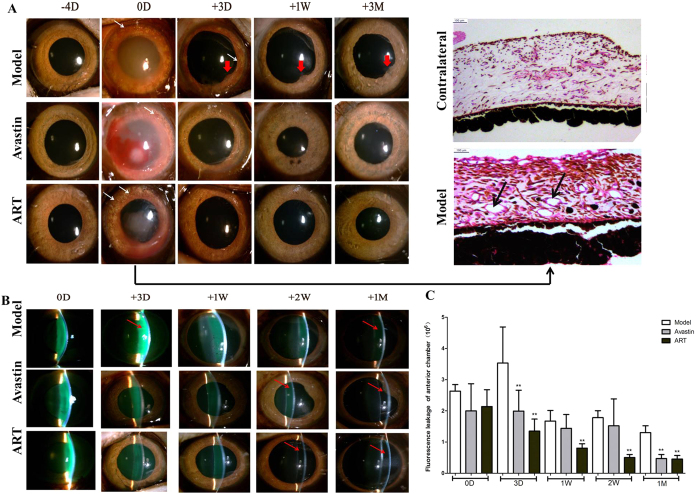
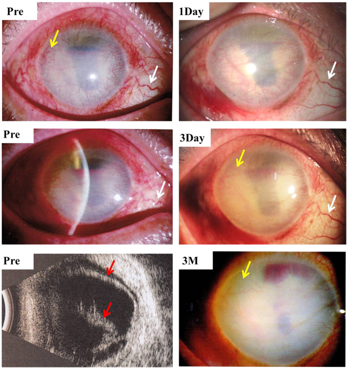
Ocular neovascularization (NV) is the primary cause of blindness in many ocular diseases. Large molecular weight anti- vascular endothelial growth factor (VEGF) protein drugs, such as Avastin and Lucentis, have saved the vision of millions. However, approximately 20-30% of patients respond poorly to anti-VEGF treatment. We found that artesunate (ART), a small molecular derivative of artemisinin, had a significant inhibitory effect on ocular NV by downregulating the expression of VEGFR2, PKCα, and PDGFR. ART significantly inhibited retinal NV in rabbits and macular edema in monkeys with greater anterior chamber penetrability and more durable efficacy than Avastin. Our pilot study showed that intravitreal injection of 80 μg ART significantly inhibited iris and corneal NV in a severe retinal detachment case. Our results suggest that ART might be a potential persistent small-molecule drug to manage ocular NV via multi-targets.


Figures

Figure 1
Figure 1. Fundus and FFA of the model group at different time points.
At day 0, vascular dilatation and tortuosity (white arrows in A), NV at the margin of medullary wings (thin black arrows in A), and fluorescein leakage (thick black arrows in D) from the disc and medullary were found in rabbit eyes injected with VEGF165+bFGF. Tractional folds of the medullary fibers (thick black arrows in A and B) appeared at 1 week, and tractional retinal detachments occurred at 2 weeks (yellow arrows in C), along with vascular crowding (red arrows in C).
Figure 2
Figure 2. The effect of ART on rabbit retinal vascular disorders induced by VEGF165+bFGF.
(A–C) ART and Avastin significantly diminished vascular dilatation and tortuosity (white arrows in A) and NV (black arrows in A) at 1 week. However, NV and fluorescein leakage recurred in the Avastin group at 2 weeks and finally resulted in tractional detachment of the retina (thick black arrows in A and C, red arrows in B). The tractional retinal detachments occurred at 2 weeks in the model group (★ in A). No visible neovascular membrane occurred in the ART group for 6 months. (D) H&E staining of cross-sections of the medullary wings (medullary) and superior to the optic disc (juxtapapillary). ART and Avastin significantly inhibited retinal edema (black arrows) at 1 week compared to the model group but did not noticeably reduce epiretinal fibrovascular membranes (red arrows). After 6 months, epiretinal fibrosis (▲) and retinal folds (black arrows) appeared in the model and Avastin groups, along with vascular crowding, which was not found in the ART group. The inner retina was disorganized (black arrows) in the model group; this did not occur in the ART or Avastin groups. (E) Grade score for retinal vascular disorders. All fundus and FFA photographs of rabbits were evaluated independently and in a masked manner by two observers and were scored using the five grades of retinal NV. The grade scores were used for ANOVA analysis, and differences with a value of p < 0.05 were considered significant.
Figure 3
Figure 3. The effect of ART on rabbit retinal vascular leakage induced by VEGF165+bFGF.
(A) ART and Avastin significantly reduced NV (thin red arrows) and fluorescein leakage (thick red arrows) at 1 week. However, NV and fluorescein leakage reccurred in the Avastin group at 2 weeks. No visible neovascular membrane occurred in the ART group for 6 months. (B,C) Diagram for analysis on the fluorescein leakage intensity from optic disc and both medullary wings in the late phase of FFA. The fluorescein leakage intensity in the selected areas was measured by Image J, and the mean fluorescein leakage intensity was used for statistical analysis. (D,E) Fluorescein leakage intensity from optic disc and medullary wings. Significant differences were determined using an ANOVA and SPSS 20.0 software (* and **denote p < 0.05 and p < 0.01 compared to the model group, and #and ##denote p < 0.05 and p < 0.01 compared to the baseline).
Figure 4
Figure 4. ART inhibited the expression of VEGFR2, PKCα, and PDGFR in rabbit retina.
(A) IHC of rabbit retina tissue. The retina tissue in the treated eyes of groups Model and Avastin showed stronger expression of VEGFR2, PKCα and PDGFR than in the contralateral eyes; however, the expression was significantly lower in the treated eyes of group ART. (BD) Western Blotting of rabbit retina tissue. The names of proteins are shown on the left, and the molecular weight sizes of proteins are shown on the right, respectively. The expression of VEGFR2, PKCα, and PDGFR in the treated eyes of the Model and Avastin groups was stronger compared that in the contralateral eyes; however, the expression was significantly lower in the ART group. Three rabbits from each group were harvested for IHC analysis, and another three rabbits from each group for Western blotting at 1 month. Each test was repeated three times and the mean intensity was used in the analysis. The blots were representative of three independently performed experiments. (One-way ANOVA, * and **denote P  <  0.05 and P  <  0.01 compared with left eye of the same rabbit)
Figure 5
Figure 5. The effect of ART on iris NV and fluorescein leakage in the anterior chamber.
(A) Iris NV (black arrows) lasted for 1 month in the model group, with the presence of posterior synechia (thick black arrows). Both gradually vanished within 1 week in the ART and Avastin groups. (B–D) The intensity of fluorescein leakage in the anterior chamber (white arrows in B) was significantly stronger compared to baseline levels in the model group, and returned to baseline levels within 1week in the ART group and within 2 weeks in the Avastin group (D). (C) Principle diagram for quantitative evaluation of the intensity of fluorescence leakage in the anterior chamber. * and **denote p < 0.05 and p < 0.01 compared with baseline levels.
Figure 6
Figure 6. ART inhibited fluorescein leakage and molecular edema in monkey models.
(A) FFA images of the model group. Retinal vascular dilatation and tortuosity (thick red arrows) and fluorescein leakage (thin red arrows) from the macula occurred at day 0 in the model group. (B) FFA images of all groups. The fluorescein leakage in the macular area lasted for 3 months in the model and Avastin groups (thin red arrows) and decreased gradually in the ART group. (C) Principle diagram for quantitative evaluation of the intensity of fluorescence leakage of the macula. Three FFA images of each monkey were taken at 15 min, a square-shaped area of the macula in the same size was selected, and the fluorescence intensity in this area was measured after background elimination. (D) Fluorescein leakage intensity of the macula. * and **denote p < 0.05 and p < 0.01 compared to the baseline level. (E) OCT. The macular retinal tear (red arrows), epiretinal membranes (red arrows), and retinal detachment (red arrows) appeared in the model group and were not found in the Avastin and ART groups. (F) Retinal thickness. The mean retinal thickness of the macular significantly increased in the model group compared to the contralateral eye and decreased in the ART and Avastin groups. (G,H) RNFL and C/D. Significant differences were determined using a t test or an ANOVA with SPSS 20.0 software. * and **denote p < 0.05 and p < 0.01, compared to the control group; #and ##denote p < 0.05 and p < 0.01compared to the model group; $ and $$denote p < 0.05 and p < 0.01 compared to the Avastin group.
Figure 7
Figure 7. Effect of ART on iris NV and fluorescein leakage into the anterior chamber in monkey models.
(A) Iris NV appeared at day 0, as revealed by a photograph of the iris (white arrows) and H&E staining (black arrows), lasted for 3 days in the model group, with the presence of posterior synechia (thick red arrows). It vanished completely in the ART and Avastin groups by day 3. (B,C) Fluorescein leakage (red arrows) in the anterior chamber lasted for 1 month in the model group and was essentially eliminated within 2 weeks in the ART group and within 1 month in the Avastin group. * and **denote p < 0.05 and p < 0.01 compared to the model group.
Figure 8
Figure 8. Intravitreal injection of 80μg ART inhibited corneal NV in a case of severe retinal detachment (red arrows) and corneal NV (yellow arrows).
Dilatation and tortuosity of conjunctival vessels were noticeably alleviated (white arrows) 1 day after treatment, and corneal NV (yellow arrows) decreased after 3 days and vanished mostly after 3 months.

Similar articles

Cited by

References

    1. Campochiaro P. A. Ocular neovascularization. J Mol Med 91, 311–321, doi: 10.1007/s00109-013-0993-5 (2013). - DOI - PMC - PubMed
    1. Aiello L. P. et al.. Vascular endothelial growth factor in ocular fluid of patients with diabetic retinopathy and other retinal disorders. N Engl J Med 331, 1480–1487, doi: 10.1056/NEJM199412013312203 (1994). - DOI - PubMed
    1. Ferrara N. & Kerbel R. S. Angiogenesis as a therapeutic target. Nature 438, 967–974, doi: Doi 10.1038/Nature04483 (2005). - DOI - PubMed
    1. Rosenfeld P. J. et al.. Ranibizumab for neovascular age-related macular degeneration. N Engl J Med 355, 1419–1431, doi: 10.1056/NEJMoa054481 (2006). - DOI - PubMed
    1. Brown D. M. et al.. Ranibizumab versus verteporfin for neovascular age-related macular degeneration. N Engl J Med 355, 1432–1444, doi: 10.1056/NEJMoa062655 (2006). - DOI - PubMed

Publication types

MeSH terms

Substances

LinkOut - more resources