. 2016 Nov;30(11):3733-3744.
doi: 10.1096/fj.201600428R. Epub 2016 Aug 1.

Profibrotic up-regulation of glucose transporter 1 by TGF-β involves activation of MEK and mammalian target of rapamycin complex 2 pathways

Affiliations

Profibrotic up-regulation of glucose transporter 1 by TGF-β involves activation of MEK and mammalian target of rapamycin complex 2 pathways

Mahefatiana Andrianifahanana et al. FASEB J. 2016 Nov.

Abstract

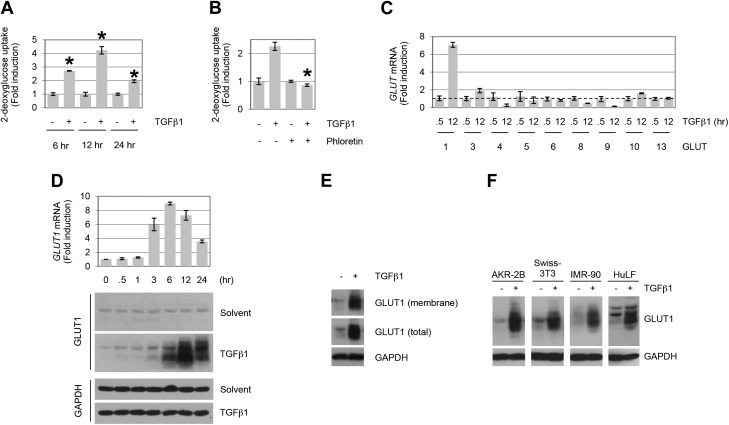
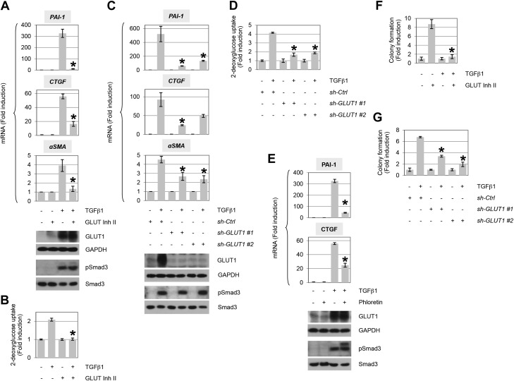
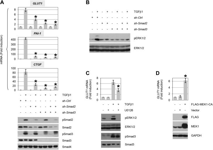
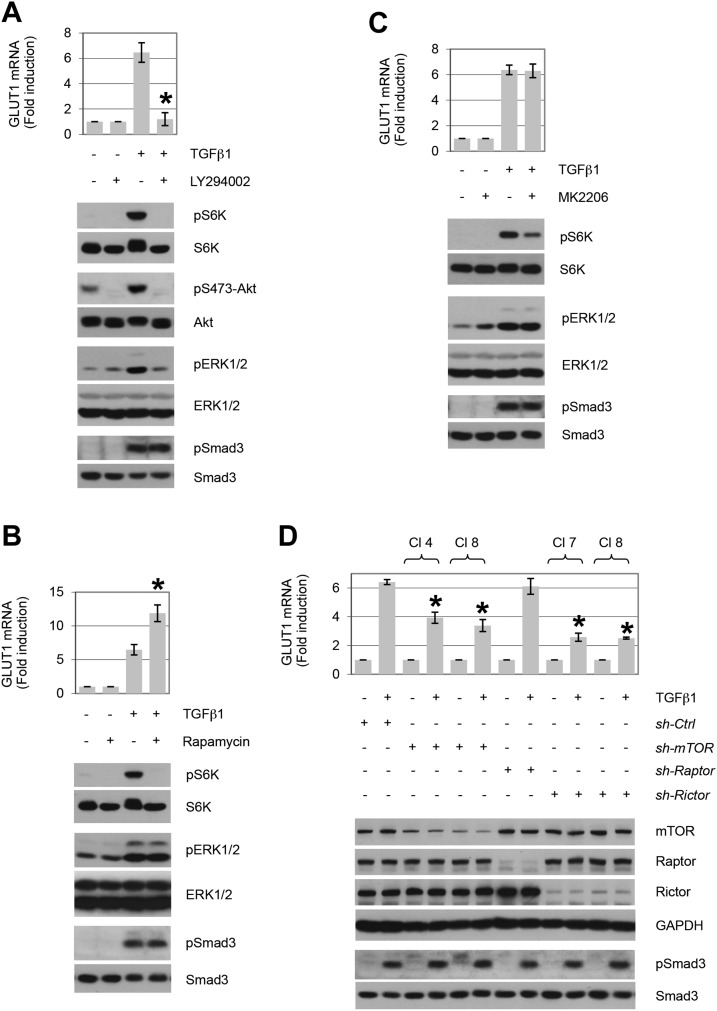
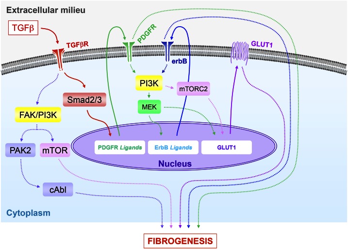
TGF-β plays a central role in the pathogenesis of fibroproliferative disorders. Defining the exact underlying molecular basis is therefore critical for the development of viable therapeutic strategies. Here, we show that expression of the facilitative glucose transporter 1 (GLUT1) is induced by TGF-β in fibroblast lines and primary cells and is required for the profibrotic effects of TGF-β. In addition, enhanced GLUT1 expression is observed in fibrotic areas of lungs of both patients with idiopathic pulmonary fibrosis and mice that are subjected to a fibrosis-inducing bleomycin treatment. By using pharmacologic and genetic approaches, we demonstrate that up-regulation of GLUT1 occurs via the canonical Smad2/3 pathway and requires autocrine activation of the receptor tyrosine kinases, platelet-derived and epidermal growth factor receptors. Engagement of the common downstream effector PI3K subsequently triggers activation of the MEK and mammalian target of rapamycin complex 2, which cooperate in regulating GLUT1 expression. Of note, inhibition of GLUT1 activity and/or expression is shown to impair TGF-β-driven fibrogenic processes, including cell proliferation and production of profibrotic mediators. These findings provide new perspectives on the interrelation of metabolism and profibrotic TGF-β signaling and present opportunities for potential therapeutic intervention.-Andrianifahanana, M., Hernandez, D. M., Yin, X., Kang, J.-H., Jung, M.-Y., Wang, Y., Yi, E. S., Roden, A. C., Limper, A. H., Leof, E. B. Profibrotic up-regulation of glucose transporter 1 by TGF-β involves activation of MEK and mammalian target of rapamycin complex 2 pathways.

Keywords: fibrosis; metabolism; signaling.


Figures

Figure 1.
Figure 1.
TGF-β stimulates GLUT1 up-regulation and glucose uptake in fibroblasts. A) AKR-2B cells were treated in the absence (−) or presence (+) of TGF-β1 (10 ng/ml), and 2-deoxyglucose uptake was determined at the indicated times (n = 3). B) Glucose uptake assay in TGF-β1–treated (12 h) AKR-2B fibroblast cells. Results show an increase in 2-deoxyglucose uptake, which is inhibited by the GLUT-specific inhibitor, phloretin (100 μM; n = 3). C) Quantitative RT-PCR analyses of GLUT induction by TGF-β1 in AKR-2B cells (n = 3). GLUT7 expression was not examined, whereas GLUTs 2 and 12 displayed no detectable expression. Dotted line indicates 1 unit on y axis. GLUTs 11 and 14 were not included, as no corresponding murine genes have been reported. The profiles of GLUT expression indicate that GLUT1 and GLUT3 exhibit >2-fold induction by TGF-β1, although only GLUT1 protein showed a parallel increase (data not shown for GLUT3 protein). D) AKR-2B cells were treated for the indicated times with TGF-β1 (10 ng/ml) or solvent (4 mM HCl, 1.0 mg/ml bovine serum albumin), and at the indicated times whole-cell lysates were prepared for quantitative PCR (top) or Western blot analysis (bottom) and blotted for GLUT1 or glyceraldehyde 3-phosphate dehydrogenase (GAPDH). Data show a time-dependent up-regulation of GLUT1 mRNA and protein by TGF-β1 (n = 3). E) Subcellular localization of GLUT1 in AKR-2B fibroblasts. Cell-surface biotinylation reveals the presence of GLUT1 in the membrane fraction under basal and TGF-β–stimulated (12 h) conditions. F) Profiles of GLUT1 protein expression in human (IMR-90 and HuLF) and murine (AKR-2B and Swiss-3T3) fibroblasts stimulated with TGF-β1 (18 or 24 h for HuLF). Results demonstrate that TGF-β1 (10 ng/ml) triggers GLUT1 up-regulation in various fibroblast cells. Asterisks denote statistical significance. Supplemental Tables 2 and 3 provide P values for indicated data points.
Figure 2.
Figure 2.
Up-regulation of GLUT1 mediates TGF-β–induced profibrotic phenotype. A) AKR-2B cells were treated in the presence (+) or absence (−) of TGF-β1 (10 ng/ml) and/or the GLUT-specific GLUT inhibitor II (Inh II; 10 μM) for 12 h, and quantitative RT-PCR was performed for PAI-1, CTGF, or α−SMA (n = 3). Western blot of the indicated proteins (bottom). B) TGF-β1–stimulated 2-deoxyglucose uptake in presence or absence of GLUT Inh II (n = 3). C) Analogous study as in panel A, except AKR-2B cells were infected with nontargeting control (ctrl) or shRNA (1 or 2) to GLUT1 (n = 3). D) As in panel C, 2-deoxyglucose uptake in presence or absence of control or GLUT1 shRNA (n = 3). E) Similar study as in panel A, using the GLUT inhibitor phloretin (100 μM; n = 3). F, G) GLUT Inh II (F) or knockdown of GLUT1 by RNA interference (G) reduces soft agar colony formation induced by TGF-β1 (n = 3). GAPDH, glyceraldehyde 3-phosphate dehydrogenase. Asterisks denote statistical significance. Supplemental Tables 2 and 3 provide P values for indicated data points.
Figure 3.
Figure 3.
GLUT1 expression in mouse and human lung tissue. AC) Histologic sections of nonfibrotic [saline:saline (SS), top], fibrotic [bleomycin:saline (BS), middle], and fibrosis dual-treated with imatinib (Imat) and lapatinib (Lap) [bleomycin:Imat + Lap (BIL), bottom] (26). Mouse lungs were stained with Masson’s trichrome (A), and GLUT1 antibodies were costained with hematoxylin (B). Color deconvoluted images of mouse lungs stained with GLUT1 (C) indicate the area and intensity of GLUT1 staining. Color-coded legend indicates intensity of GLUT1 expression. D) Quantification of GLUT1 expression in mouse lung tissues was significantly greater in fibrosis vs. normal or dual-treated lungs with Imat + Lap. Mean values of mouse tissues analyzed per condition ± sem (n = 3). EG) Histologic sections of normal (top) and fibrotic human lungs (bottom) were stained with Masson’s trichrome (E), and GLUT1 antibodies were costained with hematoxylin (F). Arrows indicate the magnified section showing fibroblasts found in normal human lung (top) or fibrotic foci observed in IPF (bottom). Color deconvoluted images are shown of GLUT1-stained human lungs (G). H) Strong GLUT1 expression observed in fibrotic foci was significantly greater than fibroblasts in normal human lung. Mean values of human cases (normal, n = 7; fibrotic, n = 12) ± sem. Asterisks denote statistical significance. Supplemental Tables 2 and 3 provide P values for indicated data points.
Figure 4.
Figure 4.
Autocrine activation of receptor tyrosine kinases (RTKs) is required for induction of GLUT1 by TGF-β. Quantitative RT-PCR analysis of 10 ng/ml TGF-β1–stimulated GLUT1 expression (12 h post-treatment) in AKR-2B cells in the presence of the PDGFR-specific inhibitor, CP673451 (A; 2 µM), the ErbB1/2-specific inhibitor, lapatinib (B; 5 µM), shRNA specific for PDGFRα+β (C), or shRNA specific for ErbB1+2 (D; n = 3). Data indicate that both RTK pathways are important for GLUT1 induction by TGF-β. Western blots for the indicated protein to document drug or knockdown efficacy and no effect on Smad3 phosphorylation or expression (bottom). Ctrl, control; GAPDH, glyceraldehyde 3-phosphate dehydrogenase; IP, immunoprecipitation. Asterisks denote statistical significance. Supplemental Tables 2 and 3 provide P values for indicated data points.
Figure 5.
Figure 5.
TGF-β induces GLUT1 expression dependent upon Smad pathway activation of MEK. A) Quantitative RT-PCR analysis of GLUT1 and profibrotic gene induction by TGF-β1 (12 h post-treatment) in AKR-2B cells (pools) that stably express control (nontargeting; ctrl) or shRNA targeting Smad2 and/or Smad3 (n = 3). Results show that both Smad2 and Smad3 are required for TGF-β1–dependent GLUT1, PAI-1, and CTGF up-regulation. Western blot analyses confirm efficient knockdown of Smad2/Smad3 and uniform activation of Smad2 and/or Smad3 in appropriate controls (5 h). B) Western blot analysis of ERK1/2 activation by TGF-β1 (5 h post-treatment) in AKR-2B cells (pools) that stably express control (nontargeting) or shRNA targeting Smad2 and/or Smad3. Results indicate that TGF-β1 activates ERK1/2 downstream of the Smad2/3 pathway. C) Quantitative RT-PCR analysis of GLUT1 induction by TGF-β1 (12 h post-treatment) in AKR-2B cells in the absence (−) or presence (+) of the MEK-ERK1/2 inhibitor U0126 (3 μM; n = 3). Prevention of ERK1/2 phosphorylation (5 h) confirms the efficacy of U0126, whereas the lack of effect on pSmad3 documents specificity. D) Quantitative RT-PCR analysis of GLUT1 induction by ectopic expression of a constitutively active MEK1 (54 h post-transfection) in AKR-2B cells (n = 3). Constitutive activation of MEK is shown to trigger up-regulation of GLUT1. TGF-β1 was used at 10 ng/ml. GAPDH, glyceraldehyde 3-phosphate dehydrogenase. Asterisks denote statistical significance. Supplemental Tables 2 and 3 provide P values for indicated data points.
Figure 6.
Figure 6.
Up-regulation of GLUT1 by TGF-β requires the cooperation of multiple PI3K-dependent signaling pathways. Quantitative RT-PCR analysis of TGF-β1–stimulated GLUT1 expression (12 h post-treatment) in AKR-2B cells in the presence (+) of the PI3K-specific inhibitor, LY294002 (A; 20 µM), mTORC1-specific inhibitor, rapamycin (B; 10 nM), the Akt-specific inhibitor, MK22006 (C; 300 nM), or shRNA specific clones (CI) for mTOR, Raptor, or Rictor (D; n = 3). Western blot analyses were performed on samples harvested at 5 h (AC) or 12 h (D) post–TGF-β treatment. Data show that inhibition of PI3K abrogates induction of GLUT1 by TGF-β (A). Whereas interfering with mTORC1 (B, D; sh-Raptor) or Akt (C) activity does not prevent TGF-β1–induced GLUT1 expression, knockdown of mTOR and Rictor is inhibitory. TGF-β1 was used at 10 ng/ml. Ctrl, control; GAPDH, glyceraldehyde 3-phosphate dehydrogenase. Asterisks denote statistical significance. Supplemental Tables 2 and 3 provide P values for indicated data points.
Figure 7.
Figure 7.
Proposed model for the profibrotic regulation of GLUT1 expression by TGF-β. This model incorporates the cooperative action of multiple signaling pathways in regulating GLUT1 induction by TGF-β and their subsequent integration in profibrotic TGF-β signaling. Engagement of the TGF-β receptor complex activates the canonical Smad pathway, which leads to up-regulation of PDGFR ligands. Subsequent PDGFR phosphorylation promotes activation of MEK/ERK and induction of ErbB ligand expression and cognate receptor activation (26, 27). Activation of the MEK/ERK and mTORC2 pathways downstream of PI3K promotes up-regulation of GLUT1, whose activity is necessary for the profibrotic effects of TGF-β. The Smad-dependent pathways (PDGFR, ErbB, and GLUT1), in concert with the noncanonical TGF-β–activated signaling modules, p21-activated kinase 2 (PAK2)/cAbl and PI3K/AKT/mTOR (–23, 25, 85), all contribute to the fibrogenic program directed by TGF-β.

Similar articles

Cited by

References

    1. Pinzani M., Vizzutti F. (2008) Fibrosis and cirrhosis reversibility: clinical features and implications. Clin. Liver Dis. 12, 901–913 - PubMed
    1. Sivakumar P., Ntolios P., Jenkins G., Laurent G. (2012) Into the matrix: targeting fibroblasts in pulmonary fibrosis. Curr. Opin. Pulm. Med. 18, 462–469 - PubMed
    1. Todd N. W., Luzina I. G., Atamas S. P. (2012) Molecular and cellular mechanisms of pulmonary fibrosis. Fibrogenesis Tissue Repair 5, 11 - PMC - PubMed
    1. Wynn T. A. (2007) Common and unique mechanisms regulate fibrosis in various fibroproliferative diseases. J. Clin. Invest. 117, 524–529 - PMC - PubMed
    1. Wynn T. A., Ramalingam T. R. (2012) Mechanisms of fibrosis: therapeutic translation for fibrotic disease. Nat. Med. 18, 1028–1040 - PMC - PubMed

Publication types

MeSH terms

Substances

LinkOut - more resources