. 2016 Aug 2:6:30749.
doi: 10.1038/srep30749.

The differential short- and long-term effects of HIV-1 latency-reversing agents on T cell function

Affiliations

The differential short- and long-term effects of HIV-1 latency-reversing agents on T cell function

G Clutton et al. Sci Rep. .

Erratum in

Abstract

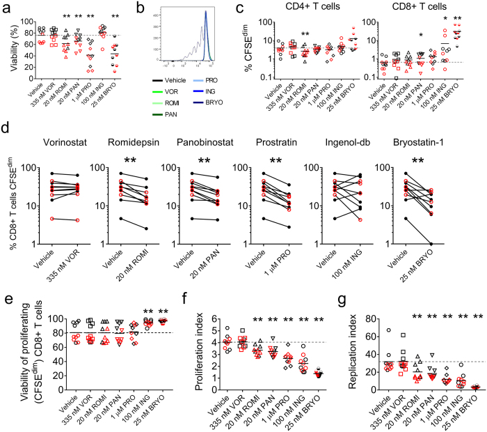
Despite the extraordinary success of HIV-1 antiretroviral therapy in prolonging life, infected individuals face lifelong therapy because of a reservoir of latently-infected cells that harbor replication competent virus. Recently, compounds have been identified that can reverse HIV-1 latency in vivo. These latency- reversing agents (LRAs) could make latently-infected cells vulnerable to clearance by immune cells, including cytolytic CD8+ T cells. We investigated the effects of two leading LRA classes on CD8+ T cell phenotype and function: the histone deacetylase inhibitors (HDACis) and protein kinase C modulators (PKCms). We observed that relative to HDACis, the PKCms induced much stronger T cell activation coupled with non-specific cytokine production and T cell proliferation. When examining antigen-specific CD8+ T cell function, all the LRAs except the HDACi Vorinostat reduced, but did not abolish, one or more measurements of CD8+ T cell function. Importantly, the extent and timing of these effects differed between LRAs. Panobinostat had detrimental effects within 10 hours of drug treatment, whereas the effects of the other LRAs were observed between 48 hours and 5 days. These observations suggest that scheduling of LRA and CD8+ T cell immunotherapy regimens may be critical for optimal clearance of the HIV-1 reservoir.


Figures

Figure 1
Figure 1. HDACis differentially activate T cells.
(a) Experimental design. PBMC from HIV-1-seropositive participants durably suppressed on cART (n = 7) were exposed to vehicle (0.5% DMSO) or LRA for 3, 6, 12, or 24 hours, washed to remove extracellular drug, and maintained in vehicle for the remaining culture period. Activation marker expression was assessed at 24 hours. (b) Expression of activation markers on CD4+ and CD8+ T cells at 24 hours. Measurements were compared between vehicle and HDACi treatment using an exact Wilcoxon Signed Rank test. *p < 0.05; **p ≤ 0.016.
Figure 2
Figure 2. PKCms potently activate T cells.
Expression of activation markers at 24, 48, and 72 hours on CD4+ and CD8+ T cells. PBMC from HIV-1-seropositive participants durably suppressed on cART (n = 8) were exposed to vehicle (0.5% DMSO), HDACis or PKCms for 3 hours, washed to remove extracellular drug, and maintained in vehicle for the remainder of the 72 hour culture period. Measurements were compared between vehicle and LRA treatment using an exact Wilcoxon Signed Rank test. *p < 0.05; **p ≤ 0.016.
Figure 3
Figure 3. Toxicity of LRAs in vitro.
Viability of PBMC from HIV-1-seropositive donors durably suppressed on cART (n = 8). PBMC were exposed to vehicle (0.5% DMSO) or LRAs for 3 (left) or 6 (right) hours, washed to remove extracellular drug, and maintained in vehicle for the remainder of the culture period. Viability was measured at 24, 48, and 72 hours. Measurements were compared between vehicle and HDACi treatment using an exact Wilcoxon Signed Rank test. *p < 0.05; **p ≤ 0.016.
Figure 4
Figure 4. Cytokine release following PBMC exposure to LRAs.
Cytokine concentrations in culture supernatants of PBMC from HIV-1-seropositive participants durably suppressed on cART (n = 5–8). PBMC were exposed to vehicle (0.5% DMSO) or LRAs for 3 hours, washed to remove extracellular drug, and maintained in vehicle for the remainder of the culture period. Supernatants were collected at 24, 48, and 72 hours. *p < 0.05; **p ≤ 0.016.
Figure 5
Figure 5. Effects of LRAs on non-specific and antigen-specific T cell function.
PBMC from HIV-1-seropositive participants durably suppressed on cART (red symbols; n = 5) and HIV-1-seronegative participants (black symbols; n = 5) were exposed to vehicle (0.5% DMSO) or a LRA for 4 hours, washed and then cultured for a further 6 hours in either vehicle (to assess non-specific responses) or in the presence of peptides (to assess antigen-specific responses). (a,b) Non-specific cytokine production by CD4+ and CD8+ T cells. Dashed lines indicate mean responses in vehicle-exposed cultures. (c) The effect of 4 hours pre-exposure to LRAs on the frequency of memory CD8+ T cells degranulating (perforinlow CD107a+) and producing IFN-γ in response to peptide stimulation. PBMC from HIV-1-seropositive participants were exposed to HIV-1 CD8+ optimal epitopes, and PBMC from HIV-1-seronegative participants were exposed to a pool of ‘Flu, EBV, and CMV (FEC) CD8+T cell epitopes. Due to sample availability, not all LRAs were tested in all participants. Measurements were compared between vehicle and LRA treatment using an exact Stratified Wilcoxon Signed Rank test. *p < 0.05; **p ≤ 0.016.
Figure 6
Figure 6. Effects of LRAs on T cell proliferation.
PBMC from HIV-1-seropositive participants durably suppressed on cART (red symbols; n = 5) and HIV-1-seronegative participants (black symbols; n = 5) were exposed to vehicle (0.5% DMSO) or a LRA for 4 hours, washed and then cultured for 5 days in either vehicle (to assess non-specific proliferation) or in the presence of peptides (to assess antigen-specific proliferation). (a) PBMC viability measured at 5 days. (b,c) Representative histograms (b) and combined data (c) showing non-specific proliferation by CD4+ and CD8+ T cells exposed to vehicle or LRA for 4 hours. The frequency of proliferating (CFSEdim) cells was measured at 5 days. (d) The effect of 4 hours pre-exposure to LRA on (live) CD8+ T cell proliferation (% CFSEdim) in response to peptide, measured at 5 days. (e) Viability of all CD8+ T cells that had proliferated one or more times in response to peptide after 5 days. (f) The effect of 4 hours pre-exposure to LRA on the mean number of cycles of proliferation undergone by (live) antigen-stimulated cells over 5 days (proliferation index). (g) The effect of 4 hours pre-exposure to LRA on the mean number of (live) daughter cells generated by each antigen-specific parent cell over 5 days (replication index). Dashed lines indicate mean responses in vehicle-pre-exposed cultures. Measurements were compared between vehicle and LRA-treatment using an exact Stratified Wilcoxon Signed Rank test. *p < 0.05; **p ≤ 0.016.
Figure 7
Figure 7. Effects of an oral dose of Vorinostat on T cell phenotype and function ex vivo.
Participants received a single 400 mg oral dose of Vorinostat. (a) Vorinostat plasma pharmacokinetics in three participants 30 minutes to 10 hours after dosing. (b) Quantification of HIV-1 RNA in resting CD4+ T cells at baseline and following oral dosing with Vorinostat (data not available for participant C). (c) Expression of activation markers on peripheral blood CD4+ and CD8+ T cells immediately prior to dosing and 4, 7, 10 and 24 hours post-dose. (d) Frequency of memory CD8+ T cells in peripheral blood degranulating (perforinlow CD107a+) and producing IFN-γ in response to peptide stimulation pre-dose and 4, 7, 10, and 24 hours post-dose. Measurements of HIV-1 RNA in resting CD4+ T cells were compared between baseline and post-dose using a Mann-Whitney test.

References

    1. Archin N. M. et al. Expression of latent HIV induced by the potent HDAC inhibitor suberoylanilide hydroxamic acid. AIDS Res Hum Retroviruses 25, 207–212, doi: 10.1089/aid.2008.0191 (2009). - DOI - PMC - PubMed
    1. Shirakawa K., Chavez L., Hakre S., Calvanese V. & Verdin E. Reactivation of latent HIV by histone deacetylase inhibitors. Trends in microbiology 21, 277–285, doi: 10.1016/j.tim.2013.02.005 (2013). - DOI - PMC - PubMed
    1. Jiang G. et al. Reactivation of HIV latency by a newly modified Ingenol derivative via protein kinase Cdelta-NF-kappaB signaling. AIDS 28, 1555–1566, doi: 10.1097/QAD.0000000000000289 (2014). - DOI - PMC - PubMed
    1. Mehla R. et al. Bryostatin modulates latent HIV-1 infection via PKC and AMPK signaling but inhibits acute infection in a receptor independent manner. Plos One 5, e11160, doi: 10.1371/journal.pone.0011160 (2010). - DOI - PMC - PubMed
    1. Williams S. A. et al. Prostratin antagonizes HIV latency by activating NF-kappaB. J Biol Chem 279, 42008–42017, doi: 10.1074/jbc.M402124200 (2004). - DOI - PubMed

Publication types

MeSH terms