. 2016 Aug 22;213(9):1655-62.
doi: 10.1084/jem.20160061. Epub 2016 Aug 1.

GPR91 senses extracellular succinate released from inflammatory macrophages and exacerbates rheumatoid arthritis

Affiliations

GPR91 senses extracellular succinate released from inflammatory macrophages and exacerbates rheumatoid arthritis

Amanda Littlewood-Evans et al. J Exp Med. .

Abstract

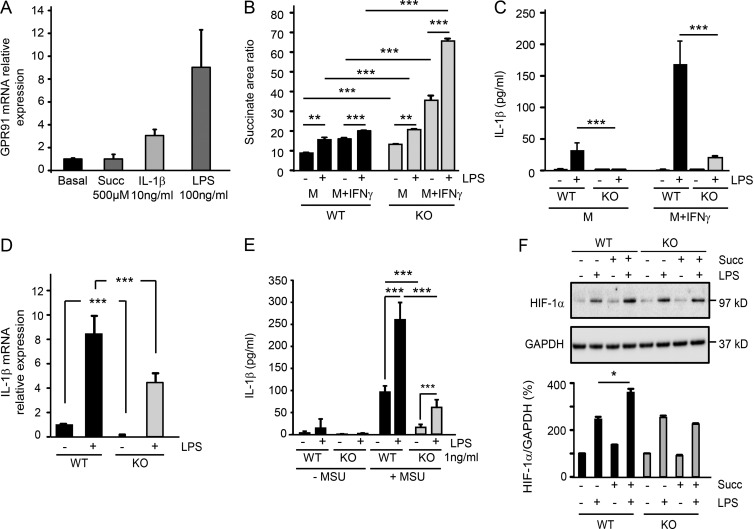
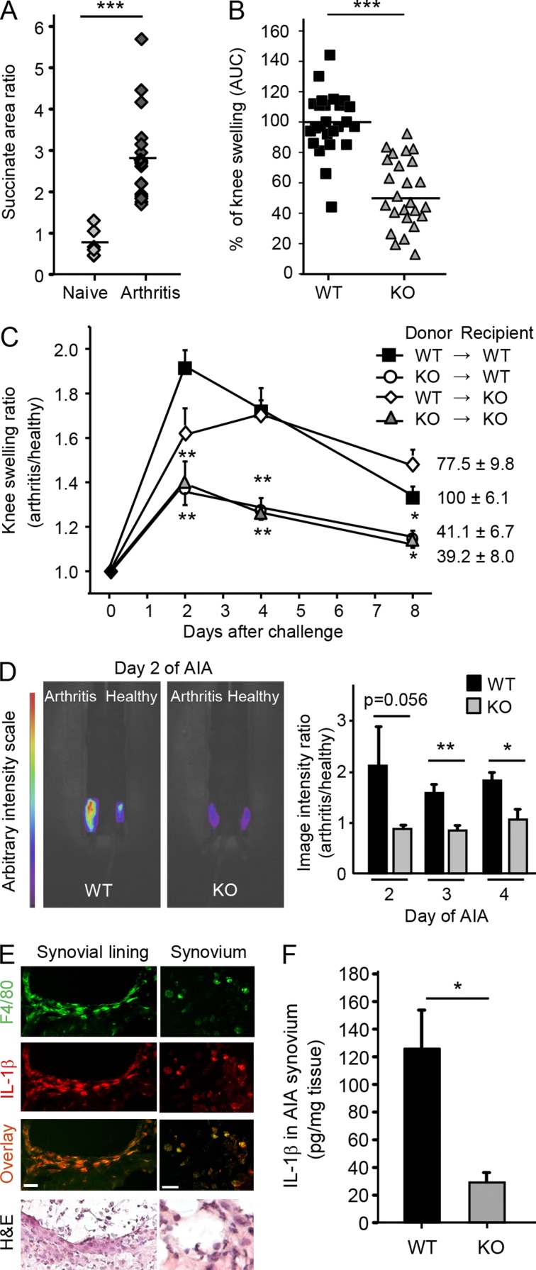
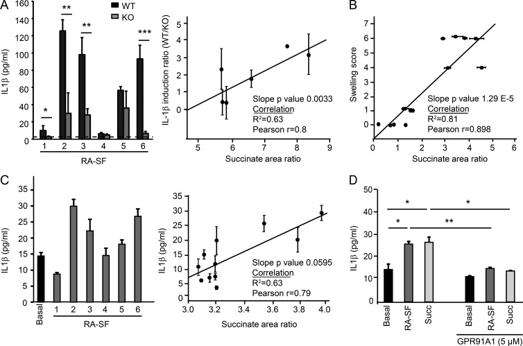
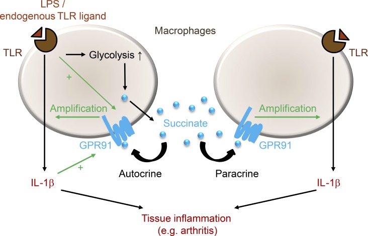
When SUCNR1/GPR91-expressing macrophages are activated by inflammatory signals, they change their metabolism and accumulate succinate. In this study, we show that during this activation, macrophages release succinate into the extracellular milieu. They simultaneously up-regulate GPR91, which functions as an autocrine and paracrine sensor for extracellular succinate to enhance IL-1β production. GPR91-deficient mice lack this metabolic sensor and show reduced macrophage activation and production of IL-1β during antigen-induced arthritis. Succinate is abundant in synovial fluids from rheumatoid arthritis (RA) patients, and these fluids elicit IL-1β release from macrophages in a GPR91-dependent manner. Together, we reveal a GPR91/succinate-dependent feed-forward loop of macrophage activation and propose GPR91 antagonists as novel therapeutic principles to treat RA.


Figures

Figure 1.
Figure 1.
Extracellular succinate signals via GPR91 to stimulate macrophages to release IL-1β. (A) GPR91 mRNA expression in WT (Janvier C57BL/6J) inflammatory BMDMs (M-CSF + IFN-γ) ± 100 ng/ml LPS, 500 µM succinate, or 10 ng/ml IL-1β for 24 h. n = 3 of Ct values. Succinate (Succ), IL-1β, and LPS related to basal (=1). Data are representative of three experiments. (B) Succinate levels (mass spectrophotometry area ratio) in medium from cultured BMDMs. Extracellular succinate from WT (littermates; black bars) and Sucnr1−/− (gray bars), neutral (M, M-CSF), or inflammatory (M + IFN-γ) BMDMs ± 100 ng/ml LPS for 24 h is shown. n = 6 wells. Data are representative of three experiments. (C) IL-1β in supernatants of WT (Janvier C57BL/6J) and Sucnr1−/− neutral or inflammatory BMDMs ± 100 ng/ml LPS for 24 h. n = 3 wells and are representative of seven experiments. (D) IL-1β mRNA levels from cell lysates from WT (Janvier C57BL/6J) or Sucnr1−/− inflammatory BMDMs ± 100 ng/ml LPS at 4 h (related to WT basal = 1). n = 2–3 of Ct values. Data are representative of two experiments. (E) IL-1β levels measured in the supernatant of WT (Janvier C57BL/6J) and Sucnr1−/− inflammatory BMDMs stimulated with 1 ng/ml LPS and 180 µg/ml MSU. n = 5–6 wells. Data are representative of two experiments. (F) Western blot of HIF-1α (representative blot of two experiments) and quantification (two experiments; 100% for no stimulus, WT, and Sucnr1−/−) at 6 h after stimulation with 500 µM succinate, 100 ng/ml LPS, or a combination of the two in WT littermate controls and Sucnr1−/− inflammatory BMDMs. *, P < 0.05; **, P < 0.01; ***, P < 0.001, unpaired Student’s t test. Data are means ± SEM.
Figure 2.
Figure 2.
Extracellular succinate levels within the SF of arthritic joints activate macrophages via GPR91 to up-regulate IL-1β production. (A) Succinate area ratio (mass spectrophotometry) in SF from knees of Janvier C57BL/6J mice on day 7–8 of AIA (n = 19; black diamonds) versus naive controls (n = 5; gray diamonds) pooled from five independent experiments. The lines depict means. A Mann-Whitney rank sum test was used, as the normality test failed (Shapiro-Wilk). (B) AUCs of knee swelling ratio curves (arthritis/healthy) over time (n = 25 per genotype), pooled from five experiments. The lines depict means. For each experiment, the WT (both Janvier C57BL/6J and littermate controls) mice were accorded a mean of 100%. WT (black squares) and Sucnr1−/− (gray triangles) were compared by unpaired Student’s t test. (C) Knee swelling ratio (arthritis/healthy) over time in reciprocal BM chimeras of WT (littermates or congenic CD45.1 SJL-Ptprca/BoyAiTac; Taconic) and Sucnr1−/− mice. (Right) AUC (percentage) expressed as means ± SEM. n = 5 per group and n = 4 in the KO–KO group. One-way ANOVA and Tukey’s posttest were used. The experiment was performed once. (D, left) NIR intensity images of folate-positive activated macrophages in knees of WT littermates and Sucnr1−/− animals on day 2 of AIA. n = 10 per group. (Right) Quantification of the NIR signal in knees of WT and Sucnr1−/− animals over 72 h after probe injection. Bars show means of NIR intensity ratios in knees (arthritis/healthy) ± SEM. n = 10 per bar, unpaired Student’s t test. Data are representative of two experiments. (E) Colocalization of F4/80-positive macrophages (green) with IL-1β (red) from the synovial lining and synovium of WT mice (littermates). Hematoxylin and eosin (H&E) staining shows tissue morphology. Bars, 25 µm. Images are representative of 30 sections from five mice. (F) IL-1β levels in synovial tissue homogenates of WT littermate or Sucnr1−/− mice on day 7 of AIA. Data are representative of two experiments. Error bars represent means ± SEM of five mice per group. *, P < 0.05; **, P < 0.01; ***, P < 0.001, unpaired Student’s t test.
Figure 3.
Figure 3.
IL-1β release is suppressed in Sucnr1−/− or GPR91 antagonist-treated human macrophages incubated with RA SF. (A, left) IL-1β production from WT (littermates) and Sucnr1−/− (gray) BMDMs incubated for 24 h with 10% human RA SF from six patients (RA SF 1–6). Error bars represent means ± SEM of triplicates. Data are representative of two experiments (unpaired Student’s t test). Where normality failed (Shapiro-Wilk), a Mann-Whitney rank sum test was used. The dashed line is the limit of detection. (Right) IL-1β induction ratios (from WT littermate/Sucnr1−/− BMDMs) by the six RA SFs were correlated with concentration of succinate in the SFs. Means ± range of the induction ratio for each SF from the two experiments (Pearson’s correlation) are shown. (B) Correlation of paw swelling with SF succinate concentrations within the same paws, measured in duplicates (collagen-induced arthritis, DBA/1; n = 13; Janvier). Pearson’s correlation was used. (C, left) IL-1β production from U937 cells incubated for 24 h with 10% RA SF from six RA patients (gray bars, RA-SF 1–6) or medium only (basal). Data are means ± SEM of triplicates and representative of two experiments. (Right) The amount of IL-1β elicited by 11 RA SFs was correlated with the concentration of succinate in the SF. Means of IL-1β ± the range for each SF from the two experiments (Pearson’s correlation) are shown. (D) IL-1β production from U937 cells incubated for 24 h with 10% RA SF or 1 mM succinate (Succ) in the presence or absence of 5 µM GPR91 antagonist GPR91A1. Error bars represent means ± SEM of triplicates. Data are representative of five experiments. *, P < 0.05; **, P < 0.01; ***, P < 0.001, unpaired Student’s t test.
Figure 4.
Figure 4.
Proposed mechanism of GPR91-driven autocrine and paracrine enhancement of IL-1β release from activated macrophages. Endogenous TLR ligands in the SF of RA patients activate macrophages locally. This leads to an enhancement of glycolysis and an increase of intracellular succinate. At the same time, succinate is released to the extracellular milieu where it binds to GPR91 and amplifies IL-1β production from either the same or a neighboring GPR91-expressing cell. Both LPS and IL-1β up-regulate GPR91 in a further feed-forward action to perpetuate inflammation.

References

    1. Ariza A.C., Deen P.M.T., and Robben J.H.. 2012. The succinate receptor as a novel therapeutic target for oxidative and metabolic stress-related conditions. Front. Endocrinol. (Lausanne). 3:22 10.3389/fendo.2012.00022 - DOI - PMC - PubMed
    1. Bhuniya D., Umrani D., Dave B., Salunke D., Kukreja G., Gundu J., Naykodi M., Shaikh N.S., Shitole P., Kurhade S., et al. . 2011. Discovery of a potent and selective small molecule hGPR91 antagonist. Bioorg. Med. Chem. Lett. 21:3596–3602. 10.1016/j.bmcl.2011.04.091 - DOI - PubMed
    1. Borenstein D.G., Gibbs C.A., and Jacobs R.P.. 1982. Gas-liquid chromatographic analysis of synovial fluid. Succinic acid and lactic acid as markers for septic arthritis. Arthritis Rheum. 25:947–953. 10.1002/art.1780250806 - DOI - PubMed
    1. Bresnihan B., Pontifex E., Thurlings R.M., Vinkenoog M., El-Gabalawy H., Fearon U., Fitzgerald O., Gerlag D.M., Rooney T., van de Sande M.G., et al. . 2009. Synovial tissue sublining CD68 expression is a biomarker of therapeutic response in rheumatoid arthritis clinical trials: consistency across centers. J. Rheumatol. 36:1800–1802. 10.3899/jrheum.090348 - DOI - PubMed
    1. Chouchani E.T., Pell V.R., Gaude E., Aksentijević D., Sundier S.Y., Robb E.L., Logan A., Nadtochiy S.M., Ord E.N., Smith A.C., et al. . 2014. Ischaemic accumulation of succinate controls reperfusion injury through mitochondrial ROS. Nature. 515:431–435. 10.1038/nature13909 - DOI - PMC - PubMed