Review
. 2016 Aug 1;8(8):a005926.
doi: 10.1101/cshperspect.a005926.

Second Messengers

Affiliations
Review

Second Messengers

Alexandra C Newton et al. Cold Spring Harb Perspect Biol. .

Abstract

Second messengers are small molecules and ions that relay signals received by cell-surface receptors to effector proteins. They include a wide variety of chemical species and have diverse properties that allow them to signal within membranes (e.g., hydrophobic molecules such as lipids and lipid derivatives), within the cytosol (e.g., polar molecules such as nucleotides and ions), or between the two (e.g., gases and free radicals). Second messengers are typically present at low concentrations in resting cells and can be rapidly produced or released when cells are stimulated. The levels of second messengers are exquisitely controlled temporally and spatially, and, during signaling, enzymatic reactions or opening of ion channels ensure that they are highly amplified. These messengers then diffuse rapidly from the source and bind to target proteins to alter their properties (activity, localization, stability, etc.) to propagate signaling.


Figures

Figure 1.
Figure 1.
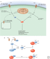
Second messengers disseminate information received from cell-surface receptors. Indicated are three examples of a receptor activating an effector to produce a second messenger that modulates the activity of a target. On the right, binding of agonists to a GPCR (the receptor) can activate adenylyl cyclase (the effector) to produce cAMP (the second messenger) to activate protein kinase A (PKA; the target). On the left, binding of growth factors to a receptor tyrosine kinase (RTK; the receptor) can activate PI3K (the effector) to generate PIP3 (the second messenger), which activates Akt (the target). In the center, binding of ligands to a GPCR (receptor) activates phospholipase C (PLC; the effector), to generate two second messengers, DAG and IP3, which activate protein kinase C (PKC; the target) and release calcium from intracellular stores, respectively.
Figure 2.
Figure 2.
(A) cAMP is the archetypical second messenger. Its levels increase rapidly following receptor-mediated activation of adenylyl cyclase (AC), which catalyzes the conversion of adenosine monophosphate (AMP) to cAMP. This small second messenger activates PKA at specific cellular locations as a result of anchoring of PKA to A-kinase-anchoring proteins (AKAPs). In addition, cAMP activates EPACs. (B) AC activity is controlled by the opposing actions of the Gs and Gi proteins. The cAMP produced by AC activates PKA by binding to its R subunit. This releases the C subunit. The signal can be terminated by the action of phosphodiesterase (PDE) enzymes.
Figure 3.
Figure 3.
Lipid-derived second messengers. (A) Hydrolysis of glycerophospholipids results in the production of lysophospholipids, free fatty acids (arachidonic acid is shown), diacylglycerol, phosphatidic acid, and, in the case of hydrolysis of PIP2, the water soluble inositol 3,4,5-trisphosphate. (B) Hydrolysis of sphingolipids yields ceramide, sphingosine, and sphingosine 1-phosphate.
Figure 4.
Figure 4.
(A) PKC transduces signals that promote phospholipid hydrolysis. Signals that cause phospholipase-C-mediated hydrolysis of PIP2 activate conventional PKC isozymes by a two-step mechanism. First, calcium (whose levels increase following IP3 production) binds to the C2 domain of PKC. This increases the affinity of the module for the plasma membrane, causing PKC to translocate to the membrane. Here, it binds its allosteric activator, DAG. This binding produces a conformational change that expels the autoinhibitory pseudosubstrate segment from the active site, allowing substrate phosphorylation and downstream signaling. (B) Activation of PI3K following engagement of growth factor receptors such as insulin receptor generates the phospholipid PIP3, which recruits the kinases PDK1 and Akt to the membrane. Subsequent phosphorylation events lead to activation of Akt.
Figure 5.
Figure 5.
(A) Calcium and magnesium signals. Both ions can enter cells via channels in the plasma membrane. In addition, calcium is stored in organelles such as the ER. Calcium exerts its effects by binding to numerous cellular protein targets, including calmodulin, whereas magnesium may function as a calcium mimetic or have magnesium-specific effects. (B) The various ways in which calmodulin can function to alter cellular targets. It is generally thought that cellular calmodulin is largely bound to proteins even when the calcium concentration is low, and that there is a relatively small pool of cytosolic calcium-free calmodulin (apocalmodulin). However, even apocalmodulin can regulate specific cellular processes (e.g., IP3 receptors, IP3Rs). When the calcium concentration is elevated, calcium ions bind to calmodulin. This causes calmodulin to be displaced from some targets and associate with others. In some cases (e.g., phosphorylase kinase), calmodulin is a constitutively bound subunit that binds calcium and activates the enzyme when the calcium concentration is elevated. Other calcium-binding proteins, such as neuronal calcium sensors, may also display complex interactions with their various targets. *CaM-dependent inhibition of IP3R channels.

References

    1. Baumgartner HK, Gerasimenko JV, Thorne C, Ferdek P, Pozzan T, Tepikin AV, Petersen OH, Sutton R, Watson AJ, Gerasimenko OV. 2009. Calcium elevation in mitochondria is the main Ca2+ requirement for mitochondrial permeability transition pore (mPTP) opening. J Biol Chem 284: 20796–20803. - PMC - PubMed
    1. Beavo JA, Brunton LL. 2002. Cyclic nucleotide research—Still expanding after half a century. Nat Rev Mol Cell Biol 3: 710–718. - PubMed
    1. Belevych AE, Radwanski PB, Carnes CA, Gyorke S. 2013. “Ryanopathy”: Causes and manifestations of RyR2 dysfunction in heart failure. Cardiovasc Res 98: 240–247. - PMC - PubMed
    1. Berridge MJ. 2004. Calcium signal transduction and cellular control mechanisms. Biochim Biophys Acta 1742: 3–7. - PubMed
    1. Berridge MJ. 2006. Calcium microdomains: Organization and function. Cell Calcium 40: 405–412. - PubMed

LinkOut - more resources