Review
. 2016 Aug 1;8(8):a022053.
doi: 10.1101/cshperspect.a022053.

Signaling Receptors for TGF-β Family Members

Affiliations
Review

Signaling Receptors for TGF-β Family Members

Carl-Henrik Heldin et al. Cold Spring Harb Perspect Biol. .

Abstract

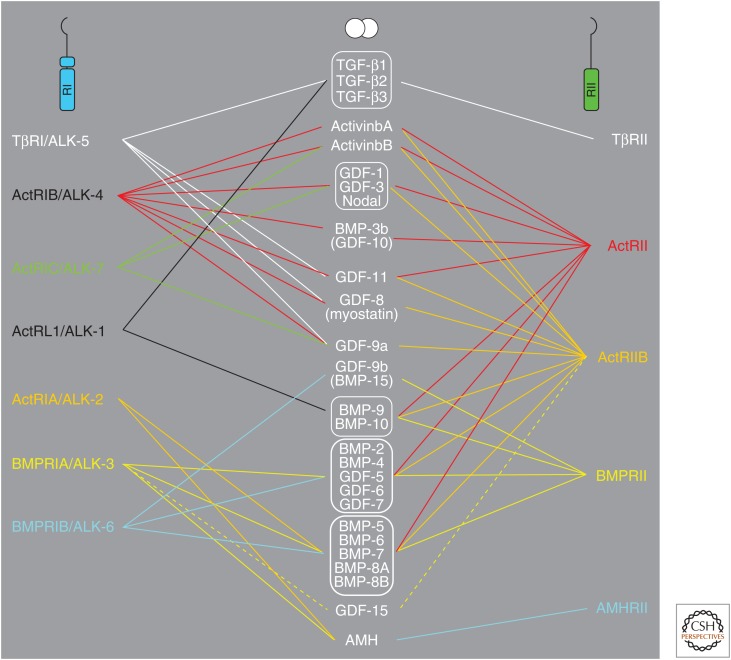
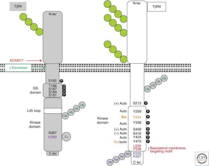
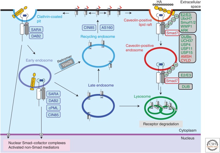
Transforming growth factor β (TGF-β) family members signal via heterotetrameric complexes of type I and type II dual specificity kinase receptors. The activation and stability of the receptors are controlled by posttranslational modifications, such as phosphorylation, ubiquitylation, sumoylation, and neddylation, as well as by interaction with other proteins at the cell surface and in the cytoplasm. Activation of TGF-β receptors induces signaling via formation of Smad complexes that are translocated to the nucleus where they act as transcription factors, as well as via non-Smad pathways, including the Erk1/2, JNK and p38 MAP kinase pathways, and the Src tyrosine kinase, phosphatidylinositol 3'-kinase, and Rho GTPases.


Figures

Figure 1.
Figure 1.
Schematic illustration of the selective binding of members of the transforming growth factor β (TGF-β) family to type I and type II serine/threonine kinase receptors.
Figure 2.
Figure 2.
Schematic illustration of the characteristics of TβRI and TβRII. Structural motifs, as well as posttranslationally modified residues and proteolytic cleavage sites are indicated. Green chains of circles, N-linked glycosylations; black circles with white P, phosphorylation sites (auto, receptor autophosphorylation; other kinases indicated: + or − symbols indicate positive or negative impact on receptor kinase activity); gray circles with green Ub, ubiquitin chains (the exact locations of the acceptor lysines are not known); gray circles with purple Su, sumoylation sites; gray circles with blue Ne, neddylation sites. Ubiquitin and NEDD8 are shown as polymeric chains, whereas sumo is illustrated by a monomer, in accordance with the knowledge about these modifications. TβRI is also autophosphorylated on serine/threonine and tyrosine residues, but their exact locations are not known (not shown). Binding of the adaptor protein Shc to a specific phosphotyrosine is also indicated. N-ter, Amino terminal; C-ter, carboxy terminal.
Figure 3.
Figure 3.
Internalization and intracellular sorting of transforming growth factor β (TGF-β) receptors. Ligand-induced TGF-β receptor complexes in an active, signaling form are shown. Single receptor subunits or oligomeric receptor complexes without ligand are not shown for simplicity. Complex N-glycans attached to the extracellular domain are shown by red chains. TGF-β signaling via Smads or non-Smad mediators can be initiated at the cell surface in clathrin-coated pits or in early endosomes. The transmembrane metalloproteinase ADAM12 promotes TGF-β receptor endocytosis. The membrane-bound adaptor protein SARA, and the adaptors DAB2, cPML, AS160, and CIN85 participate in the early steps of endocytosis and recycling, positively regulate receptor signaling. Receptors can recycle via the recycling endosome back to the cell surface (possibly in the absence of ligand; not shown), a process that is promoted by CIN85. The retromer associates with TβRII via its subunit Vps26 and promotes recycling. Moreover, AS160 promotes translocation of intracellularly located receptors to the cell surface. Receptors can enter the late endosome and either recycle or continue to the lysosomes where final receptor degradation takes place. Receptors internalized by lipid rafts enter caveolin-positive vesicles and eventually reach to lysosomes. TβRI binds Smad7, which carries several ubiquitin ligases (E2, E3) and deubiquitylases (DUBs) that regulate the rate and degree of ubiquitylation of the receptor (green molecules). Smad7 can also associate with ubiquitin ligases and DUBs that control its own ubiquitylation, and thus indirectly affect receptor internalization and degradation (red molecules). Proteasomal degradation of receptors and regulatory proteins occur during the internalization and sorting processes (not shown). Some of the molecules depicted in the figure have not been discussed in the text, such as UbcH7, WWP1, ARK, UCH37, AMSH, and CYLD. For authoritative discussion of the mechanisms of ubiquitylation and deubiquitylation during TGF-β family signaling, the reader is addressed to recent review articles (De Boeck and ten Dijke 2012; Herhaus and Sapkota 2014).

References

    1. Abdalla SA, Cymerman U, Rushlow D, Chen N, Stoeber GP, Lemire EG, Letarte M. 2005. Novel mutations and polymorphisms in genes causing hereditary hemorrhagic telangiectasia. Hum Mutat 25: 320–321. - PubMed
    1. Abdollah S, Macías-Silva M, Tsukazaki T, Hayashi H, Attisano L, Wrana JL. 1997. TβRI phosphorylation of Smad2 on Ser465 and Ser467 is required for Smad2–Smad4 complex formation and signaling. J Biol Chem 272: 27678–27685. - PubMed
    1. Akhurst RJ, Hata A. 2012. Targeting the TGFβ signalling pathway in disease. Nat Rev Drug Discov 11: 790–811. - PMC - PubMed
    1. Al-Salihi MA, Herhaus L, Macartney T, Sapkota GP. 2012. USP11 augments TGF β signalling by deubiquitylating ALK5. Open Biol 2: 120063. - PMC - PubMed
    1. Arthur HM, Ure J, Smith AJ, Renforth G, Wilson DI, Torsney E, Charlton R, Parums DV, Jowett T, Marchuk DA, et al. 2000. Endoglin, an ancillary TGFβ receptor, is required for extraembryonic angiogenesis and plays a key role in heart development. Dev Biol 217: 42–53. - PubMed

Publication types

Substances