Review
. 2016 Sep 1;6(9):a025361.
doi: 10.1101/cshperspect.a025361.

Bacterial Protein Synthesis as a Target for Antibiotic Inhibition

Affiliations
Review

Bacterial Protein Synthesis as a Target for Antibiotic Inhibition

Stefan Arenz et al. Cold Spring Harb Perspect Med. .

Abstract

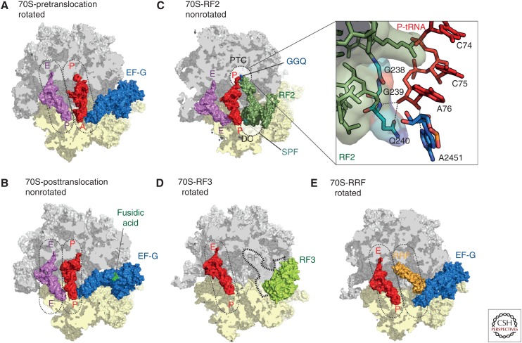
Protein synthesis occurs on macromolecular machines, called ribosomes. Bacterial ribosomes and the translational machinery represent one of the major targets for antibiotics in the cell. Therefore, structural and biochemical investigations into ribosome-targeting antibiotics provide not only insight into the mechanism of action and resistance of antibiotics, but also insight into the fundamental process of protein synthesis. This review summarizes the recent advances in our understanding of protein synthesis, particularly with respect to X-ray and cryoelectron microscopy (cryo-EM) structures of ribosome complexes, and highlights the different steps of translation that are targeted by the diverse array of known antibiotics. Such findings will be important for the ongoing development of novel and improved antimicrobial agents to combat the rapid emergence of multidrug resistant pathogenic bacteria.


Figures

Figure 1.
Figure 1.
The prokaryotic ribosome. (A) Overview of the Escherichia coli 70S ribosome (Dunkle et al. 2011) with 30S subunit colored in yellow and 50S subunit in gray. (B) Table of assembly components of the 70S ribosome as well as the 30S and 50S subunits. (C) Schematic representation of the prokaryotic ribosome bound with three transfer RNAs (tRNAs) showing the 30S subunit (yellow), 50S subunit (gray), A-tRNA (green), P-tRNA (red), E-tRNA (pink), nascent polypeptide chain (violet), and mRNA (black). The peptidyltransferase center (PTC) on the 50S subunit is depicted as a dashed sphere.
Figure 2.
Figure 2.
Overview of antibiotics inhibiting the prokaryotic translation cycle. Overview of antibiotics, inhibiting translation initiation (green), translation elongation (yellow), and translation termination/recycling (red) of the prokaryotic translation cycle (modified from Sohmen et al. 2009). tRNA, Transfer RNA; mRNA, messenger RNA; IF, initiation factor; EF-Tu, elongation factor Tu; GDP, guanosine diphosphate; EF-G, elongation factor G; GTP, guanosine triphosphate; RRF, ribosome recycling factor.
Figure 3.
Figure 3.
Initiation of translation. Schematic assembly of the (A) 30S preinitiation complex (PIC), (B) 70S-PIC, and (C) 70S-IC during translation initiation with 30S subunit (yellow), 50S subunit (gray), messenger RNA (mRNA) (dark gray), initiator transfer RNA (tRNA) (red), initiation factors (IFs): IF1 (brown), IF2 (purple), and IF3 (green). (D) Crystal structure of the prokaryotic ribosome with zoom onto the interaction of the Shine–Dalgarno (SD) sequence of canonical mRNAs (orange) with the anti-SD at the 3′ end of the 16S ribosomal RNA (rRNA) (green), including P-site tRNA (red) and E-site tRNA (pink) (Yusupova et al. 2006). (E) Crystal structure of IF1 (brown) bound to the 30S subunit (yellow) with highlighted h44 (blue) of the 16S rRNA and ribosomal protein S12 (red) (Carter et al. 2001). (F) Binding sites of dityromycin (PDB 4NVU) (Bulkley et al. 2014), gentamicin (PDB 4V53) (Borovinskaya et al. 2007a), thermorubin (PDB 3UXT) (Bulkley et al. 2012), viomycin (PDB 3KNH) (Stanley et al. 2010), neomycin (PDB 2QAL) (Borovinskaya et al. 2007a), negamycin (PDB 4RBH) (Polikanov et al. 2014c), tetracycline (PDB 4G5K) (Jenner et al. 2013), tigecycline (PDB 4G5T) (Jenner et al. 2013), amicoumacin A (PDB 4RB5) (Polikanov et al. 2014a), edeine (PDB 1I95) (Pioletti et al. 2001), kasugamycin (PDB 1VS5) (Schuwirth et al. 2006), pactamycin (PDB 4RBB) (Polikanov et al. 2014c), and emetine (PDB 3J7A) (Wong et al. 2014), on the small subunit (SSU) (yellow). P/I, Peptidyl/initiation.
Figure 4.
Figure 4.
Decoding and peptide bond formation on the ribosome. (A) Crystal structure of the ribosome in complex with elongation factor Tu (EF-Tu) (purple), A/T-transfer RNA (tRNA) (green), P-tRNA (red), E-tRNA (pink), kirromycin (light green), messenger RNA (mRNA) (brown), 30S subunit (yellow), and 50S subunit (gray) (Voorhees et al. 2010) with superimposed binding positions of thiostrepton (orange) (Harms et al. 2008) and tetracycline (blue) (Jenner et al. 2013). The decoding center (DC) on the 30S subunit is depicted as dashed-lined sphere. (B) DC within the small ribosomal subunit with A-tRNA (green), mRNA (brown), and 16S ribosomal RNA (rRNA) nucleotides G530, A1492, and A1493 (yellow) (Demeshkina et al. 2012). (C) Positions of the A-tRNA (green) and P-tRNA (red) within the peptidyltransferase center (PTC) of the 50S subunit. The nucleophilic attack of the A-tRNA α-amino group (Nα) onto the P-tRNA carbonyl-carbon (Ccarbonyl) is indicated with an arrow. (D) Overview and (E) zoom onto the binding sites of Onc112 (PDB 4ZER) (Seefeldt et al. 2015), hygromycin A (PDB 4Z3R) (Polikanov et al. 2015), A201A (PDB 4Z3S) (Polikanov et al. 2015), chloramphenicol (PDB 3OFC) (Dunkle et al. 2010), linezolid (PDB 3DLL) (Wilson et al. 2008), clindamycin (PDB 3OFZ) (Dunkle et al. 2010), erythromycin (PDB 3OFR) (Dunkle et al. 2010), bactobolin A (PDB 4WWE) (Amunts et al. 2015), and blasticidin S (PDB 4L6J) (Svidritskiy et al. 2013) within the PTC (dashed lines in D) of the 50S subunit (gray) with A-tRNA (green) and P-tRNA (red).
Figure 5.
Figure 5.
The prokaryotic ribosome. Crystal structures of the ribosome bound with elongation factor G (EF-G) in the (A) pre- (Brilot et al. 2013), and (B) posttranslocation state (Lin et al. 2015), with 30S subunit (yellow), 50S subunit (gray), EF-G (blue), P-transfer RNA (tRNA) (red), E-tRNA (pink), and fusidic acid (green). (C) Crystal structure of RF2 (dark green) bound to the poststate ribosome with P-tRNA (red) and E-tRNA (pink) (Weixlbaumer et al. 2008). The peptidyltransferase center (PTC) on the 50S (gray) and the DC on the 30S subunit (yellow) are indicated as dashed lines. The insert zooms onto the PTC showing the GGQ motif of RF2 interacting with the CCA-end of the P-tRNA. (D) Crystal structure of RF3 (pale green) bound to the rotated 70S ribosome (Zhou et al. 2012b) with superimposed position of the P/E hybrid tRNA (red) (Brilot et al. 2013). The position of class I release factors is indicated with dashed lines. (E) Crystal structure of ribosome recycling factor (RRF, orange) bound to the rotated ribosome (Borovinskaya et al. 2007a) with superimposed positions of P/E hybrid tRNA (red) and prestate EF-G (blue) (Brilot et al. 2013).

References

    1. Allen G, Zavialov A, Gursky R, Ehrenberg M, Frank J. 2005. The cryo-EM structure of a translation initiation complex from Escherichia coli. Cell 121: 703–712. - PubMed
    1. Amunts A, Fiedorczuk K, Truong TT, Chandler J, Peter Greenberg E, Ramakrishnan V. 2015. Bactobolin A binds to a site on the 70S ribosome distinct from previously seen antibiotics. J Mol Biol 427: 753–755. - PMC - PubMed
    1. Barat C, Datta PP, Raj VS, Sharma MR, Kaji H, Kaji A, Agrawal RK. 2007. Progression of the ribosome recycling factor through the ribosome dissociates the two ribosomal subunits. Mol Cell 27: 250–261. - PubMed
    1. Belova L, Tenson T, Xiong LQ, McNicholas PM, Mankin AS. 2001. A novel site of antibiotic action in the ribosome: Interaction of evernimicin with the large ribosomal subunit. Proc Natl Acad Sci 98: 3726–3731. - PMC - PubMed
    1. Blanchard SC, Gonzalez RL, Kim HD, Chu S, Puglisi JD. 2004a. tRNA selection and kinetic proofreading in translation. Nat Struct Mol Biol 11: 1008–1014. - PubMed

MeSH terms