. 2016 Sep 1;197(5):1843-51.
doi: 10.4049/jimmunol.1600556. Epub 2016 Aug 1.

Innate Invariant NKT Cell Recognition of HIV-1-Infected Dendritic Cells Is an Early Detection Mechanism Targeted by Viral Immune Evasion

Affiliations

Innate Invariant NKT Cell Recognition of HIV-1-Infected Dendritic Cells Is an Early Detection Mechanism Targeted by Viral Immune Evasion

Dominic Paquin-Proulx et al. J Immunol. .

Abstract

Invariant NKT (iNKT) cells are innate-like T cells that respond rapidly with a broad range of effector functions upon recognition of glycolipid Ags presented by CD1d. HIV-1 carries Nef- and Vpu-dependent mechanisms to interfere with CD1d surface expression, indirectly suggesting a role for iNKT cells in control of HIV-1 infection. In this study, we investigated whether iNKT cells can participate in the innate cell-mediated immune response to HIV-1. Infection of dendritic cells (DCs) with Nef- and Vpu-deficient HIV-1 induced upregulation of CD1d in a TLR7-dependent manner. Infection of DCs caused modulation of enzymes in the sphingolipid pathway and enhanced expression of the endogenous glucosylceramide Ag. Importantly, iNKT cells responded specifically to rare DCs productively infected with Nef- and Vpu-defective HIV-1. Transmitted founder viral isolates differed in their CD1d downregulation capacity, suggesting that diverse strains may be differentially successful in inhibiting this pathway. Furthermore, both iNKT cells and DCs expressing CD1d and HIV receptors resided in the female genital mucosa, a site where HIV-1 transmission occurs. Taken together, these findings suggest that innate iNKT cell sensing of HIV-1 infection in DCs is an early immune detection mechanism, which is independent of priming and adaptive recognition of viral Ag, and is actively targeted by Nef- and Vpu-dependent viral immune evasion mechanisms.


Figures

FIGURE 1.
FIGURE 1.
Elevated CD1d levels in response to TLR7 signals are antagonized by Nef and Vpu. mdDCs were infected with DHIV3 for 6–7 d and stained for surface CD11c, CD1d, and intracellular p24 and analyzed by flow cytometry. (A) Flow cytometry histogram of CD1d expression in mdDCs infected with wt DHIV3. (B) CD1d mean fluorescence intensity (MFI) on p24 and p24+ mdDCs infected with wt (n = 10). (C) Flow cytometry histogram of CD1d expression in mdDCs infected with ∆nefvpu DHIV3. (D) CD1d MFI on p24 and p24+ mdDCs infected with ∆nefvpu DHIV3 (n = 12). (E) Change in CD1d MFI was determined in p24+ mdDCs in comparison with p24 mdDCs (n = 8) infected with BaL virus, or founder virus strains pCH077.t/2627 (F1), pRHPA.c/2635 (F2), and pTHRO.c/2626 (F3). mdDC donors where p24+ infection rates in the 0.1–0.5% range were detected were included in the analysis. (F) mdDCs were infected with DHIV3 for 3 d (n = 7), 10 μM chloroquine was added for 24 h, and cells were stained for surface CD11c, CD1d, and intracellular p24 and analyzed by flow cytometry. (G) mdDCs were treated with TLR3 agonist polyinosinic-polycytidylic acid, TLR7 agonist imiquimod, or TLR8 agonist ssRNA40/LyoVec for 24 h and levels of surface CD1d were analyzed by flow cytometry (n = 6); box and whiskers, minimum to maximum. (H) mdDCs from eight independent donors were infected with DHIV3 ∆nefvpu, treated with TLR7 oligonucleotide antagonist IRS954 or control oligonucleotide on day 4, and stained for surface CD11c, CD1d, and intracellular p24 and analyzed by flow cytometry on day 5. *p < 0.05, **p < 0.01 by Wilcoxon signed rank test. MFI, mean fluorescence intensity.
FIGURE 2.
FIGURE 2.
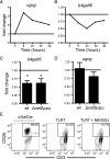
TLR7 stimulation induces changes in ugcg and b4galt6 expression and iNKT cell activation. mdDCs were stimulated with imiquimod for 8–48 h and expression levels of (A) ugcg and (B) b4galt6 were assessed by quantitative PCR; data are representative of three independent experiments. mdDCs were infected with DHIV3 wt or DHIV3 ∆nefvpu for 6 d and expression of (C) b4galt6 (n = 7) and (D) ugcg (n = 8) was assessed by quantitative PCR; mean and SD from independent experiments is shown. (E) The human iNKT cell clone HDD3 was cocultured with mdDCs in the presence of imiquimod for 24 h. As a positive control, αGalCer (100 ng/ml) was added to one coculture condition. mdDCs were left untreated or preincubated with 50 μM NB-DGJ for 24 h. FACS plots show one representative out of six independent experiments. *p < 0.05 by t test.
FIGURE 3.
FIGURE 3.
GlcCer 24:1 accumulates in HIV-1–infected DCs. mdDCs were infected with DHIV3 eGFP for 6 d and sorted based on eGFP expression. Pellets of sorted cells were extracted and then subjected to mass spectrometry analysis for glycolipid content. (A) Representative selected reaction monitoring (SRM) UPLC-MS/MS chromatograms of GlcCer 24:1. (B) Relative GlcCer 24:1 levels in eGFP mdDCs compared with eGFP+ mdDCs (n = 7). *p < 0.05 by Wilcoxon signed rank test.
FIGURE 4.
FIGURE 4.
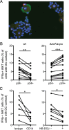
Innate targeting of HIV-1–infected cells by iNKT cells is inhibited by Nef and Vpu. (A) mdDCs were infected with ∆nefvpu DHIV3 for 6 d before coculture with iNKT cells for 4 h in the presence of brefeldin A. Following cytospinning, cells were fixed, permeabilized, and stained with anti–IFN-γ mAb, anti-p24 mAb, and DAPI. IFN-γ, red; p24, green; DAPI, blue. +, IFN-γ+ iNKT cells in contact with mdDCs; white circles, DCs. Image captured using a ×60 objective. (B) Quantification of iNKT cell IFN-γ production after coculture with DHIV3 wt or DHIV3 ΔnefΔvpu–infected mdDCs (n = 13). (C) mdDCs infected with DHIV3 ΔnefΔvpu were treated with anti-CD1d mAb (n = 6) or NB-DGJ inhibitor for 24 h (n = 7) before coculture with iNKT cells and quantification of iNKT cell IFN-γ production. *p < 0.05, **p < 0.01, ***p < 0.001 by Wilcoxon signed rank test.
FIGURE 5.
FIGURE 5.
DCs that express CD1d, CD4, and CCR5 are present in the female genital mucosa. (A) Representative FACS plots for identification of iNKT cells in the female genital mucosa. (B) Frequency of DCs, CD1d expression on DCs, and frequency of CD1d+ DCs in the endometrium and cervix (n = 11). Where paired samples were available, those data points are connected with a solid line. (C) Comparison of CD4, CCR5, and DC-SIGN expression mean fluorescence intensity (MFI) on CD1d and CD1d+ DCs in the endometrium (n = 10). (D) In situ staining for DAPI (blue), CD1d (red), and HLA-DR (green) in the endometrium, Scale bar, 120 μm. Images were collected with a ×20 objective. *p < 0.05, **p < 0.01 by Mann–Whitney U test in (B) and by Wilcoxon signed rank test in (C).
FIGURE 6.
FIGURE 6.
Proposed model of iNKT cell immune surveillance in the genital mucosa. Effectiveness of iNKT cell immune surveillance depends on competition between the immuno-evasive capacity of infecting HIV-1 strains as well as the TLR7-mediated upregulation of the CD1d Ag presentation pathway.

References

    1. Van Rhijn I., Moody D. B. 2015. Donor unrestricted T cells: a shared human T cell response. J. Immunol. 195: 1927–1932. - PMC - PubMed
    1. Mori L., Lepore M., De Libero G. 2016. The immunology of CD1- and MR1-restricted T cells. Annu. Rev. Immunol. 34: 479–510. - PubMed
    1. Brennan P. J., Brigl M., Brenner M. B. 2013. Invariant natural killer T cells: an innate activation scheme linked to diverse effector functions. Nat. Rev. Immunol. 13: 101–117. - PubMed
    1. Van Kaer L., Parekh V. V., Wu L. 2013. Invariant natural killer T cells as sensors and managers of inflammation. Trends Immunol. 34: 50–58. - PMC - PubMed
    1. Kinjo Y., Wu D., Kim G., Xing G. W., Poles M. A., Ho D. D., Tsuji M., Kawahara K., Wong C. H., Kronenberg M. 2005. Recognition of bacterial glycosphingolipids by natural killer T cells. Nature 434: 520–525. - PubMed

Publication types

MeSH terms