. 2016 Sep 1;197(5):1832-42.
doi: 10.4049/jimmunol.1600143. Epub 2016 Aug 1.

Induction of Th1-Biased T Follicular Helper (Tfh) Cells in Lymphoid Tissues during Chronic Simian Immunodeficiency Virus Infection Defines Functionally Distinct Germinal Center Tfh Cells

Affiliations

Induction of Th1-Biased T Follicular Helper (Tfh) Cells in Lymphoid Tissues during Chronic Simian Immunodeficiency Virus Infection Defines Functionally Distinct Germinal Center Tfh Cells

Vijayakumar Velu et al. J Immunol. .

Abstract

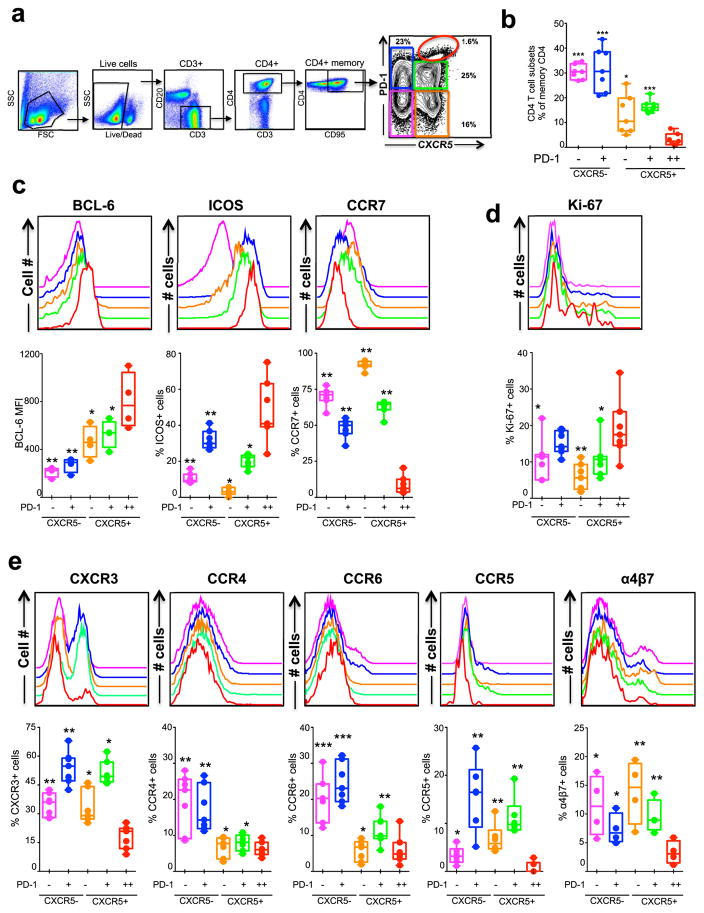
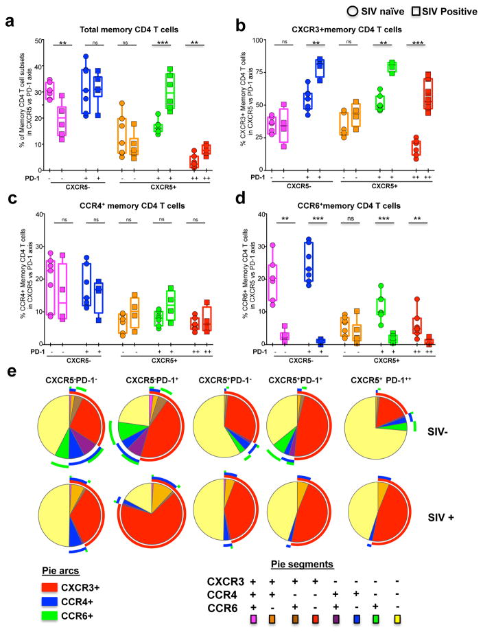
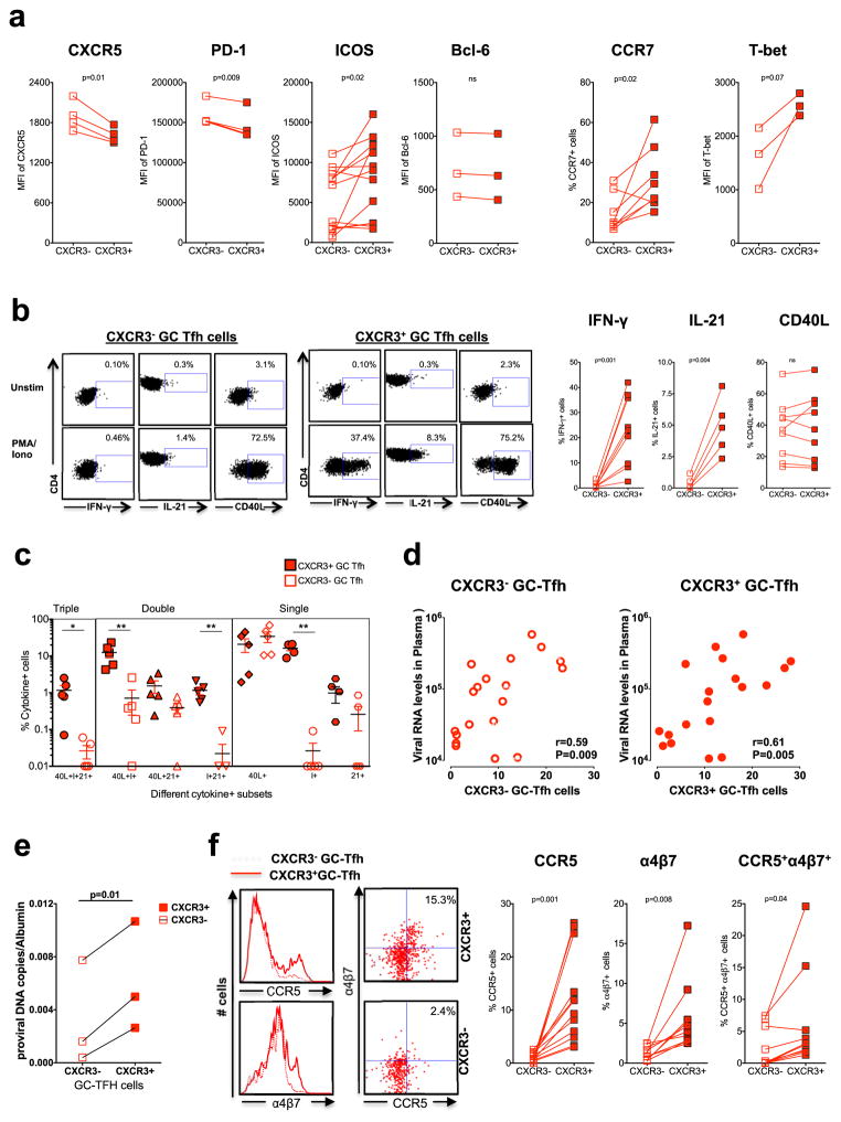
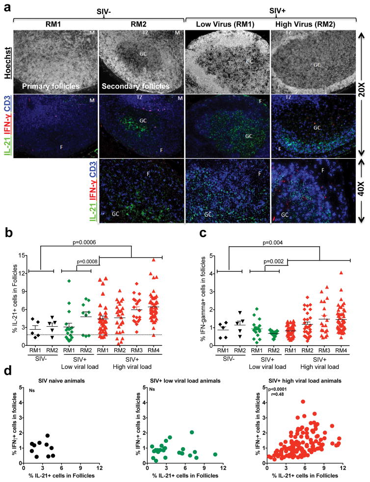
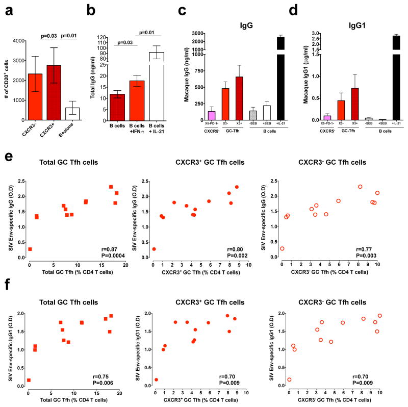
Chronic HIV infection is associated with accumulation of germinal center (GC) T follicular helper (Tfh) cells in the lymphoid tissue. The GC Tfh cells can be heterogeneous based on the expression of chemokine receptors associated with T helper lineages, such as CXCR3 (Th1), CCR4 (Th2), and CCR6 (Th17). However, the heterogeneous nature of GC Tfh cells in the lymphoid tissue and its association with viral persistence and Ab production during chronic SIV/HIV infection are not known. To address this, we characterized the expression of CXCR3, CCR4, and CCR6 on GC Tfh cells in lymph nodes following SIVmac251 infection in rhesus macaques. In SIV-naive rhesus macaques, only a small fraction of GC Tfh cells expressed CXCR3, CCR4, and CCR6. However, during chronic SIV infection, the majority of GC Tfh cells expressed CXCR3, whereas the proportion of CCR4(+) cells did not change, and CCR6(+) cells decreased. CXCR3(+), but not CXCR3(-), GC Tfh cells produced IFN-γ (Th1 cytokine) and IL-21 (Tfh cytokine), whereas both subsets expressed CD40L following stimulation. Immunohistochemistry analysis demonstrated an accumulation of CD4(+)IFN-γ(+) T cells within the hyperplastic follicles during chronic SIV infection. CXCR3(+) GC Tfh cells also expressed higher levels of ICOS, CCR5, and α4β7 and contained more copies of SIV DNA compared with CXCR3(-) GC Tfh cells. However, CXCR3(+) and CXCR3(-) GC Tfh cells delivered help to B cells in vitro for production of IgG. These data demonstrate that chronic SIV infection promotes expansion of Th1-biased GC Tfh cells, which are phenotypically and functionally distinct from conventional GC Tfh cells and contribute to hypergammaglobulinemia and viral reservoirs.


Figures

Figure 1
Figure 1. Characterization of five memory CD4 T cell subsets defined based on CXCR5 and PD-1 expression in the lymph node (LN) of SIV naïve rhesus macaques (RM)
(a, b) Relative distribution of five memory CD4 T cell subsets (CD95+ CD4+ cells) in the LN defined based on CXCR5 and PD-1 expression. Representative flow plot is shown in (a) and summary for a group (n=7) of SIV-naïve RM is shown in (b). (c) Expression of Tfh markers Bcl-6 and ICOS, and non-Tfh marker CCR7 (n=7). (d) Expression of proliferation marker Ki-67 (n=7). (e) Expression of different chemokine receptors CXCR3, CCR4, CCR6, CCR5 and α4β7 (n=7 except for α4β7 for which n=4). P values shown are for CXCR5+ PD-1++ (GC-Tfh) subset vs other subsets. *p < 0.05, **p < 0.01, ***p < 0.001, ****p < 0.0001. The legend for histogram plots shown in c–e is shown in b.
Figure 2
Figure 2. Enrichment of CXCR3+ CD4 T cells in the LN during chronic SIV infection
(a) Relative distribution of five memory CD4 T cell subsets in the LN defined based on CXCR5 and PD-1 expression before and after (24 weeks) SIV infection. Expression of CXCR3 (b), CCR4 (c) and CCR6 (d) on five memory CD4 T cell subsets defined based on CXCR5 and PD-1 expression before and 24 weeks after SIV infection. (e) Pie charts summarizing the co-expression of chemokine receptors CXCR3, CCR4 and CCR6 on different memory CD4 T cell subsets. Red arcs identify CXCR3+ populations, blue arcs identify CCR4+ population, green arcs identify CCR6+ population. Data for seven SIV and SIV+ animals are shown.
Figure 3
Figure 3. CXCR3+ GC-Tfh cells are phenotypically and functionally distinct from CXCR3 GC-Tfh cells
(a) Expression of Tfh and Th1 markers on CXCR3+ and CXCR3 GC-Tfh cells (b) Expression of IFN-γ, IL-21 and CD40L by sorted CXCR3+ and CXCR3 GC-Tfh cells following stimulation with PMA/Ionomycin. Representative flow plots are shown on the left. (c) Boolean analysis determining co-expression of IFN-γ, IL-21 and CD40L. (d) Correlation between CXCR3+ GC-Tfh or CXCR3 GC-Tfh and plasma viral RNA levels at 24 weeks post SIV infection. (e) Copies of SIV proviral DNA in sorted CXCR3+ and CXCR3 GC-Tfh cells. (f) Expression of CCR5 and α4β7 on CXCR3+ and CXCR3 GC-Tfh cells.
Figure 4
Figure 4. Chronic SIV infection results in accumulation of IL-21+ and IFN-γ+ cells within the follicles
(a) Representative IHC images showing IFN-γ+ and IL-21+ cells in the follicles, T cell zone, and medulla of LN of SIV-naïve and SIV+ RMs with low (<100 copies) and high (>10,000 copies) viral load. (b) Expression of IL-21 in follicles. (c) Expression of IFNγ in follicles. Each dot indicates the percentage of cytokine intensity in one follicle. (d) Correlation between the expression of IFN-γ+ and IL-21+ cells in follicles.
Figure 5
Figure 5. Both CXCR3+ and CXCR3 GC-Tfh cells help B cells equally irrespective of their functional difference
Sorted CXCR3+ GC-Tfh and CXCR3 GC-Tfh cells from chronic SIV infected macaques were co-cultured with autologous sorted B cells. (a) Total numbers of viable B cells at day 5 from the T and B co-culture experiment. (b) IgG production by sorted B cells (B cell alone) from SIV infected RM in the presence of IFN-γ (5ng/ml) or IL-21 (10ng/ml). (c) IgG (n=4) and (d) IgG1 (n=3) concentrations at day 9 in T and B co-culture experiments. (e) Correlation between SIV-envelop specific IgG (at 1: 9000 dilution of sera) antibody levels and total, CXCR3+ or CXCR3 GC-Tfh cells at 24 weeks post SIV infection. (f) Correlation between SIV-envelop specific IgG1 (at 1: 450 dilution of sera) antibody levels and total, CXCR3+ or CXCR3 GC-Tfh cells at 24 weeks post SIV infection.

References

    1. O’Shea JJ, Paul WE. Mechanisms underlying lineage commitment and plasticity of helper CD4+ T cells. Science. 2010;327:1098–1102. - PMC - PubMed
    1. Pepper M, Jenkins MK. Origins of CD4(+) effector and central memory T cells. Nature immunology. 2011;12:467–471. - PMC - PubMed
    1. Bluestone JA, Mackay CR, O’Shea JJ, Stockinger B. The functional plasticity of T cell subsets. Nature reviews Immunology. 2009;9:811–816. - PMC - PubMed
    1. Sallusto F, Geginat J, Lanzavecchia A. Central memory and effector memory T cell subsets: function, generation, and maintenance. Annual review of immunology. 2004;22:745–763. - PubMed
    1. Kim CH, Rott LS, Clark-Lewis I, Campbell DJ, Wu L, Butcher EC. Subspecialization of CXCR5+ T cells: B helper activity is focused in a germinal center-localized subset of CXCR5+ T cells. The Journal of experimental medicine. 2001;193:1373–1381. - PMC - PubMed

MeSH terms