. 2016 Sep 23;291(39):20353-71.
doi: 10.1074/jbc.M116.736967. Epub 2016 Aug 1.

Deciphering the Molecular and Functional Basis of RHOGAP Family Proteins: A SYSTEMATIC APPROACH TOWARD SELECTIVE INACTIVATION OF RHO FAMILY PROTEINS

Affiliations

Deciphering the Molecular and Functional Basis of RHOGAP Family Proteins: A SYSTEMATIC APPROACH TOWARD SELECTIVE INACTIVATION OF RHO FAMILY PROTEINS

Ehsan Amin et al. J Biol Chem. .

Abstract

RHO GTPase-activating proteins (RHOGAPs) are one of the major classes of regulators of the RHO-related protein family that are crucial in many cellular processes, motility, contractility, growth, differentiation, and development. Using database searches, we extracted 66 distinct human RHOGAPs, from which 57 have a common catalytic domain capable of terminating RHO protein signaling by stimulating the slow intrinsic GTP hydrolysis (GTPase) reaction. The specificity of the majority of the members of RHOGAP family is largely uncharacterized. Here, we comprehensively investigated the sequence-structure-function relationship between RHOGAPs and RHO proteins by combining our in vitro data with in silico data. The activity of 14 representatives of the RHOGAP family toward 12 RHO family proteins was determined in real time. We identified and structurally verified hot spots in the interface between RHOGAPs and RHO proteins as critical determinants for binding and catalysis. We have found that the RHOGAP domain itself is nonselective and in some cases rather inefficient under cell-free conditions. Thus, we propose that other domains of RHOGAPs confer substrate specificity and fine-tune their catalytic efficiency in cells.

Keywords: GTPase; GTPase-activating protein (GAP); RAC (RAC GTPase); Ras homolog gene family, member A (RHOA); arginine finger; crystal structure; deleted in liver cancer 1 (DLC1); protein-protein interaction; signal transduction; substrate specificity.


Figures

FIGURE 1.
FIGURE 1.
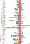
Evolutionary conservation of domains of the RHOGAP family. Domain composition of 66 RHOGAPs is presented according to their phylogenetic categorization based on GAP domain alignment. In addition to a catalytic GAP domain (red), most RHOGAPs have multiple other functional domains, which are probably involved in lipid and membrane binding (blue), protein interaction (green), or enzymatic activities (red and orange). A scale of amino acid numbers in increments of 200 is shown at the bottom; the total number of the amino acids of the respective RHOGAPs is listed in Table 1. Domain properties and statistics are compiled in supplemental Tables S4 and S5.
FIGURE 2.
FIGURE 2.
tamraGTP and cy3GTP but not mant-GTP as fluorescent sensors for monitoring GAP-stimulated GTP hydrolysis of RHO proteins in real time. A, chemical structure of fluorescent reporter groups (mant, TAMRA-ethylenediamine, and cy3-ethylenediamine) coupled to GTP and its non-hydrolyzable analog GppNHp. B, stimulated GTP hydrolysis of CDC42 and RHOA (0.2 μm) was measured using fluorescent GTP and p50GAP (10 μm) in a Hi-Tech Scientific (SF-61) stopped-flow instrument and a buffer containing 30 mm Tris, pH 7.5, 10 mm potassium phosphate, 10 mm MgCl2, and 3 mm dithiothreitol at 25 °C as described (2). In contrast to mantGTP, which did not provide any significant change in fluorescence, cy3GTP turned out to be most suitable for the RHO isoforms and tamraGTP the most suitable for CDC42-and RAC-like proteins (CDC42 is shown as a representative) as well as for RHOD and RIF. Observed rate constants (kobs) for GAP-catalyzed GTPase reactions can be obtained by single exponential fitting of the fluorescence decay using GraFit program.
FIGURE 3.
FIGURE 3.
Varying activity and broad selectivity of the RHOGAP family proteins. Individual GTP hydrolysis reaction rates (kobs; values on the bar charts) of 12 RHO proteins (0.2 μm, respectively) in the absence (no GAP) and in the presence of 10 RHOGAPs (10 μm, respectively) are plotted as bar charts. All data shown are an average of 4–5 different experiments. Color coding is the same as in Table 2 and changes from green for very high to yellow for middle and to red for no GAP activity.
FIGURE 4.
FIGURE 4.
Statistical diagram of the catalytic efficiency of the RHOGAPs. Values of fold activation are plotted against respective RHOGAP-RHO protein pairs in numeric order. This diagram illustrates the broad spectrum of catalytic efficiencies and substrate-specific properties of various RHOGAPs for the different RHO proteins, which are divided into six efficiency groups as indicated. Color codes are the same as used in Fig. 3 and Table 2.
FIGURE 5.
FIGURE 5.
Binding affinity of RHOGAPs to CDC42. Real time monitoring of the association reaction rates of GAPs (10 μm) with mantGppNHp-bound CDC42 (0.2 μm) has been measured and represented as bar diagram (left panel) in direct comparison with the reaction rates obtained for the respective GAP-stimulated GTP hydrolysis of CDC42 (right panel).
FIGURE 6.
FIGURE 6.
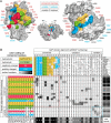
Interaction interface between RHO and RHOGAP proteins. A, interacting residues (<4 Å in distance; color-coded) of the RHO-RHOGAP complex are indicated using an open book representation (rotated 90° along a horizontal axis) of the crystal structure of the RHOA-p50 complex (PDB code 1TX4; RHOA, aa 1–181; p50, aa 236–431). Conserved and variable residues are shown in red, blue, and black, respectively. Coloring criteria were taken from the interaction matrix in B. B, interaction matrix of RHO and RHOGAP proteins. The interacting residues (<4 Å in distance) were determined using the six available crystal structures of RHO-RHOGAP proteins complexes counting eight distinct RHO-GAP pairs (supplemental Table S1). They are shown with corresponding residues from the alignment of the GAP and G domains of RHOGAPs and RHO proteins (supplemental Figs. S1 and S2) used in this study, respectively. Orientation numbers for interacting residues correspond to the numbering of p50 and RHOA, respectively. Residues sharing more than 60% sequence similarity in alignments (supplemental Figs. S1 and S2) are colored as follows: yellow, hydrophobic residues; green, hydrophilic residues; blue, positively charged residues; red, negatively charged residues. Numbers (0–8) in the black-gray-white gradient-colored boxes illustrate the respective contacts found in eight RHO-GAP pairs. Red and blue boxes represent contacts between conserved and variable regions, respectively.
FIGURE 7.
FIGURE 7.
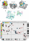
Interaction matrix adapted for the crystal structure of p190 in complex with RHOA. A, crystal structure of the RHOA-p190 complex (PDB code 5IRC) is illustrated in close representation as surface and ribbon (middle panel) and as open book representation rotated 90° along a horizontal axis. The interacting residues (<4 Å in distance) are color-coded as in Fig. 6. B, overlay of p190, p50, and ARHGAP20 structures. Overlay of p190 (PDB code 5IRC), p50 (PDB code 1TX4), and GAP20 (PDB code 3MSX) reveals high structural conservation except for the zoomed variable 1 region. C, interaction matrix calculated solely for the RHOA-p190 structure. Corresponding residues from p50 GAP are left as reference. Hot spots are highlighted in agreement with general interaction matrix in Fig. 6. Contacts missing or excessive in the RHOA-p190 structure are shown as red or blue, respectively.
FIGURE 8.
FIGURE 8.
p190-FL acts on RHOA in cells but not on RHOD. Transiently expressed Myc-tagged RHOD (A and B) and RHOA (C and D) were pulled down from HEK293T cell lysates with RHO-binding domains of DIA1 and RTKN, respectively, as GST-fused proteins in the presence of either HA-tagged p190-FL (A and C) or FLAG-tagged p190GAP domain (B and D). R1284A and Q63L variants of p190 and RHOA were used in addition to wild type (WT). Immunoblots of pulled down samples and the cell lysates were performed using antibodies against the respective tags. WB, Western blot.

References

    1. Dvorsky R., and Ahmadian M. R. (2004) Always look on the bright site of Rho: structural implications for a conserved intermolecular interface. EMBO Rep. 5, 1130–1136 - PMC - PubMed
    1. Jaiswal M., Dubey B. N., Koessmeier K. T., Gremer L., and Ahmadian M. R. (2012) in Rho GTPases Methods and Protocols (Rivero F., ed) pp. 37–58, Springer, Germany - PubMed
    1. Zhang B., and Zheng Y. (1998) Regulation of RhoA GTP hydrolysis by the GTPase-activating proteins p190, p50RhoGAP, Bcr, and 3BP-1. Biochemistry 37, 5249–5257 - PubMed
    1. Fidyk N. J., and Cerione R. A. (2002) Understanding the catalytic mechanism of GTPase-activating proteins: demonstration of the importance of switch domain stabilization in the stimulation of GTP hydrolysis. Biochemistry 41, 15644–15653 - PubMed
    1. Haeusler L. C., Blumenstein L., Stege P., Dvorsky R., and Ahmadian M. R. (2003) Comparative functional analysis of the RAC GTPases. FEBS Lett. 555, 556–560 - PubMed

Publication types

MeSH terms

Substances

LinkOut - more resources