. 2016 Aug 2;134(5):405-21.
doi: 10.1161/CIRCULATIONAHA.116.021311.

Beneficial Role of Erythrocyte Adenosine A2B Receptor-Mediated AMP-Activated Protein Kinase Activation in High-Altitude Hypoxia

Affiliations

Beneficial Role of Erythrocyte Adenosine A2B Receptor-Mediated AMP-Activated Protein Kinase Activation in High-Altitude Hypoxia

Hong Liu et al. Circulation. .

Abstract

Background: High altitude is a challenging condition caused by insufficient oxygen supply. Inability to adjust to hypoxia may lead to pulmonary edema, stroke, cardiovascular dysfunction, and even death. Thus, understanding the molecular basis of adaptation to high altitude may reveal novel therapeutics to counteract the detrimental consequences of hypoxia.

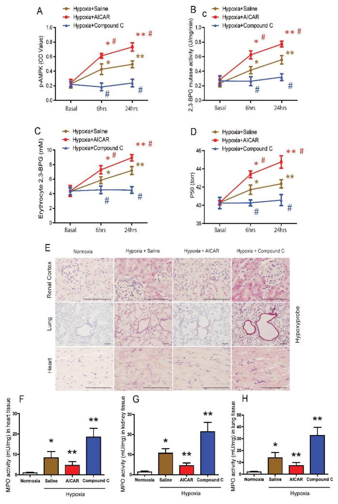
Methods: Using high-throughput, unbiased metabolomic profiling, we report that the metabolic pathway responsible for production of erythrocyte 2,3-bisphosphoglycerate (2,3-BPG), a negative allosteric regulator of hemoglobin-O2 binding affinity, was significantly induced in 21 healthy humans within 2 hours of arrival at 5260 m and further increased after 16 days at 5260 m.

Results: This finding led us to discover that plasma adenosine concentrations and soluble CD73 activity rapidly increased at high altitude and were associated with elevated erythrocyte 2,3-BPG levels and O2 releasing capacity. Mouse genetic studies demonstrated that elevated CD73 contributed to hypoxia-induced adenosine accumulation and that elevated adenosine-mediated erythrocyte A2B adenosine receptor activation was beneficial by inducing 2,3-BPG production and triggering O2 release to prevent multiple tissue hypoxia, inflammation, and pulmonary vascular leakage. Mechanistically, we demonstrated that erythrocyte AMP-activated protein kinase was activated in humans at high altitude and that AMP-activated protein kinase is a key protein functioning downstream of the A2B adenosine receptor, phosphorylating and activating BPG mutase and thus inducing 2,3-BPG production and O2 release from erythrocytes. Significantly, preclinical studies demonstrated that activation of AMP-activated protein kinase enhanced BPG mutase activation, 2,3-BPG production, and O2 release capacity in CD73-deficient mice, in erythrocyte-specific A2B adenosine receptor knockouts, and in wild-type mice and in turn reduced tissue hypoxia and inflammation.

Conclusions: Together, human and mouse studies reveal novel mechanisms of hypoxia adaptation and potential therapeutic approaches for counteracting hypoxia-induced tissue damage.

Keywords: AMP-activated protein kinases; adenosine; altitude; hypoxia, brain; oxygen; signal transduction.


Figures

Figure 1
Figure 1. Metabolomic profiling reveals high altitude hypoxia increases erythrocyte specific Rapoport-Luebering Shunt to induce 2,3-BPG production and P50 levels in humans
(A) Table of human volunteer characteristics including age, height (HT), weight (WT), body mass index (BMI), and schema for high altitude human studies. (n=21) (B) Schematic drawing of erythrocyte-specific Rapoport-Luebering shunt occurring at a branch point in the pathway of anaerobic glycolysis for 2,3-bisphophoglycerate (2,3-BPG) production. (C–F) Metabolomic profiling reveals the significant changes in the relative erythrocyte concentration of glyceraldehyde-3-phosphate (G3P) (C), bisphosphoglycerate (BPG) (D), the monophosphoglycerates, 2-phosphoglycerate (2-PG) and 3-phosphoglycerate (3-PG) (E) and phophoenopyruvate (PEP) (F) at sea level (SL) and at high altitude on day 1 and day 16. (G–J) Erythrocyte 2,3-BPG concentration, P50 level, plasma adenosine concentration and soluble CD73 activity (sCD73) were elevated at high altitude hypoxia on ALT1 and ALT16 over SL. Erythrocyte 2,3-BPG concentration (G), P50 level (H), plasma adenosine concentration (I), and sCD73 (J). Data are expressed as mean ± SEM; *P<0.05 vs SL; **P<0.05 vs ALT1. AU (area under the peak)
Figure 2
Figure 2. Elevated CD73 is essential for hypoxia-induced plasma adenosine, erythrocyte 2,3-BPG and oxygen release capacity to prevent tissue hypoxia, inflammation and lung damage in mice
(A–D) CD73 is essential for hypoxia-induced plasma adenosine, erythrocyte 2,3-BPG concentration and P50 levels in mice. Plasma adenosine concentration (A), soluble CD73 activity (B), Erythrocyte 2,3-BPG (C), and P50 (D) in WT mice and Cd73−/− mice under normoxia or hypoxia (10% O2 1 week). Data are expressed as mean ± SEM; *P<0.05 vs WT under normoxia; **P<0.05 vs WT under hypoxia (n=10). (EJ) Deletion of Cd73−/− results in elevated tissue hypoxia and inflammation infiltration in multiple tissues, as well as pulmonary dysfunction under hypoxia (10% O2 1 week). Immunohistochemical analysis of tissue hypoxia by hypoxyprobe (E) and Myeloperoxidase activity (F) in kidney, lung and heart. Bronchoalveolar lavage fluid (BALF) total cell count (G), BALF albumin concentration (H), BALF interleukin 6 concentration (I) in WT mice and Cd73−/− mice. Data are expressed as mean ± SEM; *P<0.05 vs WT under normoxia; **P<0.05 vs WT under hypoxia (n=10; Scale bar=200μm).
Figure 3
Figure 3. Erythrocyte ADORA2B activation induces erythrocyte 2,3-BPG production and oxygen release capacity to counteract tissue hypoxia, inflammation and lung damage in mice
(A and B) Hypoxia induces 2,3-BPG production (A) and P50 (B) levels in cultured WT, Adora1−/−, Adora2a−/−, Adora3−/− but not Adora2b−/− mouse erythrocytes in a time-dependent manner. *P<0.05 for 3 hours hypoxia vs normoxia; **P<0.05 for 6 hours hypoxia vs 3 hours hypoxia (n=8). (CE) Erythrocyte ADORA2B contributes to hypoxia-induced 2,3-BPG production and P50 levels. Plasma adenosine (C), erythrocyte 2,3-BPG (D) and P50 (E) of EpoR-Cre+ mice and Adora2bf/f/EpoR-Cre+ mice under normoxia or hypoxia (10% O2 1 week). Data are expressed as mean ± SEM; *P<0.05 vs EpoR-Cre+ mice under normoxia; **P<0.05 vs EpoR-Cre+ mice under hypoxia (n=10). Targeted deletion of erythrocyte ADORA2B results in elevated tissue hypoxia and inflammation infiltration in multiple tissues, as well as pulmonary dysfunction under hypoxia (10% O2, 1 week). Immunohistochemical analysis of tissue hypoxia by hypoxyprobe (F) and Myeloperoxidase activity (G) in kidney, lung and heart. Bronchoalveolar lavage fluid (BALF) total cell count (H), BALF albumin concentration (I), BALF interleukin 6 concentration (J) in EpoR-Cre+ mice and Adora2bf/f/EpoR-Cre+ mice. Data are expressed as mean ± SEM; *P<0.05 vs EpoR-Cre+ mice under normoxia; **P<0.05 vs EpoR-Cre+ mice under hypoxia (n=10; Scale bar=200μm).
Figure 4
Figure 4. AMPK functions downstream of erythrocyte ADORA2B and underlies hypoxia-induced 2,3-BPG production by phosphorylation and activation of 2,3-BPG mutase in mice
(AD) Erythrocyte ADORA2B is essential for hypoxia-induced p-AMPK, 2,3-BPG mutase activity, 2,3-BPG concentration and P50 levels in vivo. Erythrocyte p-AMPKα (quantified by ELISA) (A), 2,3-BPG mutase activity (B), 2,3-BPG concentration (C) and P50 (D) of EpoR-Cre+ mice and Adora2bf/f/EpoR-Cre+ mice under normoxia or hypoxia (10% O2, 90% N2) for 1 day, 3 days, and 7 days. Data are expressed as mean ± SEM; *P<0.05 vs EpoR-Cre+ mice under normoxia; **P<0.05 vs EpoR-Cre+ mice under hypoxia for 1 day; #P<0.05 Adora2bf/f/EpoR-Cre+ mice vs EpoR-Cre+ mice at the same time point (n=10). (E) Representative western blot and relative image quantification analysis of p-AMPKα in erythrocytes of EpoR-Cre+ mice and Adora2bf/f/EpoR-Cre+ mice under normoxia or hypoxia (10% O2, 90% N2) for 1 week. (F) Representative western blot and relative image quantification analysis of p-AMPK phosphorylated 2,3-BPG mutase levels in the erythrocyte lysates in EpoR-Cre+ mice and Adora2bf/f/EpoR-Cre+ mice under normoxia or hypoxia (n=3). Data are expressed as mean ± SEM; *P<0.05 vs EpoR-Cre+ mice under normoxia; **P<0.05 vs EpoR-Cre+ mice under hypoxia. (GJ) AICAR treatment significantly stimulated hypoxia-induced erythrocyte AMPK phosphorylation (G), 2,3-BPG mutase activity (H), 2,3-BPG production (I) and P50 levels (J) compared to saline-treated group in Cd73−/− mice and Adora2bf/f/EpoR-Cre+ mice under hypoxia (10% O2, 90% N2) for 3 days. Data are expressed as mean ± SEM; *P<0.05 for AICAR-treated mice vs saline-treated mice (n=8).
Figure 5
Figure 5. In vivo effects of AICAR and Compound C treatment under hypoxia
(AD) AICAR treatment significantly stimulated erythrocyte p-AMPK (A), 2,3-BPG mutase activity (B), 2,3-BPG production (C) and P50 levels (D) in WT mice compared to saline-treated WT mice under hypoxia (8% O2) in a time-dependent manner, while Compound C treatment significantly attenuated erythrocyte p-AMPK (A), 2,3-BPG mutase activity (B), 2,3-BPG production (C) and P50 levels (D) in WT mice compared to saline-treated WT mice under hypoxia (8% O2). Data are expressed as mean ± SEM; *P<0.05 for 6-hour vs basal level, **P<0.05 for 24-hour vs 6-hour. #P<0.05 for AICAR-treated group vs saline group, and Compound C-treated group vs saline group at the same time point (n=8). (EH) AICAR treatment prevented tissue hypoxia and inflammation infiltration, while Compound C treatment aggravated tissue hypoxia and MPO activity in WT mice under hypoxia for 24 hours. IHC analysis of tissue hypoxia by hypoxyprobe in kidney, lung and heart (E) (Scale bar=200μm). MPO activity in heart (F), kidney (G) and lung (H). AICAR or Compound C-treated WT mice after 24 hours hypoxia treatment (n=8). Data are expressed as mean ± SEM; *P<0.05 vs WT mice under normoxia; **P<0.05 vs WT mice with saline treatment under hypoxia.
Figure 6
Figure 6. Erythrocyte p-AMPK and 2,3-BPG mutase activity are induced in humans at high altitude, and AMPK activation induces erythrocyte 2,3-BPG mutase activity, 2,3-BPG production and oxygen release in cultured human erythrocytes
(A) Representative western blot and relative image quantification analysis (n=3 per group) of p-AMPKα subunit phosphorylated at threonine 172 (T172), total AMPKα subunit, and β-actin in the erythrocytes of humans at sea level (SL) and high altitude on day 1 and day 16. Erythrocyte p-AMPK levels quantified by ELISA (B) and 2,3-BPG mutase activity (C) at high altitude ALT1 and ALT16 over SL. (D) Representative western blot and relative image quantification analysis (n=3 per group) of p-AMPK phosphorylated 2,3-BPG mutase at SL and high altitude on day 1 and day 16 in erythrocyte lysates. Data are expressed as mean ± SEM; *P<0.05 for high altitude on day 1 vs SL; **P<0.05 for high altitude on day 16 vs day 1. (EH) AMPK activator AICAR-induced phosphorylation of AMPK (E), 2,3-BPG mutase activity (F), 2,3-BPG concentration (G) and P50 levels (H) in cultured normal human erythrocytes under normoxia in a time-dependent manner. Compound C significantly attenuated the effect of AICAR treatment. Data are expressed as mean ± SEM; *P<0.05 for AICAR-treated 2-hour group vs 1-hour group, and for AICAR-treated 2-hour group vs DMSO-treated 2-hour group, **P<0.05 for AICAR-treated 4-hour group vs 2-hour group, and for AICAR-treated 4-hour group vs DMSO-treated 4-hour group. #P<0.05 for AICAR plus Compound C vs AICAR at the same time point (n=8). (I) Working model: CD73 is essential for high altitude hypoxia-induced plasma adenosine production. Elevated plasma adenosine prevents hypoxia-induced tissue inflammation and damage by activating ADORA2B on erythrocytes to induce 2,3-BPG mutase activity, 2,3-BPG production, and subsequently increased P50 and increased O2 release to peripheral hypoxic tissues. AMPK is a key enzyme that functions downstream of ADORA2B to activate 2,3-BPG mutase, promote 2,3-BPG production and O2 release from erythrocytes. Thus, enhancing the CD73-ADORA2B-AMPK signaling pathway is a promising therapeutic strategy to treat or prevent hypoxia-induced tissue damage.

References

    1. Semenza GL. Hypoxia-inducible factor 1 and cardiovascular disease. Annu Rev Physiol. 2014;76:39–56. - PMC - PubMed
    1. Giordano FJ. Oxygen, oxidative stress, hypoxia, and heart failure. J Clin Invest. 2005;115:500–508. - PMC - PubMed
    1. Anderson JD, Honigman B. The effect of altitude-induced hypoxia on heart disease: Do acute, intermittent, and chronic exposures provide cardioprotection? High Alt Med Biol. 2011;12:45–55. - PubMed
    1. Weissmann N, Sommer N, Schermuly RT, Ghofrani HA, Seeger W, Grimminger F. Oxygen sensors in hypoxic pulmonary vasoconstriction. Cardiovasc Res. 2006;71:620–629. - PubMed
    1. Tuder RM, Yun JH, Bhunia A, Fijalkowska I. Hypoxia and chronic lung disease. J Mol Med (Berl) 2007;85:1317–1324. - PubMed

Publication types

MeSH terms