. 2016 Aug 1;214(3):293-308.
doi: 10.1083/jcb.201605090.

BLOC-1 and BLOC-3 regulate VAMP7 cycling to and from melanosomes via distinct tubular transport carriers

Affiliations

BLOC-1 and BLOC-3 regulate VAMP7 cycling to and from melanosomes via distinct tubular transport carriers

Megan K Dennis et al. J Cell Biol. .

Abstract

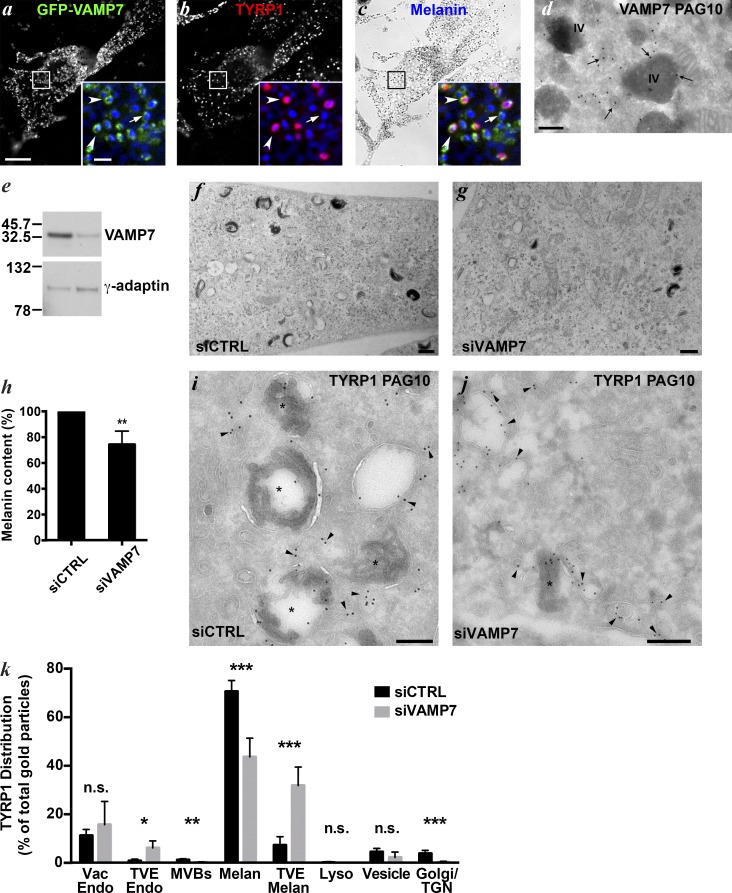
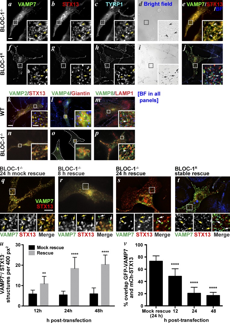
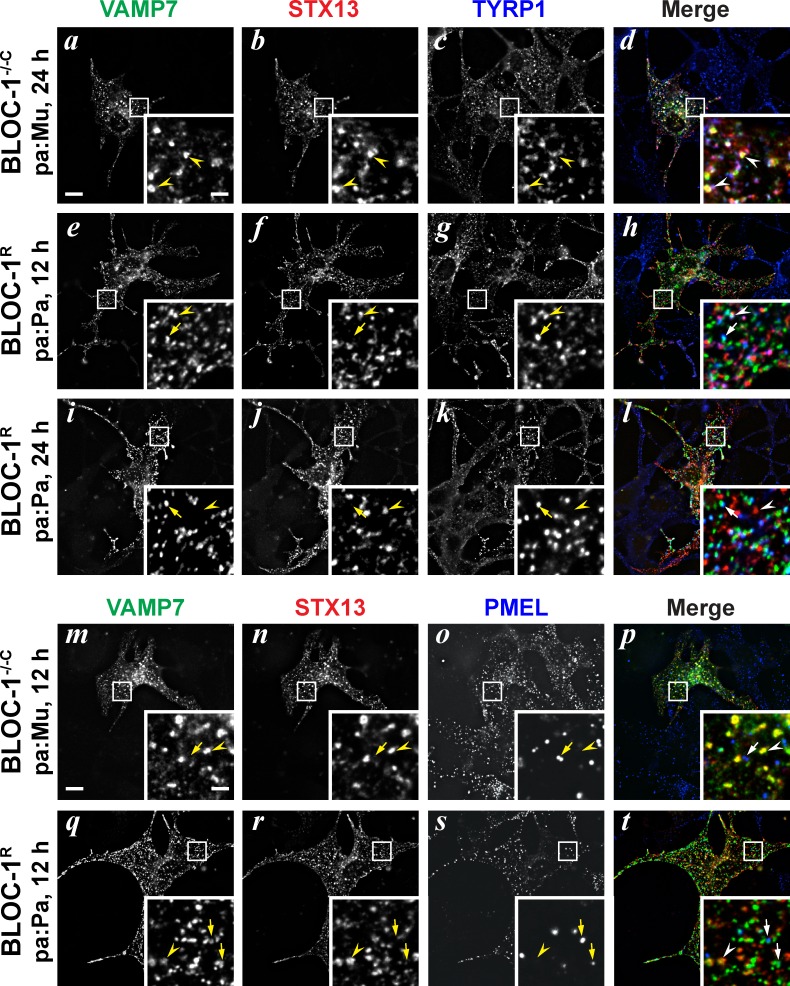
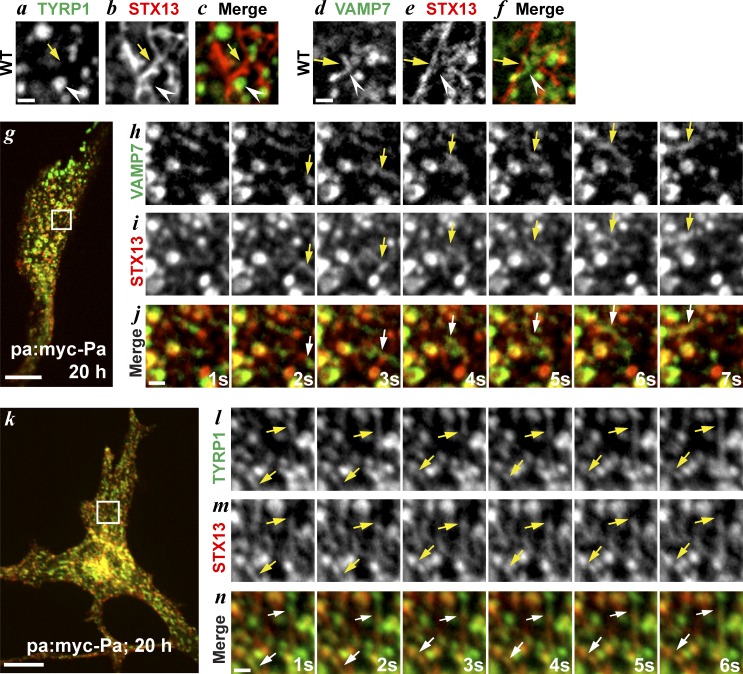
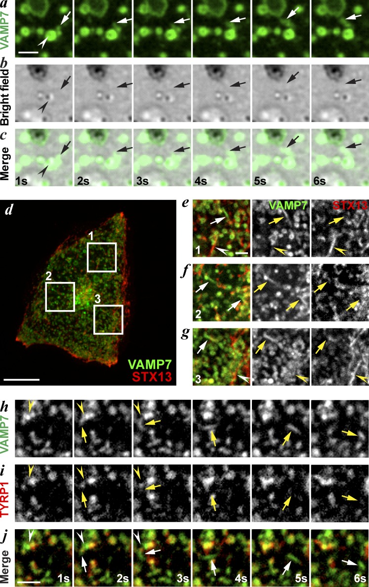
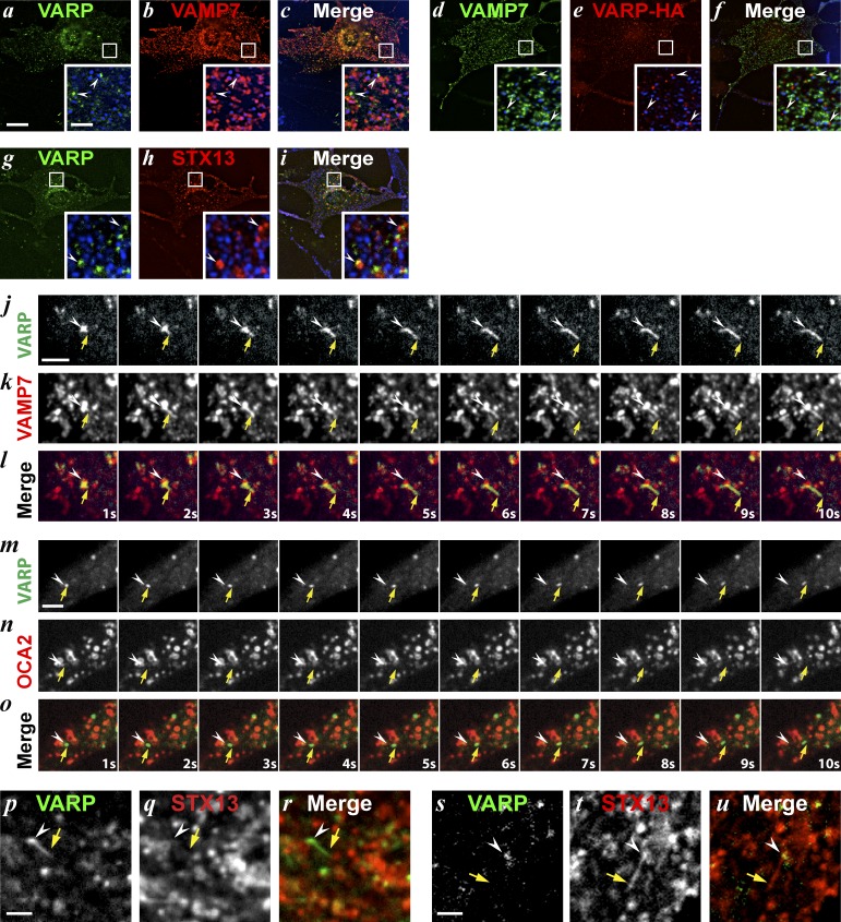
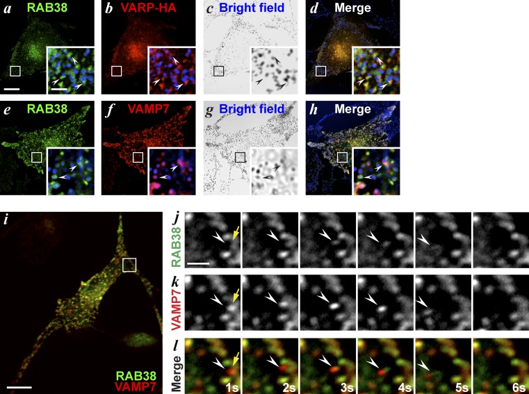
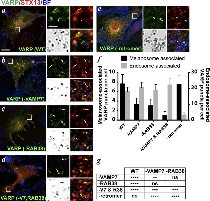
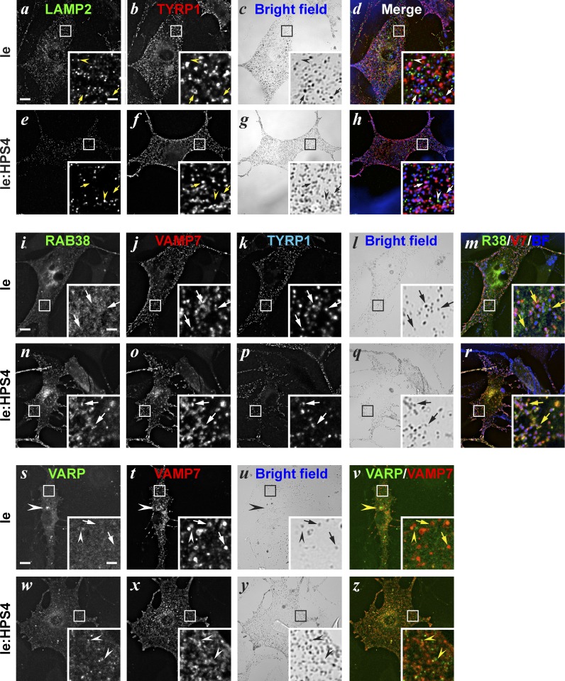
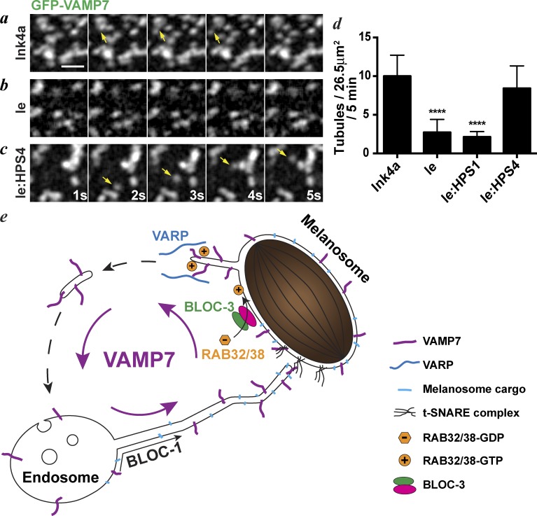
Endomembrane organelle maturation requires cargo delivery via fusion with membrane transport intermediates and recycling of fusion factors to their sites of origin. Melanosomes and other lysosome-related organelles obtain cargoes from early endosomes, but the fusion machinery involved and its recycling pathway are unknown. Here, we show that the v-SNARE VAMP7 mediates fusion of melanosomes with tubular transport carriers that also carry the cargo protein TYRP1 and that require BLOC-1 for their formation. Using live-cell imaging, we identify a pathway for VAMP7 recycling from melanosomes that employs distinct tubular carriers. The recycling carriers also harbor the VAMP7-binding scaffold protein VARP and the tissue-restricted Rab GTPase RAB38. Recycling carrier formation is dependent on the RAB38 exchange factor BLOC-3. Our data suggest that VAMP7 mediates fusion of BLOC-1-dependent transport carriers with melanosomes, illuminate SNARE recycling from melanosomes as a critical BLOC-3-dependent step, and likely explain the distinct hypopigmentation phenotypes associated with BLOC-1 and BLOC-3 deficiency in Hermansky-Pudlak syndrome variants.


Figures

Figure 1.
Figure 1.
VAMP7 localizes to melanosomes and is required for pigmentation and cargo trafficking. (a–c) WT melan-Ink4a melanocytes transiently expressing GFP-VAMP7 (green) were fixed and labeled for TYRP1 (red) and analyzed by deconvolution immuno-FM. Bar, 10 µm. BF (melanin) image in c is pseudocolored blue in insets. Insets of boxed regions are magnified five times; bar, 1 µm. Melanosomes labeled by both VAMP7 and TYRP1 (arrowheads) or VAMP7 only (arrow) are indicated. (d) Ultrathin cryosection of WT melan-a melanocytes transfected with GFP-VAMP7 and labeled for VAMP7 with 10 nm protein A gold (PAG10). Arrows show GFP-VAMP7 on stage IV melanosomes (IV) and adjacent vesicles. Bar, 200 nm. (e–k) MNT-1 melanoma cells treated with control (siCTRL) or VAMP7-specific siRNA (siVAMP7) were analyzed 5 d later. (e) Whole-cell lysates fractionated by SDS-PAGE were immunoblotted for VAMP7 or the AP-1 subunit γ-adaptin as a control. Relevant bands and positions of nearby molecular weight markers are shown. (f and g) Thin sections of fixed cells in epon were analyzed by conventional EM. Bars, 500 nm. (h) Melanin content in cell lysates was assayed by spectrophotometry. Data are normalized to siCTRL and represent mean ± SD from at least three experiments. (i and j) Ultrathin cryosections of fixed cells were labeled for TYRP1 with PAG10. Asterisks show pigmented melanosomes, and arrowheads show TYRP1 labeling on melanosomal membrane or closely adjacent vesicular structures. Bars, 200 nm. (k) Quantification of TYRP1 localization in siCTRL and siVAMP7 cells (mean ± SD from three measurements). Golgi/TGN, Golgi and trans-Golgi network; Lyso, lysosomes; Melan, melanosomes; MVBs, multivesicular bodies; TVE endo, tubulovesicular endosomes associated with endosomes; TVE Melan, tubulovesicular endosomes associated with melanosomes; Vac Endo, vacuolar endosomes; Vesicle, other vesicular structures. *, P < 0.05; **, P < 0.01; ***, P < 0.005; n.s., no significant difference.
Figure 2.
Figure 2.
VAMP7 is a BLOC-1 cargo. (a–j) BLOC-1–deficient melan-pa (BLOC-1−/−; a–e) or “rescued” melan-pa cells stably expressing myc-Pallidin (BLOC-1R; f–j) and transiently expressing GFP-VAMP7 (green) and mCh-STX13 (red) were fixed and labeled for TYRP1 (cyan) and analyzed by deconvolution immuno-FM. Arrowheads show GFP-VAMP7 and TYRP1 retained in mCh-STX13–positive endosomes, and arrows show melanosomes with GFP-VAMP7 and TYRP1 but lacking mCh-STX13. Insets of boxed regions are magnified five times. (k–m) WT melan-Ink4a (n–p) and BLOC-1–deficient melan-pa (BLOC-1−/−) transiently expressing GFP-VAMP2 (green) and mCh-STX13 (red, k and n), VAMP4-HA (green, l and o), or GFP-VAMP8 (green, m and p) were fixed, labeled for HA (green) and Giantin (red, l and o) or for LAMP1 (red, m and p), and analyzed by deconvolution immuno-FM. Merged images are shown with BF images pseudocolored blue. Dashed line in o indicates cell borders. Insets are boxed regions magnified six times. (q–v) BLOC-1−/− melan-pa melanocytes transiently transfected with GFP-VAMP7 (green), mCh-STX13 (red), and either myc-Pallidin (“rescue”) or myc-Muted (“mock rescue”) for indicated times. Arrowheads show GFP-VAMP7 retained in mCh-STX13 endosomes; and arrows point to GFP-VAMP7 in melanosome precursors. Note that pigmentation is not detected until ∼72 h after transfection with myc-Pallidin. Insets are boxed regions magnified 3.5 times. (u) Quantification (mean ± SD from 15 regions of at least nine cells per time point representing three independent experiments) of GFP-VAMP7–labeled structures that lack mCh-STX13 in rescue and mock rescue cells at 12, 24, and 48 h after transfection. (v) Area (mean ± SD from at least 15 cells per time point representing four independent experiments) of overlap between GFP-VAMP7 and mCh-STX13 in the periphery of mock rescue and rescue cells quantified at 12, 24, and 48 h after transfection. **, P < 0.01; ****, P < 0.0001. BF images in d, I, and k–t are pseudocolored blue in merge. Bars: (a–t) 10 µm; (insets) 2 µm.
Figure 3.
Figure 3.
VAMP7 traffics with TYRP1 to maturing melanosomes upon BLOC-1 rescue. (a–t) BLOC-1−/− (melan-pa) cells transiently transfected with GFP-VAMP7 (green), mCh-STX13 (red), and either myc-Pallidin (BLOC-1R) or myc-Muted (BLOC-1−/−C) were fixed 12 or 24 h after transfection. Cells were then immunolabeled for TYRP1 (blue, a–l) or PMEL (blue, m–t) and analyzed by deconvolution immuno-FM. Arrowheads show GFP-VAMP7 and TYRP1 (a–l) or GFP-VAMP7 alone (m–t) retained in mCh-STX13–labeled endosomes, and arrows point to GFP-VAMP7 and either TYRP1 (a–l) or PMEL (m–t) colabeled in melanosomal precursors that lack mCh-STX13. Insets are boxed regions magnified five times. Bars: (main) 10 µm; (insets) 2 µm.
Figure 4.
Figure 4.
VAMP7 and TYRP1 traffic to melanosomes in BLOC-1dependent membrane tubules. (a–f) WT melan-Ink4a cells transiently transfected with mCh-STX13 (red) and either TYRP1-GFP (green, a–c; cell shown in Fig. S2 a) or GFP-VAMP7 (green; d-f; cell shown in Figs. 5 d and S2 b) were analyzed 24 h later by spinning-disk confocal microscopy at 1 fps. Regions from a single frame are shown. Arrows, mCh-STX13-labeled endosomal tubules; arrowheads, TYRP1-GFP-labeled melanosome (a–c) or GFP-VAMP7-labeled tubule (d-f). Note that tubular mCh-STX13–labeled endosomes in live-cell analyses appear punctate upon fixation (e.g., Fig. 2, g, s, and t). Bar, 1 µm. (g–n) melan-pa melanocytes were transiently transfected with myc-Pallidin, mCh-STX13 and either GFP-VAMP7 (g–j) or TYRP1-GFP (k-n) and analyzed 20 h later by spinning-disk confocal microscopy at ∼1 fps. (g and k) Single frames of representative cells showing overlap of mCh-STX13 with GFP-VAMP7 (g) or TYRP1-GFP (k). Bars, 10 µm. Image sequences from the boxed regions in g and k are magnified five times in h–j and l–n, respectively. Arrows show mCh-STX13–labeled tubules containing GFP-VAMP7 (h–j) or TYRP1-GFP (l–n). Elapsed time (in seconds) is indicated at the lower right. Bars, 1 µm.
Figure 5.
Figure 5.
GFP-VAMP7 exits melanosomes in STX13-independent tubules lacking TYRP1. (a–f) WT melan-Ink4a melanocytes transiently transfected with GFP-VAMP7 alone (green, a–c) or with mCh-STX13 (red, d–g) or TYRP1-mRFP (red, h–j) were analyzed 24 h later by spinning-disk confocal microscopy at ∼1 fps. (a–c) Image sequence from a cell (shown in Fig. S2 i) expressing GFP-VAMP7 relative to melanosomes visualized by BF microscopy. Arrow shows a GFP-VAMP7 tubule, and the arrowhead points to a melanosome in the BF image. Bar, 2 µm. (d) Single frame from representative cell expressing GFP-VAMP7 and mCh-STX13. Bar, 10 µm. (e–g) Single frames from boxed regions in d with single channels and merged image. Arrows show tubules labeled by GFP-VAMP7 (green), and arrowheads show tubules labeled by mCh-STX13 (red). Bar, 1 µm. Insets are magnified two times from d. (h–j) Image sequence from a cell (shown in Fig. S2 j) expressing GFP-VAMP7 (green) and TYRP1-mRFP (red). A GFP-VAMP7–labeled tubule (arrow) emerges from a TYRP1-mRFP/GFP-VAMP7–labeled melanosome (arrowhead). Elapsed time is shown in seconds (s) at the bottom. Bar, 2 µm.
Figure 6.
Figure 6.
A cohort of VARP localizes to melanosomes and accompanies GFP-VAMP7 on departing tubules. (a–i) WT melan-Ink4a melanocytes transiently transfected with VARP-GFP and mCh-VAMP7 (a–c), GFP-VAMP7 and VARP-HA (d–f), or VARP-GFP and mCh-STX13 (g–i) were fixed 48 h later, labeled with anti-HA (d–f), and analyzed by deconvolution immuno-FM. BF images are pseudocolored blue and shown in merge and insets (boxes magnified five times). Arrowheads show VARP puncta adjacent to melanosomes (a–f) or to mCh-STX13–labeled endosomes (g–i). Bars: (main), 10 µm; (insets), 2 μm. (j–u) WT melan-Ink4a melanocytes transiently transfected with VARP-GFP and either mCh-VAMP7 (j-l; see Fig. S3 a), mRFP-OCA2 (m–o; see Fig. S3 b) or mCh-STX13 (p–u; see Fig. S3, c and d) were analyzed 48 h later by spinning-disk confocal microscopy at ∼1 fps. Elapsed time (in seconds) is indicated at lower right. (j–l) A VARP-GFP– and mCh-VAMP7–labeled tubule (arrow) extends from a melanosome (arrowhead). (m–o) A VARP-GFP–labeled vesicle (arrow) exits from an mRFP-OCA2–labeled melanosome (arrowhead). (p–r) A VARP-GFP tubule (arrow) departs from a mCh-STX13–labeled endosome (arrowhead). (s–u) A mCh-STX13 tubule (arrow) exits from a mCh-STX13/VARP-GFP double-labeled endosome (arrowhead). Bars, 2 µm.
Figure 7.
Figure 7.
RAB38 overlaps with VARP on melanosomes and accompanies GFP-VAMP7 on departing tubules. (a–l) WT melan-Ink4a cells transiently transfected with GFP-RAB38 (green) and either VARP-HA (red, a–d), or mCh-VAMP7 (red, e–l) were analyzed 48 h later. (a–h) Cells were fixed, labeled with anti-HA (a–d), and analyzed by deconvolution immuno-FM. BF images are pseudocolored blue in merge and insets (boxed regions magnified five times). (a–d) Arrowheads show VARP-HA and GFP-RAB38 puncta adjacent to melanosomes. (e–h) Arrowheads show GFP-RAB38 and mCh-VAMP7 localized to melanosomes. (i–l) Cells were analyzed by spinning-disk confocal microscopy at ∼1 fps. (j–l) Image sequence of boxed region in i, magnified five times; elapsed time (in seconds) is indicated at the lower right. A mCh-VAMP7/GFP-RAB38–labeled structure (arrowhead) emerges from a mCh-VAMP7–labeled melanosome (arrow). Bars: (main) 10 µm; (insets and j–l) 2 µm.
Figure 8.
Figure 8.
VARP localizes to melanosomes by interaction with both VAMP7 and RAB38. (a–g) WT melan-Ink4a cells were transiently transfected with mCh-STX13 (red) and either WT VARP-GFP (green, a) or site-directed mutants (green, b–e) with interfering mutations in the binding sites for VAMP7 (-VAMP7, b), RAB38 (-RAB38, c), both VAMP7 and RAB38 (-V7, RAB38, d), or the VPS29 retromer subunit (-retromer, e). Cells were fixed 48 h after transfection and analyzed by deconvolution FM; bar, 10 µm. Insets are boxed regions magnified five times. Bar, 2 µm. BF images are pseudocolored blue in merged images. Arrowheads show VARP-GFP puncta adjacent to melanosomes visualized by BF, and arrows point to VARP-GFP puncta adjacent to mCh-STX13-labeled endosomes. (f) WT or mutant VARP-GFP puncta that were associated with melanosomes (visualized by BF; black bars) or with endosomes (labeled by mCh-STX13; gray bars) were quantified as mean ± SD from 10 cells per VARP variant representing three independent experiments. (g) P-values for pairwise comparisons of melanosome-associated VARP puncta in f. For endosome-associated VARP, only -V7 & R38 (P < 0.05 vs. WT) and -retromer (P < 0.0001 vs. all others) showed significant differences.
Figure 9.
Figure 9.
VAMP7 and TYRP1, but not RAB38 and VARP, localize normally to melanosomes in BLOC-3deficient cells. (a–z) BLOC-3–deficient melan-le (le; a–d, i–m, and s–v) and “rescued” melan-le cells stably expressing HA-HPS4 (le:HPS4; e–h, n–r, and w–z) were untreated (a–h) or transiently transfected with mCh-VAMP7 (red, i–z) and either GFP-RAB38 (green, i–r) or VARP-GFP (green, s–z), fixed 24 h (a–r) or 48 h (s–z) after transfection, and immunolabeled for LAMP2 (green, a–h) and/or TYRP1 (red, a–h; cyan, i–r) and analyzed by deconvolution immuno-FM. (a–h) Arrowhead points to a LAMP2-positive lysosome, and arrows show TYRP1-positive melanosomes. (i–r) Arrows show mCh-VAMP7 and TYRP1 colocalized to melanosomes visualized by BF. Note RAB38-GFP localization to melanosomes in le:HPS4 (n), but not in le (i). (s–z) Large arrowhead in le main panel (s) shows perinuclear accumulation of VARP-GFP. Arrowheads in insets show VARP-GFP puncta associated with melanosomes, and arrows point to melanosomes lacking VARP-GFP puncta. BF images in c, g, l, and q are pseudocolored blue in the merged images in d, h, m, and r. Insets, boxed regions magnified five times. Bars: (main) 10 µm; (insets) 2 µm.
Figure 10.
Figure 10.
BLOC-3 is required to retrieve VAMP7 from melanosomes. (a–d) WT melan-Ink4a (Ink4a; a), melan-le (le; b), melan-le:HPS1 (le:HPS1; not depicted), or melan-le:HPS4 (le:HPS4; c) cells transiently transfected with GFP-VAMP7 were analyzed by spinning-disk confocal microscopy 24 h later. Images were acquired at ∼1 fps and a segment of the image sequence is shown. Elapsed time (in seconds) is indicated at lower right. Bar, 2 µm. Arrows show GFP-VAMP7–labeled structures exiting GFP-VAMP7–labeled melanosomes. (d) Fission of GFP-VAMP7 transport intermediates from melanosomes was quantified by counting events within 26.5-µm2 regions in the cell periphery during 5-min image sequences acquired at 1 fps. At least 15 regions from a minimum of six cells, representing three independent experiments, were quantified for each cell type; shown are mean values ± SD. ****, P < 0.0001. (e) Model for VAMP7 cycling during melanosome biogenesis.

Comment in

References

    1. Anderson P.D., Huizing M., Claassen D.A., White J., and Gahl W.A.. 2003. Hermansky-Pudlak syndrome type 4 (HPS-4): clinical and molecular characteristics. Hum. Genet. 113:10–17. - PubMed
    1. Barlowe C.K., and Miller E.A.. 2013. Secretory protein biogenesis and traffic in the early secretory pathway. Genetics. 193:383–410. 10.1534/genetics.112.142810 - DOI - PMC - PubMed
    1. Bennett D.C., Cooper P.J., and Hart I.R.. 1987. A line of non-tumorigenic mouse melanocytes, syngeneic with the B16 melanoma and requiring a tumour promoter for growth. Int. J. Cancer. 39:414–418. 10.1002/ijc.2910390324 - DOI - PubMed
    1. Boissy R.E., Zhao Y., and Gahl W.A.. 1998. Altered protein localization in melanocytes from Hermansky-Pudlak syndrome: support for the role of the HPS gene product in intracellular trafficking. Lab. Invest. 78:1037–1048. - PubMed
    1. Bonifacino J.S., and Glick B.S.. 2004. The mechanisms of vesicle budding and fusion. Cell. 116:153–166. 10.1016/S0092-8674(03)01079-1 - DOI - PubMed

MeSH terms

LinkOut - more resources