. 2016 Aug 16;113(33):E4784-93.
doi: 10.1073/pnas.1610179113. Epub 2016 Aug 1.

Distinct cellular properties of oncogenic KIT receptor tyrosine kinase mutants enable alternative courses of cancer cell inhibition

Affiliations

Distinct cellular properties of oncogenic KIT receptor tyrosine kinase mutants enable alternative courses of cancer cell inhibition

Xiarong Shi et al. Proc Natl Acad Sci U S A. .

Abstract

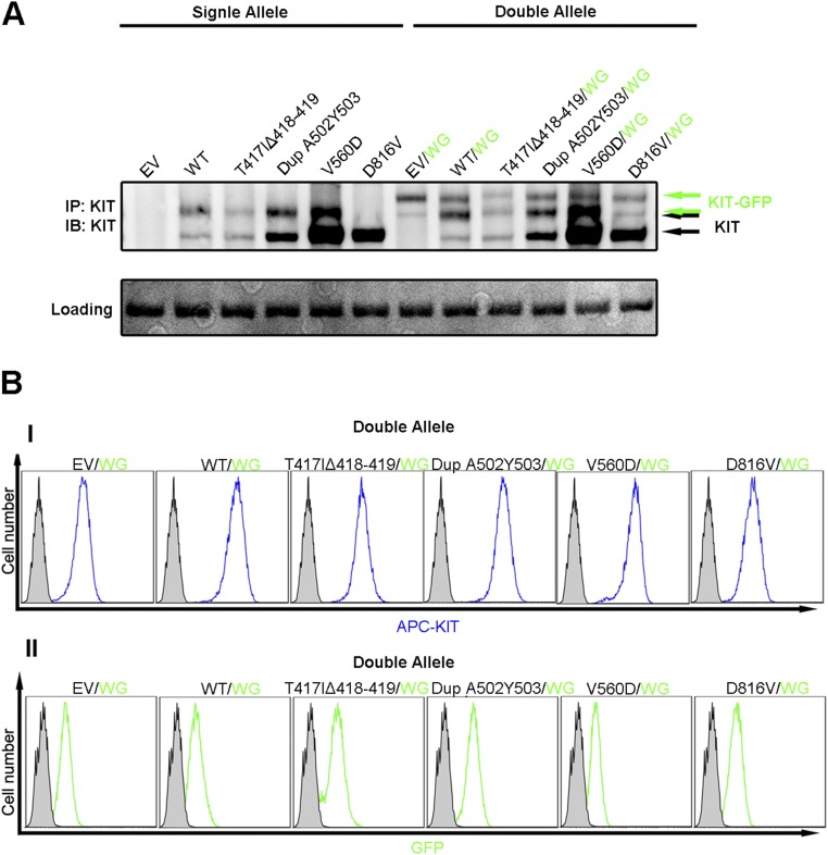
Large genomic sequencing analysis as part of precision medicine efforts revealed numerous activating mutations in receptor tyrosine kinases, including KIT. Unfortunately, a single approach is not effective for inhibiting cancer cells or treating cancers driven by all known oncogenic KIT mutants. Here, we show that each of the six major KIT oncogenic mutants exhibits different enzymatic, cellular, and dynamic properties and responds distinctly to different KIT inhibitors. One class of KIT mutants responded well to anti-KIT antibody treatment alone or in combination with a low dose of tyrosine kinase inhibitors (TKIs). A second class of KIT mutants, including a mutant resistant to imatinib treatment, responded well to a combination of TKI with anti-KIT antibodies or to anti-KIT toxin conjugates, respectively. We conclude that the preferred choice of precision medicine treatments for cancers driven by activated KIT and other RTKs may rely on clear understanding of the dynamic properties of oncogenic mutants.

Keywords: KIT; monoclonal antibody; oncogenic mutant; receptor tyrosine kinase; targeted therapy.


Conflict of interest statement

E.M.M.-B. and Y.H. are employees of Kolltan Pharmaceuticals.

Figures

Fig. 1.
Fig. 1.
Location of the major oncogenic KIT mutations. (A) Schematic presentation of KIT extracellular and cytoplasmic domains showing the positions of oncogenic KIT mutations. The mutations are located in four hot spots. Two are located in D5 of the extracellular domain, one is located in the juxtamembrane (JM) region, and another one is in the TK domain. The extracellular Ig-like domains D1–D5 are marked in blue, green, yellow, brown, and pink, respectively. The transmembrane (TM) is in black, the JM domain and kinase domains are in purple and in red, respectively. The kinase insert (KI) region and the C-terminal tail are in black. (B) Crystal structures depicting the location of the oncogenic mutations in D5, JM, and TK domains within an electron microcopy (EM) image of full-size dimeric KIT dimers fitted with X-ray crystal structures of KIT extracellular and cytoplasmic domains.
Fig. 2.
Fig. 2.
Quantification of surface expression and analysis of glycosylation patterns of WT or oncogenic KIT mutants. (A) KIT proteins expressed on the cell surface of NIH 3T3 cells were labeled with phycoerythrin (PE)-conjugated anti-KIT antibodies, and cell surface-bound antibodies were measured using FACS analysis. Black dotted curves represent FACS analyses of control NIH 3T3 cells, and red curves represent NIH 3T3-expressing WT or oncogenic KIT mutants labeled with the PE-conjugated anti-KIT antibodies. (B) The level of WT or oncogenic KIT proteins expressed on the cell surface was quantified from three independent FACS experiments and presented as the percentage of WT ± SEM. (C) Glycosylation patterns of WT or oncogenic KIT mutants. (I) KIT proteins are expressed in a form of fully (145-kDa) and partially (125-kDa) glycosylated proteins. Cell lysates of NIH 3T3 cells expressing WT or oncogenic KIT mutants were subjected to immunoprecipitation with anti-KIT D4 mAb. The washed immunoprecipitates were next treated with PNGase F, an enzyme that cleaves all N-linked sugars, followed by SDS/PAGE analysis and immunoblotting with anti-KIT antibodies. (II) NIH 3T3 cells expressing WT or oncogenic KIT mutants were treated for 16 h with tunicamycin, a drug that blocks protein glycosylation. Cell lysates prepared from tunicamycin-treated or untreated cells were subjected to immunoprecipitation with anti-KIT D4 mAb followed by SDS/PAGE analysis and immunoblotting with anti-KIT antibodies. The core KIT protein seen in the tunicamycin-treated samples migrates as a band with apparent molecular mass of 110 kDa. (D) Cell surface proteins of NIH 3T3 cells expressing WT or oncogenic KIT mutants were labeled with non–cell-permeable biotin probe. Half of the cell lysate was subjected to immunoprecipitation with anti-KIT D4 mAb to determine the total number of KIT proteins (I) and the other half of the lysate was subjected to a pull down experiment with avidin beads to determine KIT proteins located on the cell membrane (II). The samples that were treated with avidin beads were subjected to a second immunoprecipitation with anti-D4 mAb (III). All precipitations were then analyzed by SDS/PAGE and by immunoblotting with anti-KIT antibodies.
Fig. S1.
Fig. S1.
Cellular localization of WT or oncogenic KIT mutants. GFP fusion proteins of WT or oncogenic KIT mutants were transiently expressed in COS-7 cells grown on glass coverslips. Each plasmid was transfected into the cells at a similar efficiency (∼40–60% by FACS). The cells were fixed with paraformaldehyde and stained with PE-conjugated anti-KIT antibodies (Dako) to visualize the population of KIT molecules expressed on the cell surface. Images were taken using a confocal microscope. (Scale bar: 20 μm.)
Fig. S2.
Fig. S2.
Densitometric analysis of KIT protein immunoblots. (A) Five independently developed Western blots with the ChemiDoc XRS+ imaging system (Bio-Rad) were used for quantification. (B) The intensity of upper bands (145-kDa) were quantified using Image Lab software and normalized to WT; the error bar represents the SE of the mean (SEM) from the five blots. (C) The ratio of upper/lower band intensity was calculated from the five blots and expressed as mean ± SEM. A t test was performed between WT and each of the mutants. *P < 0.01, **P < 0.001, ***P < 0.0001.
Fig. 3.
Fig. 3.
The stability and ubiquitination of WT KIT or oncogenic KIT mutants in SCF-stimulated or unstimulated cells. (A) Oncogenic KIT mutants are ubiquitinated to become destined for degradation. SCF-stimulated or unstimulated NIH 3T3 cells expressing WT or KIT oncogenic mutants were subjected to immunoprecipitation with anti-KIT mAb followed by SDS/PAGE and immunoblotting with anti-ubiquitin antibodies (Top), anti-phospho tyrosine antibodies (Middle), or anti-KIT antibodies (Bottom). It is noteworthy that only the 145-kDa form is preferentially tyrosine-phosphorylated whereas the 125-kDa form undergoes a weak tyrosine phosphorylation. (B) Basal or ligand-stimulated degradation of WT or oncogenic KIT mutants. NIH 3T3 cells expressing either WT or KIT oncogenic mutants were treated with cycloheximide. Cell lysate from unstimulated cells, cells stimulated with SCF alone, cells stimulated with SCF together anti-D4 mAb, or cells stimulated with SCF together with imatinib were collected at various time points as indicated and subjected to immunoprecipitation with anti-KIT antibodies, followed by SDS/PAGE analysis and immunoblotting using the anti-KIT antibodies.
Fig. 4.
Fig. 4.
Comparison of the inhibitory effect of anti-D4 mAb, anti-D5 mAb, imatinib, sunitinib, or a combination of monoclonal antibodies with TKIs on the activation of WT or oncogenic KIT mutants. NIH 3T3 cells expressing either WT or KIT oncogenic mutants were stimulated with 25 ng/mL SCF alone, with SCF together anti-D4 mAb, SCF together anti-D5 mAb (A), SCF together with imatinib (B), or SCF together with sunitinib (C). Cell lysates were collected at various time points as indicated and subjected to immunoprecipitation with anti-KIT antibodies, followed by SDS/PAGE and immunoblotting with either anti-pTyr antibodies or anti-KIT antibodies. (A) Anti-D4 or anti-D5 mAbs inhibit the activation of WT and class I oncogenic KIT mutants but have minimal or no effect on the activation of class II oncogenic KIT mutants. The TKI imatinib and sunitinib inhibit the activation of most KIT oncogenic mutants (B and C) but are much less potent than anti-D4 or anti-D5 in inhibiting the activation of WT or class I oncogenic mutants (A). Treatment of cells expressing the Dup A502Y503 D5 mutant with a combination of anti-D4 together with the TKI imatinib (D) or sunitinib (E) shows strong inhibitory effect. Due to the large size of these experiments, some blots that are separated by vertical black lines (D and E) were taken from separate experiments.
Fig. S3.
Fig. S3.
A kinase-deficient mutant of WT or the V560D KIT mutant exhibit prolonged half-life in SCF-stimulated cells. Two different tyrosine kinase-deficient KIT mutants were generated by mutating a critical amino acid in the nucleotide binding site (K623A) or in the activation loop (Y823F) of the tyrosine kinase domain. KIT degradation in cycloheximide-treated SCF-stimulated or unstimulated cells that were treated with imatinib or anti-KIT D4 mAb was monitored for 4 h. Cell lysates collected at different time points were subjected to immunoprecipitation with anti-KIT mAb, followed by SDS/PAGE and immunoblotting with anti-KIT mAb. The experiment shows that the half-life of the V560D oncogenic mutant containing an additional mutation that compromised tyrosine kinase activity is extended in both unstimulated or SCF-stimulated cells, as well as in cells treated with imatinib or anti-KIT D4 mAb.
Fig. S4.
Fig. S4.
Oncogenic KIT D5 mutants are recognized by anti-D4 or anti-D5 mAbs. Lysates of cells stably expressing WT or oncogenic KIT mutants were subjected to immunoprecipitation with either anti-D4 or anti-D5 mAbs, followed by SDS/PAGE and immunoblotting with rabbit polyclonal anti-KIT antibodies. A similar pattern of KIT proteins was detected in the anti-D4 or anti-D5 immunoprecipitates.
Fig. 5.
Fig. 5.
Anti-D4 inhibits colony formation in soft agar of NIH 3T3 cells and proliferation of Ba/F3 cells expressing KIT D5 oncogenic mutants. (A) SCF-stimulated or unstimulated NIH 3T3 cells expressing either WT or KIT oncogenic mutants were grown in the presence or absence of anti-D4 or imatinib in six-well plates containing soft agar medium. After 3 wk, the plates were stained with crystal violet and scanned for colony counting. Only colonies larger than 50 µm were counted, using ImageJ software. Colony number: —, 0–10; *, 10–20; ***, >100. (B) Expression of WT or oncogenic KIT mutants in Ba/F3 cells. Lysates of Ba/F3 cells expressing WT or oncogenic KIT mutants were subjected to immunoprecipitation with anti-D4 mAb, followed by SDS/PAGE analysis and immunoblotting with anti-KIT antibodies. (C) Cell proliferation of Ba/F3 cells expressing WT or oncogenic KIT mutants. Ba/F3 cells expressing WT or oncogenic KIT mutants were grown for 3 d in IL-3–free growth medium supplemented without/with SCF. Cell number was counted from triplicate wells, and cell proliferation was calculated as fold increase in cell number. (D) Inhibition of proliferation of Ba/F3 cells expressing WT or KIT oncogenic mutants by anti-D4, imatinib, or a combination of anti-D4 and imatinib. SCF-stimulated or unstimulated Ba/F3 cells expressing either WT or KIT oncogenic mutants were grown for 3 d in IL-3–free growth media containing different concentrations of anti-D4 mAb, imatinib, or a combination of anti-D4 mAb and imatinib. Cell number was counted from triplicate wells, and cell proliferation was calculated as fold increase in cell number. Anti-D4 exhibits strong inhibition on the proliferation of Ba/F3 expressing WT or the oncogenic D5 mutants (IIII) but does not inhibit the proliferation of Ba/F3 cells expressing either the V560D (IV) or the D816V oncogenic mutants (V and VI). Imatinib alone inhibits the proliferation of the V560D mutant, but anti-D4 mAb has no effect (IV). Dasatinib alone can inhibit D816V, but anti-D4 does not potentiate it (VI). *P < 0.05.
Fig. 6.
Fig. 6.
Efficient killing of Ba/F3 cells expressing WT or oncogenic KIT mutants by anti-D4 toxin conjugate (αD4-toxin). (A) Killing Ba/F3 cells expressing WT, each oncogenic KIT mutant or control empty vector. (B) Killing Ba/F3 cells coexpressing WT or each oncogenic KIT mutant together with WT KIT-GFP fusion protein (WG). Ba/F3 cells were plated in 96-well plates to which either αD4-toxin or αKLH-toxin (control toxin conjugate) was added at various concentrations as indicated. Cells were then left to grow for 3 d, and the number of live cells was determined using the CellTiter Glo assay (Promega). The results are presented as raw luminescence units (RLUs), which correlates with the number of live cells, versus logarithm concentration of antibody-toxin conjugates. The IC50 (half maximal inhibitory concentration) was calculated from a sigmoidal dose–response curve fitted using GraphPad Prism software.
Fig. S5.
Fig. S5.
Anti-KIT mAb conjugated to toxin. Anti-KIT IgG1ĸ mAb conjugated to the SG3227 pyrrolo[2,1-c][1,4]benzodiazepine antitumor antibiotic. SG3227 is a DNA-interactive pyrrolo[2,1-c][1,4]benzodiazepine dimer that binds to the minor groove of DNA to cross-link DNA strands and produce highly lethal lesions. SG3227 contains a cathepsin-cleavable valine/alanine linker attached at the N10 site of the PBD dimer. Random conjugation occurs via an iodoacetamide functional group in the linker at cysteine residues of the antibody after antibody reduction, with an average drug-to-antibody (DAR) ratio of ∼2.
Fig. S6.
Fig. S6.
Ba/F3 cells expressing WT or oncogenic KIT mutants alone or together with a GFP fusion full-length KIT fused to GFP. Ba/F3 cells expressing either empty vector (EV), WT, or oncogenic KIT mutants were cotransfected with pMSCVhygro-WT-KIT-GFP (WG). The expression of both alleles was analyzed by immunoblotting with anti-KIT mAb (A) or by FACS analysis (B). WT or oncogenic KIT mutants were analyzed using APC-conjugated anti-KIT mAb (I), and the WG allele was visualized by directly monitoring GFP fluorescence (II).
Fig. S7.
Fig. S7.
An anti-KIT D4 antibody toxin conjugate (αD4-toxin) inhibits tumor growth in an H526 small cell lung cancer xenograft mouse model. BALB/c mice were s.c. inoculated with 5 × 10⁶ H526 tumor cells expressing WT KIT. Mice were treated with one i.p. injection of control conjugate, anti-KIT conjugate, or PBS after the growth of tumors to 150–200 mm3. Tumor volumes were determined twice weekly. Treatment of mice with 3 mg/kg αD4-toxin resulted in nearly complete tumor growth inhibition whereas treatment with 1 mg/kg resulted in significant tumor inhibition. On the other hand, treatment with αKLH-toxin had no significant impact on tumor growth compared with treatment with PBS alone. For s.c. xenograft studies, H526 small cell lung cancer tumor cells were maintained in RPMI 1640 medium supplemented with 10% (vol/vol) heat-inactivated FBS at 37 °C in 5% CO2. Once growing in an exponential growth phase, cells were harvested for tumor inoculation. Female BALB/c mice were inoculated s.c. in the right flank at 6–8 wk of age with 5 × 10⁶ H526 tumor cells in 0.1 mL of PBS supplemented 1:1 with Matrigel. Upon reaching a tumor size of 150–200 mm3, animals were randomized into treatment groups for the efficacy study. Animals were treated with a single i.p. injection of PBS or conjugated antibody at 10 µL/g. Tumor sizes were measured twice weekly in two dimensions using a caliper, and the volume is expressed in mm3 using the formula: V = 0.5 a × b2, where a and b are the long and short diameters of the tumor, respectively.
Fig. 7.
Fig. 7.
Oncogenic KIT mutations are classified into two main groups, class I and class II. Class I mutants are expressed at the cell surface, albeit at different levels, and exhibit sensitized ligand response. Class I mutations include the D5 point mutations D419A and N505I, deletion of Y418 D419, and duplication of A502Y503. Class II have constitutively activated tyrosine kinase activities and show low or negligible surface expression. Class II mutations include the T417IΔ418-419 D5 mutation and the intracellular V560D and D816V point mutants.

References

    1. Meyerson M, Gabriel S, Getz G. Advances in understanding cancer genomes through second-generation sequencing. Nat Rev Genet. 2010;11(10):685–696. - PubMed
    1. Garraway LA, Lander ES. Lessons from the cancer genome. Cell. 2013;153(1):17–37. - PubMed
    1. Vogelstein B, et al. Cancer genome landscapes. Science. 2013;339(6127):1546–1558. - PMC - PubMed
    1. Ashman LK, Griffith R. Therapeutic targeting of c-KIT in cancer. Expert Opin Investig Drugs. 2013;22(1):103–115. - PubMed
    1. Ashman LK. The biology of stem cell factor and its receptor C-kit. Int J Biochem Cell Biol. 1999;31(10):1037–1051. - PubMed

MeSH terms

Substances

LinkOut - more resources