. 2016 Aug 16;113(33):E4847-56.
doi: 10.1073/pnas.1607710113. Epub 2016 Aug 1.

CCN1/CYR61-mediated meticulous patrolling by Ly6Clow monocytes fuels vascular inflammation

Affiliations

CCN1/CYR61-mediated meticulous patrolling by Ly6Clow monocytes fuels vascular inflammation

Beat A Imhof et al. Proc Natl Acad Sci U S A. .

Erratum in

Abstract

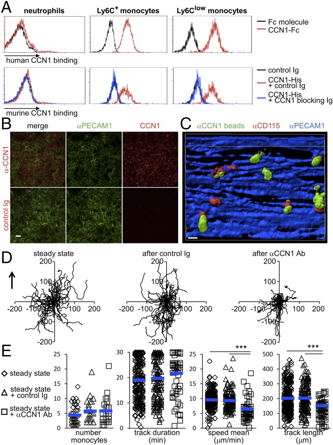
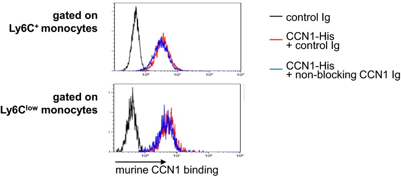
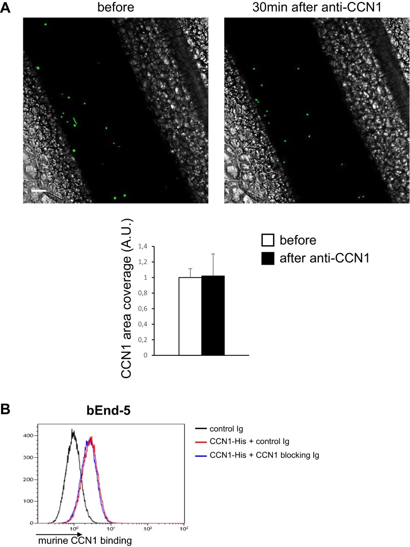
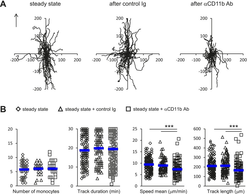
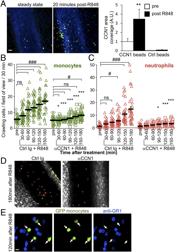
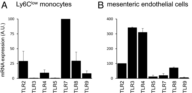
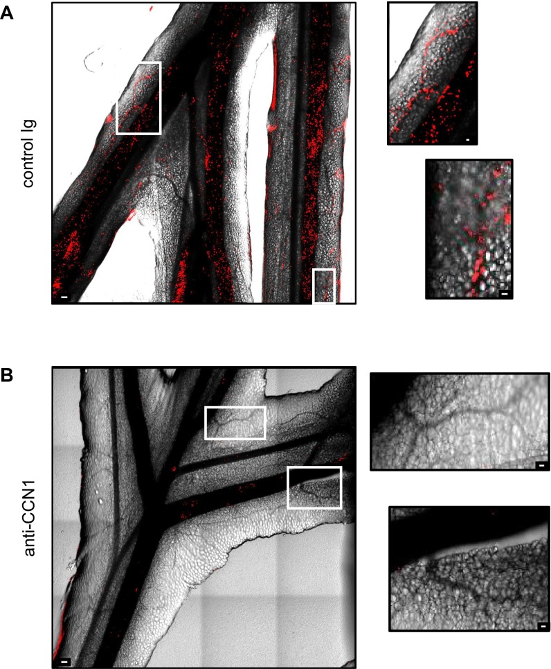
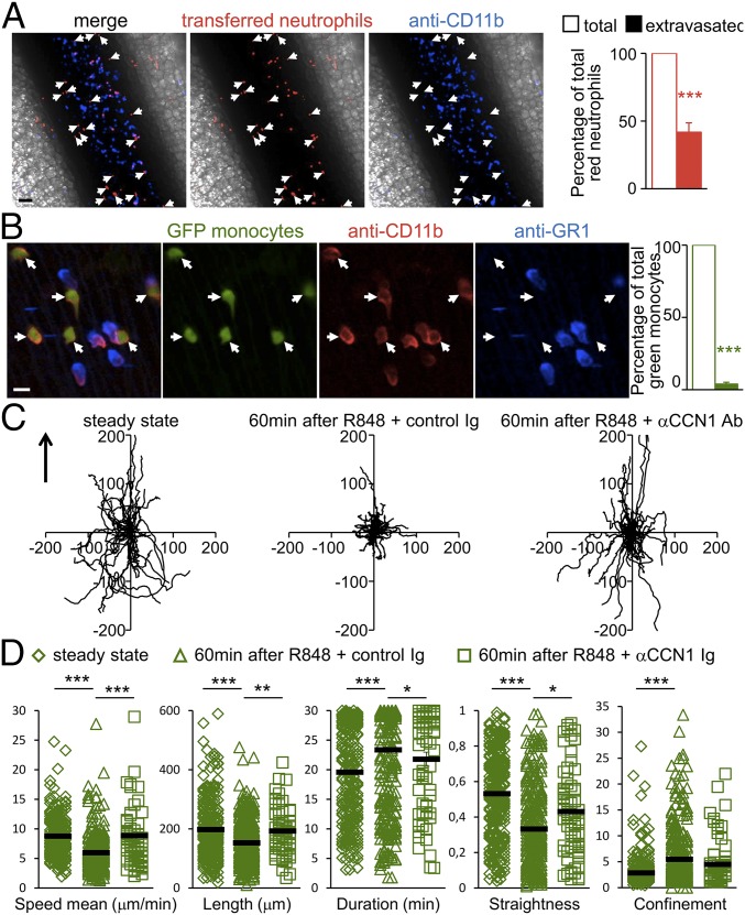
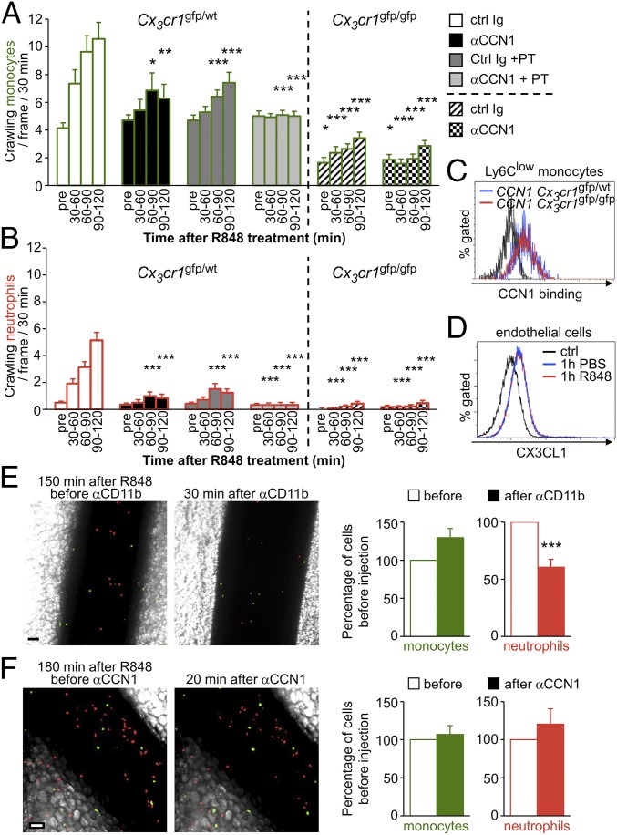
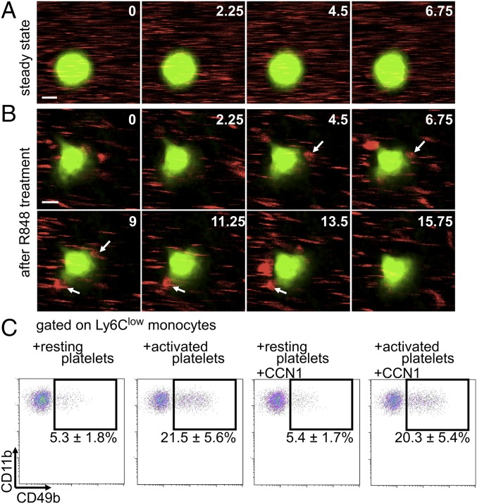
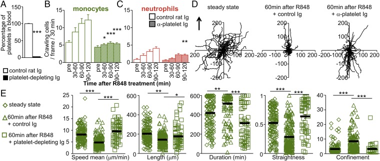
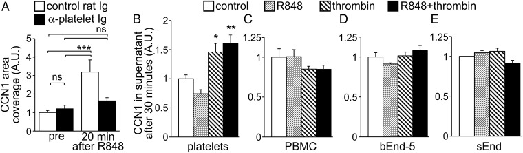
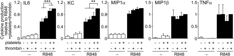
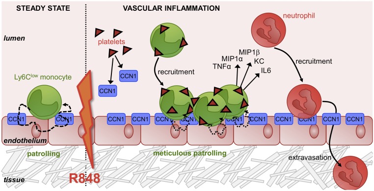
Inflammation is characterized by the recruitment of leukocytes from the bloodstream. The rapid arrival of neutrophils is followed by a wave of inflammatory lymphocyte antigen 6 complex (Ly6C)-positive monocytes. In contrast Ly6C(low) monocytes survey the endothelium in the steady state, but their role in inflammation is still unclear. Here, using confocal intravital microscopy, we show that upon Toll-like receptor 7/8 (TLR7/8)-mediated inflammation of mesenteric veins, platelet activation drives the rapid mobilization of Ly6C(low) monocytes to the luminal side of the endothelium. After repeatedly interacting with platelets, Ly6C(low) monocytes commit to a meticulous patrolling of the endothelial wall and orchestrate the subsequent arrival and extravasation of neutrophils through the production of proinflammatory cytokines and chemokines. At a molecular level, we show that cysteine-rich protein 61 (CYR61)/CYR61 connective tissue growth factor nephroblastoma overexpressed 1 (CCN1) protein is released by activated platelets and enables the recruitment of Ly6C(low) monocytes upon vascular inflammation. In addition endothelium-bound CCN1 sustains the adequate patrolling of Ly6C(low) monocytes both in the steady state and under inflammatory conditions. Blocking CCN1 or platelets with specific antibodies impaired the early arrival of Ly6C(low) monocytes and abolished the recruitment of neutrophils. These results refine the leukocyte recruitment cascade model by introducing endothelium-bound CCN1 as an inflammation mediator and by demonstrating a role for platelets and patrolling Ly6C(low) monocytes in acute vascular inflammation.

Keywords: CCN1; inflammation; monocyte; neutrophil; platelet.


Conflict of interest statement

The authors declare no conflict of interest.

Figures

Fig. 1.
Fig. 1.
Endothelium-bound CCN1 sustains the patrolling of Ly6Clow monocytes in the steady state. (A) Binding of CCN1 (1 μg/mL) was assessed by flow cytometry. For human CCN1, peripheral blood leukocytes were treated with recombinant Fc or human CCN1-Fc for 5 min at room temperature. Human CCN1 binding was detected by flow cytometry using a Dylight 488-conjugated anti-human Fc antibody. For murine CCN1, recombinant CCN1-His (1 μg/mL) was preincubated for 2 h at room temperature with control sheep Ig or blocking polyclonal sheep anti-CCN1 antibodies (50 μg/mL). Then cells were treated with recombinant murine CCN1-His for 5 min at room temperature. Murine CCN1 binding was detected by flow cytometry using a DyLight 650-conjugated anti–His-tag antibody. Data are representative of three experiments. (B) Cell surface-bound CCN1 on live bEnd-5 cells. Cells were incubated with sheep anti-CCN1 antibody or control Ig and then with Cy3-conjugated anti-sheep Ig in the presence of AF488-conjugated anti-PECAM1 to detect the vascular wall surface and the presence of CCN1. (Scale bar: 50 μm.) Representative maximal projections of four preparations are shown. (C) 3D reconstruction of mouse mesenteric veins obtained by confocal intravital microscopy depicting CD115+ patrolling monocytes located next to CCN1 hot spots on the luminal face of mesenteric veins. The view shows the luminal side of the vein. The i.v. injection of AF647-conjugated anti-PECAM1 (CD31, 10 μg) and AF594-conjugated anti-CD115 (1 μg) stained the endothelium blue and monocytes red, respectively. The luminal presence of CCN1-rich areas (green) was assessed after injection of protein A-coupled YFP beads conjugated with a nonblocking antibody to CCN1. (Scale bar: 10 μm.) (Movie S1.) (D) Representative tracks of Ly6Clow monocytes in the steady state (before) and 30 min after injection of control Ig or CCN1-blocking antibodies (50 μg/mL). Fifty monocytes per condition are represented. The arrow indicates the direction of the blood flow. (E) The number of crawling Ly6Clow monocytes and their track duration, speed, and length in D are quantified. n = 4 mice per condition; 25–26 vessels were analyzed in each condition. Data are mean ± SEM; ***P < 0.005; Kruskal–Wallis with Dunn’s multiple comparisons test.
Fig. S1.
Fig. S1.
CCN1 binding to other populations of leukocytes. Binding of human CCN1 (1 μg/mL) was assessed by flow cytometry. Peripheral blood leukocytes were treated with recombinant Fc or human CCN1-Fc for 5 min at room temperature. Human CCN1 binding was detected by flow cytometry using a Dylight 488-conjugated anti-human Fc antibody. Data are representative of three experiments.
Fig. S2.
Fig. S2.
Nonblocking rabbit anti-CCN1 antibody does not affect the binding of CCN1 to monocytes. Binding of murine CCN1 (1 μg/mL) to Ly6C+ monocytes (Upper) and Ly6Clow monocytes (Lower) was assessed by flow cytometry. Recombinant CCN1-His (1 μg/mL) was preincubated for 2 h at room temperature with control rabbit Ig or with nonblocking polyclonal rabbit anti-CCN1 antibodies (50 μg/mL). Then cells were treated with recombinant murine CCN1-His for 5 min at room temperature. Murine CCN1 binding was detected by flow cytometry using a DyLight 650-conjugated anti–His-tag antibody. The y axis represents the percentage of gated cells. Data are representative of three experiments.
Fig. S3.
Fig. S3.
Blocking sheep anti-CCN1 antibody does not affect the binding of CCN1 to endothelium. (A, Upper) Representative confocal intravital microscopy images of a mesenteric vein in the steady state (before) and 30 min after i.v. injection of sheep anti-CCN1 antibody (50 μg). Luminal CCN1 accumulation was assessed after i.v. injection of protein A-coupled YFP beads conjugated with a nonblocking antibody to CCN1. (Scale bar: 50 μm.) (Lower) Quantification in arbitrary units (A.U.). n = 8 vessels per condition. Data are mean ± SEM. (B) Binding of murine CCN1 (1 μg/mL) was assessed by flow cytometry. Recombinant CCN1-His (1 μg/mL) was preincubated for 2 h at room temperature with control rabbit Ig or with nonblocking polyclonal rabbit anti-CCN1 antibodies (50 μg/mL). Then cells were treated with recombinant murine CCN1-His for 5 min at room temperature. Murine CCN1 binding was detected by flow cytometry using a DyLight 650-coupled anti–His-tag antibody. Data are representative of three experiments.
Fig. S4.
Fig. S4.
CD11b blocking recapitulates the effect of CCN1 blocking on patrolling of Ly6Clow monocytes in the steady state. (A) Representative tracks of Ly6Clow monocytes in the steady state (before) and after injection of control Ig or blocking anti-CD11b antibodies (50 μg/mL). Fifty monocytes per condition are represented. The arrow indicates the direction of the blood flow. (B) The number of crawling Ly6Clow monocytes and track duration, speed, and length. n = 3 or 4 mice per condition; 27–35 vessels per condition were analyzed. Data are mean ± SEM; ***P < 0.005; Kruskal–Wallis with Dunn’s multiple comparisons test.
Fig. 2.
Fig. 2.
CCN1 mediates luminal recruitment of Ly6Clow monocytes, which precedes the arrival of neutrophils during TLR7/8-mediated inflammation. (A) Luminal CCN1 accumulation during inflammation. Representative confocal intravital microscopy images of mesenteric vein in the steady state (before) and 20 min after induction of inflammation by treatment with R848 (100 μg). AF647-conjugated anti-PECAM1 (CD31, 10 μg, i.v.) stained the endothelium blue. Luminal CCN1 accumulation was assessed after i.v. injection of protein A-coupled YFP beads conjugated with a nonblocking antibody to CCN1. (Scale bar: 50 μm.) Quantification is provided on the right. “Pre” denotes steady-state conditions before R848 treatment and antibody injection. Protein A-coupled YFP beads conjugated with rabbit Ig were used as controls. Eight vessels from two mice were analyzed. **P < 0.01; Wilcoxon matched-pairs signed rank test. (B and C) Kinetics of recruitment of Ly6Clow monocytes (green) (B) and neutrophils (red) (C) in response to R848 stimulation (100 μg) in Cx3cr1gfp/wt mice transferred with CellTracker Orange-labeled neutrophils. Mice were administered control sheep Ig or CCN1-blocking antibodies (50 μg/mL, i.v.) after steady-state conditions were achieved. Simultaneously, R848 was applied to the monitored vessels to induce inflammation. n = 5–8 mice per condition; 28–50 vessels were analyzed per time point. Data are mean ± SEM; *P < 0.05, ***P < 0.005, control (Ctrl) Ig vs. αCCN1 treatment; #P < 0.05, ###P < 0.005, antibody and R848 treatment vs. the precondition; two-way ANOVA with Tukey’s multiple comparisons test. (D) Representative confocal intravital microscopy images of mesenteric vein 180 min after R848 treatment in the presence of control sheep Ig or CCN1-blocking antibodies (50 μg/mL) (from Movie S2 and experiments shown in Fig. 3 B and C). (Scale bar: 50 μm.) (E) Representative confocal intravital microscopy images of mesenteric vein from Movie S4. Mice were administered BV421-conjugated GR1 antibody (1 µg, i.v.) 120 min after R848 treatment. Endogenous neutrophils are labeled in blue, and all monocytes are green. Inflammatory Ly6C+ monocytes would appear in blue and green, and Ly6Clow monocytes only in green. All observed monocytes were GR1, indicating that they were Ly6Clow monocytes. White arrows indicate the position of monocytes. Fifteen vessels from three mice were analyzed. (Scale bar: 10 μm.)
Fig. S5.
Fig. S5.
TLR expression. Expression of TLRs in Ly6Clow monocytes (n = 4) (A) and mesenteric endothelial cells (n = 3) (B) was determined by quantitative PCR. Data are mean ± SEM.
Fig. S6.
Fig. S6.
CCN1 blocking impairs leukocyte recruitment. Representative maximal projection of stitch images were acquired by confocal intravital microscopy of mesenteric vessels 150 min after the initiation of inflammation with R848 (200 μg) and i.v. injection of control sheep Ig (A) or sheep anti-CCN1 antibody (50 μg) (B). Intravascular neutrophils were detected by i.v. injection of AF647-conjugated anti-Ly6G (0.2 μg) 5 min before starting image acquisition. Vessels are determined by phase contrast. (Scale bars: 100 μm for large images; 25 μm for Insets.) Images are representative of experiments performed on five different mice per condition.
Fig. 3.
Fig. 3.
Recruited neutrophils extravasate while accumulated Ly6Clow monocytes meticulously patrol the luminal side of the endothelium upon TLR7/8-mediated inflammation. (A) Recruited neutrophils exit the vessel and invade the surrounding tissue. Shown are representative confocal intravital microscopy images of the mesenteric vein. Mesenteric veins of mice transferred with CellTracker Orange-labeled neutrophils were treated with R848 (100 μg). After 120 min, mice were injected with AF647-conjugated anti-CD11b antibody (1 µg, i.v.). All transferred neutrophils are CD11b+ cells. Endogenous neutrophils are blue. Transferred red neutrophils crawling on the luminal side of the endothelium are in contact with circulating labeled anti-CD11b antibody (red and blue). Extravasated red neutrophils do not have contact with the labeled anti-CD11b antibody and appear only in red (white arrows). (Scale bar: 50 μm.) Quantification is provided on the right. n = 3 mice; 20 vessels were analyzed. ***P < 0.005; Wilcoxon matched-pairs signed rank test. (B) Ly6Clow monocytes patrol the luminal side of the endothelium. Shown are representative confocal intravital microscopy images of mesenteric vein. AF594-conjugated anti-CD11b and BV421-conjugated anti-GR1 antibodies were administered (1 µg, i.v.) to mice 120 min after R848 treatment. Ly6Clow monocytes are GFP+ (green) and GR1 (not blue). Arrows indicate monocytes. All observed cells were CD11b+ (red), indicating that they are located on the luminal side of the endothelium. (Scale bar: 10 μm.) Quantification is provided on the right. ***P < 0.005; Wilcoxon matched-pairs signed rank test. n = 3 mice; 16 vessels were analyzed. (C and D) Meticulous patrolling of Ly6Clow monocytes after R848 treatment. (C) Representative tracks of Ly6Clow monocytes in the steady state (before) and 60 min after R848 treatment that were injected with control sheep Ig or antibodies blocking CCN1 (from experiments in B). Fifty monocytes per condition are represented. The arrow indicates the direction of the blood flow. (D) Track speed, length, duration, linearity, and confinement ratio of recruited patrolling Ly6Clow monocytes from experiments in B. Data are mean ± SEM; *P < 0.05; **P < 0.01; ***P < 0.005; Kruskal–Wallis with Dunn’s multiple comparisons test.
Fig. 4.
Fig. 4.
Recruitment of patrolling Ly6Clow monocytes relies on chemokine receptors. (A and B) Kinetics of recruitment of Ly6Clow monocytes (bars outlined in green) (A) and neutrophils (bars outlined in red) (B) in response to R848 stimulation (100 μg) in Cx3cr1gfp/wt (Left) and Cx3cr1gfp/gfp (Right) mice transferred with CellTracker Orange-labeled neutrophils. After steady-state conditions were recorded, R848 was applied to the monitored vessels to induce inflammation. Simultaneously, mice were i.v. injected with control sheep Ig or antibodies blocking CCN1 in PBS containing PT (50 µg). “Pre” denotes steady state conditions before R848 treatment and antibody injection. n = 3 mice per condition; 14–21 vessels were analyzed per condition. *P < 0.05, **P < 0.01. ***P < 0.005; two-way ANOVA with Tukey’s multiple comparisons test. (C) FACS analysis of CCN1 binding to Ly6Clow monocytes of Cx3cr1gfp/wt and Cx3cr1gfp/gfp mice. (D) Mesenteric vasculature of C57BL/6J mice was treated with PBS or R848 (100 μg). Then mesenteric endothelial cells were analyzed by FACS for CX3CL1 surface expression. n = 3 mice per condition. (E) Representative confocal intravital microscopy images of mesenteric vein. Mice were injected with anti-CD11b antibody (50 µg, i.v.) 150 min after R848 treatment (100 μg). Thirty minutes later, attached monocytes (green) and neutrophils (red) were quantified. (Scale bar: 50 μm.) Quantification is provided on the right. n = 3 mice; 20 vessels were analyzed. ***P < 0.005; Wilcoxon matched-pairs signed rank test. (F) Representative confocal intravital microscopy images of mesenteric vein. Mice were injected with anti-CCN1 antibody (50 µg, i.v.) 180 min after R848 treatment (100 μg). Twenty minutes later, attached monocytes (green) and neutrophils (red) were quantified. (Scale bar: 50 μm.) Quantification is provided on the right. n = 2 mice; 14 vessels were analyzed. Data are mean ± SEM.
Fig. 5.
Fig. 5.
Early platelet–Ly6Clow monocyte interactions. (A and B) Intravital microscopy of interactions (arrows in B) of platelets (labeled with anti-CD49b; red) and Ly6Clow monocytes (labeled with GFP, green) in mesenteric veins in the steady state (A) and after R848 stimulation (50 µg) of the vasculature (B). Images in A are from Movie S7. (Scale bar: 5 μm.) Images in B are from Movie S8. Arrows indicate platelets interacting with a monocyte. Numbers in corners indicate time (in seconds). (Scale bar: 5 μm.) Images are representative of 71 monocytes analyzed. (C) Representative flow cytometry analysis of platelets interacting with Ly6Clow monocytes. Peripheral blood leukocytes were incubated with thrombin (0.25 U/mL)-activated platelets for 20 min in the presence of recombinant CCN1 (5 µg/mL). Plots are gated on Ly6Clow monocytes. Data are mean ± SEM from four preparations.
Fig. 6.
Fig. 6.
Platelet–Ly6Clow monocyte interactions are required for the initiation of inflammation and meticulous patrolling. (A) C57BL/6J mice were administered control rat Ig or platelet-depleting antibodies (50 µg, i.v.). Depletion of platelets from blood circulation was evaluated 1 h later by flow cytometry. Data are expressed relative to control rat Ig-treated mice. Three mice were studied per condition. ***P < 0.005; the Mann–Whitney unpaired test was used for statistical analysis. (B and C) Kinetics of recruitment of Ly6Clow monocytes (green) (B) and neutrophils (red) (C) in response to R848 stimulation (100 μg) in Cx3cr1gfp/wt mice transferred with CellTracker Orange-labeled neutrophils. Mice were administered control rat Ig or platelet-depleting antibodies (50 µg, i.v.) 1 h before R848 was applied to the monitored vessels. “Pre” denotes steady-state conditions before R848 treatment. Three mice were used, and 19–21 vessels were analyzed per condition. *P < 0.05, **P < 0.01, ***P < 0.005; two-way ANOVA with Tukey’s multiple comparisons test. (D and E) Impaired meticulous patrolling of Ly6Clow monocytes after R848 treatment (100 μg) in platelet-depleted mice. (D) Representative tracks of Ly6Clow monocytes in the steady state (before) and 60 min after R848 treatment from experiments in B in mice injected with control rat Ig or platelet-depleting antibodies. Fifty monocytes per condition are represented. The arrow indicates the direction of the blood flow. (E) Track speed, length, duration, straightness, and confinement ratio of recruited patrolling Ly6Clow monocytes from experiments in B. Data are mean ± SEM; *P < 0.05, **P < 0.01, ***P < 0.005; Kruskal–Wallis with Dunn’s multiple comparisons test.
Fig. 7.
Fig. 7.
Platelets contribute to luminal CCN1 upon TLR7/8-mediated inflammation. (A) C57BL/6J mice were administered control rat Ig or platelet-depleting antibodies (50 µg, i.v.) 1 h before starting the experiment. Luminal CCN1 accumulation in mesenteric veins in the steady state (before) and 20 min after R848 treatment (100 μg) was assessed after i.v. injection of protein A-coupled YFP beads conjugated with a nonblocking antibody to CCN1. Protein A-coupled YFP beads conjugated with rabbit Ig were used as controls. Data are normalized to the level of CCN1 before R848 treatment of control Ig-treated mice; 19–23 vessels from three mice per group were analyzed. ***P < 0.005; two-way ANOVA with Sidak’s multiple comparisons test. (BE) CCN1 release by platelets (B), PBMCs (C), and bEnd-5 (D) and sEnd cells (E) were assessed by ELISA. Cells were incubated with thrombin (0.25 U/mL) and/or R848 (3 μg/mL) for 30 min. Data are normalized to the level of CCN1 release in the control incubation. Seven independent preparations per condition were used for platelets, and four independent preparations per condition were used for PBMCs and bEnd-5 and sEnd cells. Data are mean ± SEM; *P < 0.05, **P < 0.01, ***P < 0.005; one-way ANOVA with Holm–Sidak multiple comparisons test.
Fig. 8.
Fig. 8.
Platelets potentiate the production of inflammatory cytokines and chemokines by Ly6Clow monocytes. Cytokine production by FACS-sorted Ly6Clow monocytes treated for 16 h with R848 (3 μg/mL) in the presence of platelets and thrombin (0.25 U/mL). Data are mean ± SEM normalized to cytokine production in the presence of R848, platelets, and thrombin. n = 4–8 per condition. **P < 0.01, ***P < 0.005; one-way ANOVA with Fisher’s LSD multiple comparisons test.
Fig. 9.
Fig. 9.
Model of the CCN1-mediated mechanisms of vascular inflammation. Schematic representation of Ly6Clow monocytes’ behavior with the endothelium in the steady state and after TLR7/8-mediated vascular inflammation. CCN1 sustains the patrolling of Ly6Clow monocytes in the steady state. Under inflammatory conditions, CCN1 is released by platelets. Luminal accumulation of CCN1 stimulates the recruitment of Ly6Clow monocytes. While interacting with platelets, mobilized Ly6Clow monocytes meticulously patrol the luminal face of the vasculature and orchestrate the recruitment of neutrophils via the production of inflammatory cytokines and chemokines.

Similar articles

Cited by

References

    1. Ryan GB, Majno G. Acute inflammation. A review. Am J Pathol. 1977;86(1):183–276. - PMC - PubMed
    1. Soehnlein O, Lindbom L, Weber C. Mechanisms underlying neutrophil-mediated monocyte recruitment. Blood. 2009;114(21):4613–4623. - PubMed
    1. Geissmann F, Jung S, Littman DR. Blood monocytes consist of two principal subsets with distinct migratory properties. Immunity. 2003;19(1):71–82. - PubMed
    1. Sunderkötter C, et al. Subpopulations of mouse blood monocytes differ in maturation stage and inflammatory response. J Immunol. 2004;172(7):4410–4417. - PubMed
    1. Geissmann F, et al. Development of monocytes, macrophages, and dendritic cells. Science. 2010;327(5966):656–661. - PMC - PubMed

Publication types