Imaging Characteristics and Prevalence of Pancreatic Carcinoma in Kosovo During 2011-2015 - Diagnostic Method as Choice
- PMID: 27482128
- PMCID: PMC4949018
- DOI: 10.5455/aim.2016.24.162-167
Imaging Characteristics and Prevalence of Pancreatic Carcinoma in Kosovo During 2011-2015 - Diagnostic Method as Choice
Abstract
Introduction: Pancreatic cancer is the 10(th)most common malignancy and the 4(th)largest cancer killer in adults.
Aim: The purpose of this paper is to evaluate the number of cases presented with pancreatic carcinoma during the years 2011-2015, our experience of the imaging characteristics of pancreatic carcinoma. We evaluated prevalence of the pancreatic cancers, distant metastases and other local infiltration signs among the total cases of the pancreatic cancers diagnosed in the University Clinical Center of Kosovo, with the aim to compare these research findings to similar studies made in the developed countries. This is a retrospective research study done during the period of 2011-2015.
Materials and methodology: This retrospective research study includes 362 patients recently diagnosed with pancreatic cancer, examined in the period of 2011-2015 at the University Clinical Center of Kosovo. The imaging diagnostics are performed with MSCT Sensation 64 Siemens, MSCT Emotion 6 Siemens, and 1.5T MRI Symphony Siemens, biopsy guide with MSCT Sensation 64 Siemens in the Radiologic Clinic of UCCK; while the histopathology diagnostics has been performed in Clinic of Pathology at UCCK and prevalence is taken from the number of cases Reported at the Institute of Oncology Institute of Statistics and NIPH (National Institute of Public Health of Kosovo).
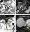
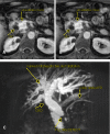
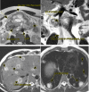
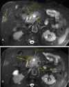
Results: Out of a total of the 362 patients diagnosed with pancreas cancer, results is female 39.5% (n=143) and male 61.5% (n=219), report M: F (1: 1.6), 286 cases resulted in head and neck 79 % (n=286), 76 cases resulted in body and tail cancers (21%), distant metastases in first imaging modality were found in(n=155) patients 43 %, local infiltration was found in patients: gastric infiltration 15 % (n=54), duodenal and papilla infiltration 26% (n=94), local infiltration spleen 16% (n=57), local infiltration mesentery 43 % (n= 155), dilated biliary tree 34 % (n=123), regional lymph node infiltration 83 % (n= 300). Out of a total of the 362 patients diagnosed with pancreas cancer, 346 cases resulted > 2 cm and 16 cases resulted < 2 cm and with component cystic was 41.2 % (n = 149), solid with component cystic - necrotic 33% (n= 119), solid 25.7% (n= 93). The prevalence is 19.9: 100,000 inhabitants.
Conclusions: Prevalence of carcinoma of the pancreas for 5 years in Kosovo has proved to be prevalence is 19.9: 100,000 inhabitants. Seventy-four percent (74 %, n= 268) of all cancers are found in Stage III and IV. From an imaging point of view, these cancers were presented in an advanced stage, mainly due to their late clinical symptoms and limited access to imaging methods in our country.
Keywords: Distant Metastasis; Kosovo; Local Infiltration; MRI; MSCT; Pancreatic Cancer; Prevalence; UCCK Pristina.
Conflict of interest statement
• Conflict of interest: None declared.
Figures



Similar articles
-
THE FREQUENCY OF RISK FACTORS ON TRENDS OF PANCREATIC CANCER IN KOSOVO.Mater Sociomed. 2016 Apr;28(2):108-11. doi: 10.5455/msm.2016.28.108-112. Epub 2016 Mar 25. Mater Sociomed. 2016. PMID: 27147915 Free PMC article.
-
Robotic total thyroidectomy with modified radical neck dissection via unilateral retroauricular approach.Ann Surg Oncol. 2014 Nov;21(12):3872-5. doi: 10.1245/s10434-014-3896-y. Epub 2014 Sep 17. Ann Surg Oncol. 2014. PMID: 25227305
-
More advantages in detecting bone and soft tissue metastases from prostate cancer using 18F-PSMA PET/CT.Hell J Nucl Med. 2019 Jan-Apr;22(1):6-9. doi: 10.1967/s002449910952. Epub 2019 Mar 7. Hell J Nucl Med. 2019. PMID: 30843003
-
[Prospects for standardization of surgical procedures for carcinoma of the pancreas].Nihon Geka Gakkai Zasshi. 2003 May;104(5):412-21. Nihon Geka Gakkai Zasshi. 2003. PMID: 12774526 Review. Japanese.
-
Pretherapeutic evaluation of patients with upper gastrointestinal tract cancer using endoscopic and laparoscopic ultrasonography.Dan Med J. 2012 Dec;59(12):B4568. Dan Med J. 2012. PMID: 23290296 Review.
References
-
- Pancreatic Cancer imaging:Which Method by E.Santo. 2013;3(2):113–20. http://springpublishing.org/index.php/APCI/article/view/4(Medline)
-
- Cancer of the pancreas:ESMO Clinical Practice Guidelines for diagnosis, treatment and follow-up. European Society for Medical Oncology (Medline) 2014 Jan-Feb;38(1):146–52. - PubMed
-
- Bond-Smith G, Banga N, Hammond TM, et al. Pancreatic adenocarcinoma. BMJ. 2012 May;16(344):2476. - PubMed
-
- Pancreatic cancer incidence statistics. Cancer Research UK. 2013;168:649–56.
-
- Lowenfels AB, Maisonneuve P. Epidemiology and risk factors for pancreatic cancer. Best Practice & Research Clinical Gastroenterology. 2006 Apr;20(2):195–416. - PubMed
LinkOut - more resources
Full Text Sources
Other Literature Sources
