

Study of chondrogenic potential of stem cells in co-culture with chondrons
- PMID: 27482345
- PMCID: PMC4951603
Study of chondrogenic potential of stem cells in co-culture with chondrons
Abstract
Objectives: Three-dimensional biomimetic scaffolds have widespread applications in biomedical tissue engineering due to similarity of their nanofibrous architecture to native extracellular matrix. Co-culture system has stimulatory effect on chondrogenesis of adult mesenchymal stem cells. This work presents a co-culture strategy using human articular chondrons and adipose-derived stem cells (ASCs) from infrapatellar fat pad (IPFP) for cartilage tissue production.
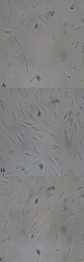
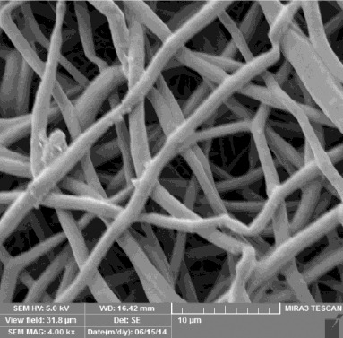
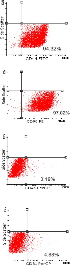
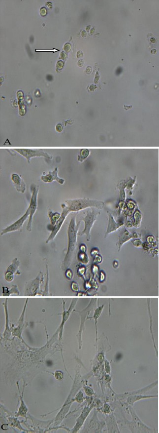
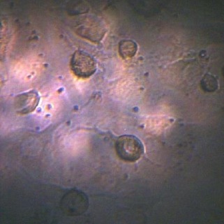
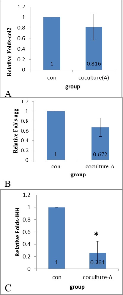
Materials and methods: Isolated stem cells were characterized by flowcytometry. Electrospun and polycaprolactone (PCL) scaffolds (900 nm fiber diameter) was obtained from Bon Yakhteh (Tehran-Iran) and human infrapatellar fat pad-derived stem cells (IPFP-ASCs) were seeded on them. IPFP-ASCs on scaffolds were co-cultured with articular chondrons using transwell. After 21 day, chondrogenic differentiation of stem cell was evaluated by determining the genes expression of collagen2, aggrecan and Indian hedgehog using real-time RT-PCR.
Results: Genes expression of collagen2, aggrecan by IPFP-ASCs did not alter significantly in comparison with control group. Howevers, expression of Indian hedgehog decreased significantly compared to control group (P< 0.05).
Conclusion: These findings indicate that chondrons obtained from osteoarthritic articular cartilage did not stimulate chondrogenic differentiation of IPFP-ASCs in co-culture.
Keywords: Chondron; Co-culture; Nanofiber; Poly-e-caprolactone scaffold.
Figures
References
-
- Buckwalter J, Mankin H. Articular cartilage: degeneration and osteoarthritis, repair, regeneration, and transplantation. Instr Course lect. 1997;47:487–504. - PubMed
-
- Buckwalter JA, Mankin HJ, Grodzinsky AJ. Articular cartilage and osteoarthritis. Instr Course Lect. 2005;54:465. - PubMed
-
- Caplan AI. Tissue engineering designs for the future: new logics, old molecules. Tissue Eng. 2000;6:1–8. - PubMed
-
- Magazine E. From UMassWiki. Tissue Eng. 1993;60:920.
LinkOut - more resources
Full Text Sources