. 2016 Jul 18;3(3):ENEURO.0109-16.2016.
doi: 10.1523/ENEURO.0109-16.2016. eCollection 2016 May-Jun.

Dorsal Medial Habenula Regulation of Mood-Related Behaviors and Primary Reinforcement by Tachykinin-Expressing Habenula Neurons

Affiliations

Dorsal Medial Habenula Regulation of Mood-Related Behaviors and Primary Reinforcement by Tachykinin-Expressing Habenula Neurons

Yun-Wei A Hsu et al. eNeuro. .

Abstract

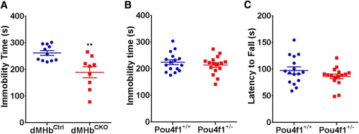
Animal models have been developed to investigate aspects of stress, anxiety, and depression, but our understanding of the circuitry underlying these models remains incomplete. Prior studies of the habenula, a poorly understood nucleus in the dorsal diencephalon, suggest that projections to the medial habenula (MHb) regulate fear and anxiety responses, whereas the lateral habenula (LHb) is involved in the expression of learned helplessness, a model of depression. Tissue-specific deletion of the transcription factor Pou4f1 in the dorsal MHb (dMHb) results in a developmental lesion of this subnucleus. These dMHb-ablated mice show deficits in voluntary exercise, a possible correlate of depression. Here we explore the role of the dMHb in mood-related behaviors and intrinsic reinforcement. Lesions of the dMHb do not elicit changes in contextual conditioned fear. However, dMHb-lesioned mice exhibit shorter immobility time in the tail suspension test, another model of depression. dMHb-lesioned mice also display increased vulnerability to the induction of learned helplessness. However, this effect is not due specifically to the dMHb lesion, but appears to result from Pou4f1 haploinsufficiency elsewhere in the nervous system. Pou4f1 haploinsufficiency does not produce the other phenotypes associated with dMHb lesions. Using optogenetic intracranial self-stimulation, intrinsic reinforcement by the dMHb can be mapped to a specific population of neurokinin-expressing habenula neurons. Together, our data show that the dMHb is involved in the regulation of multiple mood-related behaviors, but also support the idea that these behaviors do not reflect a single functional pathway.

Keywords: fear conditioning; habenula; interpeduncular nucleus; learned helplessness; substance P.


Conflict of interest statement

The authors report no conflict of interest.

Figures

Figure 1.
Figure 1.
Pou4f1 knock-out models used for analysis of dMHb function. A, Summary of genetic models used to generate dMHb lesions. B, C, Coronal sections through the habenula at bregma −1.58mm were stained with antibodies for choline acetyltransferase (Chat) and either Pou4f1 (Brn3a protein) to reveal habenula neurons expressing this factor, or for the lacZ gene product βGal, expressed by the Pou4f1tlacZ allele, which allows the identification of neurons that would normally express Pou4f1 in cells in which the gene has been deleted. B, dMHbCtrl mouse with the genotype Pou4f1flox/+/Syt6Cre. Nuclear staining for Pou4f1 shows expression in both the vMHb and dMHb, and scattered expression in the LHb. The vMHb is distinguished by the expression of Chat, and the Pou4f1-positive, Chat-negative dMHb is circled. Scale bar, 100 μm. C, dMHbCKO mouse with the genotype Pou4f1flox/tlacZ/Syt6Cre. Staining for lacZ is used to show the extent of the dMHb lesion in the absence of Pou4f1 protein. The extent of the dMHb is greatly reduced (circle) and only a few βGal-positive, Chat-negative neurons remain in the medial habenula. Neurons of the vMHb, distinguished by Chat expression in A and B, are intact in both the dMHbCtrl and dMHbCKO mice. The area within the dMHb that is not stained by any of the antibodies used consists of axon tracts of the striae medularis and/or habenula commissure.
Figure 2.
Figure 2.
Conditioned fear response in dMHbCKO mice. A, Training session: time spent in freezing behavior in 1 min intervals during 3 min of acclimation, followed by six intervals preceded by the delivery of a 1 s shock, is shown. Shaded bar shows the period of shock administration. B, Contextual conditioned fear response: on the testing day, the time spent in freezing behavior was assessed in the same environment as the training session, but without shock delivery, to evaluate the conditioned fear response. dMHbCKO and dMHbCtrl mice exhibited the same amount of freezing during both the training and testing day. C, Extinction of the conditioned fear response: time spent in freezing behavior during 3 subsequent days of testing is shown. The conditioned fear response showed gradual extinction in the absence of further shock stimuli. *p = 0.033, **p = 0.0082, ***p = 0.0003, and ****p < 0.0001, significant difference between days for these genotypes. N = 12 dMHbCtrl and 10 dMHbCKO mice.
Figure 3.
Figure 3.
Learned helplessness assessed by active avoidance in dMHbCKO mice. A, Learned helplessness response: the mean latency to escape in the shuttle box following 1 d of inescapable shock training, or exposure to the training chamber without a shock, is shown. Both the dMHbCKO and dMHbCtrl mice that received shocks during training showed increased latency to escape, but dMHbCKO mice exhibited a stronger effect. **p < 0.01, ****p < 0.0001 for difference between groups indicated. B, Persistence of the learned helplessness response: the mean latency to escape was reassessed for the cohort shown in A 3 weeks after inescapable shock training. dMHbCtrl mice that received inescapable shocks returned to near-baseline escape times. dMHbCKO mice that received inescapable shocks retained the learned helplessness behavior. **p < 0.01 and ****p < 0.0001, significant difference between groups. N = 13 no-shock dMHbCtrl, 10 shocked dMHbCtrl, 11 no-shock dMHbCKO, and 17 shocked dMHbCKO mice. C, Convergence of learned helplessness response with extended training: the mean latency to escape was assessed after 3 d of inescapable shock training in a different cohort of mice. Both the dMHbCKO and dMHbCtrl mice received shocks during the training. The prolonged training increased escape latency for both genotypes when assessed by active avoidance 1 d after the final training session. N = 11 dMHbCtrl and 9 dMHbCKO mice. D, E, Assessment of learned helplessness in a separate cohort of Pou4f1 hemizygous mice. D, Learned helplessness response: the mean latency to escape in the shuttle box following 1 d of inescapable shock training is shown. Both the Pou4f1+/- and Pou4f1+/+ (wild-type) mice received shocks during the training. Pou4f1+/- mice exhibited increased latency to escape relative to Pou4f1+/+ mice, i.e., were more susceptible to the induction of learned helplessness. **p = 0.0059, significant difference between the genotypes. E, Persistence of learned helplessness response: the mean latency to escape was reassessed for the cohort shown in D 3 weeks after inescapable shock training. The Pou4f1+/- mice showed persistently elevated escape latency relative to Pou4f1+/+ mice. ***p = 0.0007, significant difference between the genotypes. Minor modifications to the active avoidance protocol used in D and E, resulting in somewhat longer escape times, are described in Materials and Methods.
Figure 4.
Figure 4.
TST immobility time in dMHbCKO and Pou4f1 hemizygous mice, and affect of hemizygosity on rotarod performance. A, Time spent immobile in the TST is shown for dMHbCKO and dMHbCtrl mice. **p < 0.01, significance difference between the genotypes. N = 11 dMHbCtrl and 9 dMHbCKO mice. B, Time spent immobile in the TST is shown for Pou4f1+/+ and Pou4f1+/- mice. Pou4f1 gene dosage did not affect immobility time in the absence of a developmental dMHb lesion. N = 16 Pou4f1+/+ and 16 Pou4f1+/- mice. Pou4f1+/- mice have the genotype Pou4f1+/tlacZ. C, Rotarod performance is not affected in Pou4f1+/- mice. Both the Pou4f1+/- and Pou4f1+/+ mice had similar latency to fall times in this test. N = 16 of each genotype.
Figure 5.
Figure 5.
Specific expression of channelrhodopsin in tachykinin-expressing neurons in dMHb-TacChR2 mice. A, B, Comparison of Tac1 and Tac2 mRNA expression in the habenula; the axial level is approximately bregma −1.6mm. Data are derived from the Allen Mouse Brain Atlas. C, D, Conditional expression of ChR2-eYFP in Ai32 mice driven by Tac2IRESCre in the habenula. Dashed lines demarcate the extent of the habenula based on the expression of the nuclear factor Pou4f1 (C). Expression is infrequently seen in the vMHb, as defined by the expression of Chat (D). E, Conditional expression of tdTomato mRNA in Ai14 mice driven by Tac2IRESCre in the habenula (Allen Transgenic Mouse Characterization Project). FH, ChR2-eYFP labeled fibers terminate predominantly in the lateral part of the interpeduncular nucleus, which receives afferents from the dMHb (F, H); these fibers are largely excluded from the IPR/IPC, which receive fibers mainly from the vMHb (G). I, J, Colocalization of SP, product of the Tac1 gene, with ChR2-eYFP in the interpeduncular nucleus of dMHb-TacChR2 mice. K, Colocalization of neurokinin B, product of the Tac2 gene, with ChR2-eYFP. Scale bars: A, 200 μm; C, F, I, 100 μm; J, 50 μm.
Figure 6.
Figure 6.
Intrinsic reinforcement mediated by neurokinin-expressing dMHb neurons. A, B, Light entrainment of dMHb neurons expressing Tac2Cre-driven ChR2. C, Optogenetic ICSS in dMHbChR2 mice. Two nose-poke receptacles in each behavioral compartment were randomized to active and inactive at the beginning of the trial. The initial assignment was maintained for days 1–4 of training, and then reversed for days 5–8 of training (reversal trials). Mice received a 2 s light stimulation of the dMHb for each nose-poke event in the active receptacle. D, ICSS in control mice lacking a Cre-driver; no preference for the active receptacle was observed. E, ICSS in vMHbChR2 mice; no preference was observed. F, Average values for nose-poke events in the inactive and active receptacles over 4 d of trials for dMHbChR2 (N=7), vMHbChR2 (N=6) and control (N=11) mice.
Figure 7.
Figure 7.
Placement of optical fibers in ICSS experimental mice. Mice with bilateral implanted fiber optic cannulas were perfused with a fixative at the conclusion of behavioral experiments and brains were examined for cannula placement. Fiber termini are shown on the level of a standard anatomical map (Paxinos and Franklin, 2001) closest to their rostrocaudal position at bregma −1.34, 1.46, 1.58, or 1.70 mm. Nearly all of the cannulas thus were positioned within ±0.2 mm of the intended coordinates at bregma −1.6 mm. Although some cannulas were displaced laterally, at least one of the two optical fibers terminated close to the habenula in every case. Fibers for dMHbChR2 mice are shown in red, vMHbChR2 mice are shown in green, and control mice are shown in blue. Connected dots indicate the probable ventral termini of the optical fibers from each case. In some cases, the right and left optical fibers mapped most accurately to different planes of section and are shown by disconnected dots. If the cannula track could not be followed to the terminus of the optical fiber, for instance due to termination in the ventricle, the most ventral position and the direction of the cannula track observed are indicated by an arrow. In all cases, the optical fibers were intact and transmitted light efficiently when examined postmortem after the experimental protocol. Scale bar, 0.5 mm. Light was delivered through the bilateral cannula for a total output of 8 mW, corresponding to 4 mW per 100 μm fiber or 509 mW/mm2 at the fiber teriminus. Because some of the cannulas were displaced laterally, we used a published empirically derived model for the diffusion of 473 nm light in the mouse brain tissue to estimate the light intensity at target structures (Yizhar et al., 2011). The example laterally displaced cannula pair is indicated by an asterisk (bregma 1.46 view). The boundary of light penetration at 1% of that delivered at the fiber terminus is indicated by a dashed line (∼5 mw/mm2). Although one cannula is displaced laterally, the medial cannula is predicted to illuminate the entire habenula. The predicted intensity of light, 5 mW/mm2, is sufficient to elicit reliable action potentials from dMHb neurons in brain-slice preparations.
Figure 8.
Figure 8.
Real-time place preference. RTPP studies were conducted in a two-chamber place-preference box in which mice received light stimulation in one side, and could move freely between compartments. A, B, Example activity traces of control (A) and dMHbChR2 (B) mice. Data are shown for an entire 15 min trial for a single animal of each genotype. C, D, Side preference of control (C; n=8) and dMHbChR2 mice (D; n=8) displayed in 5 min bins over the course of a 15 min trial. The large variability in the side occupancy in the later intervals is likely to represent decreasing exploration during the course of the trial, with individual mice settling on one side of the chamber or the other, apparently without preference. E, Summary of side occupancy for 15 min trial. No significant effect of side or genotype was observed.

References

    1. Amat J, Sparks PD, Matus-Amat P, Griggs J, Watkins LR, Maier SF (2001) The role of the habenular complex in the elevation of dorsal raphe nucleus serotonin and the changes in the behavioral responses produced by uncontrollable stress. Brain Res 917:118–126. - PubMed
    1. Bilkei-Gorzo A, Racz I, Michel K, Zimmer A (2002) Diminished anxiety- and depression-related behaviors in mice with selective deletion of the Tac1 gene. J Neurosci 22:10046–10052. - PMC - PubMed
    1. Chourbaji S, Zacher C, Sanchis-Segura C, Dormann C, Vollmayr B, Gass P (2005) Learned helplessness: validity and reliability of depressive-like states in mice. Brain Res Brain Res Protoc 16:70–78. 10.1016/j.brainresprot.2005.09.002 - DOI - PubMed
    1. Cryan JF, Mombereau C (2004) In search of a depressed mouse: utility of models for studying depression-related behavior in genetically modified mice. Mol Psychiatry 9:326–357. 10.1038/sj.mp.4001457 - DOI - PubMed
    1. Cryan JF, Mombereau C, Vassout A (2005) The tail suspension test as a model for assessing antidepressant activity: review of pharmacological and genetic studies in mice. Neurosci Biobehav Rev 29:571–625. 10.1016/j.neubiorev.2005.03.009 - DOI - PubMed

MeSH terms

LinkOut - more resources