. 2016 Jul 7;1(10):e86673.
doi: 10.1172/jci.insight.86673.

Recognition of influenza H3N2 variant virus by human neutralizing antibodies

Affiliations

Recognition of influenza H3N2 variant virus by human neutralizing antibodies

Sandhya Bangaru et al. JCI Insight. .

Abstract

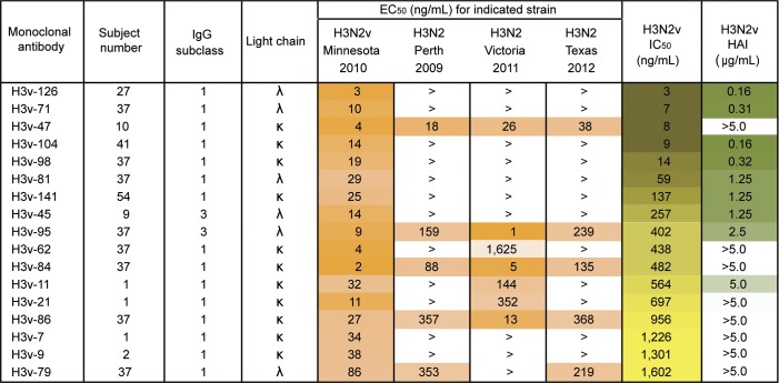
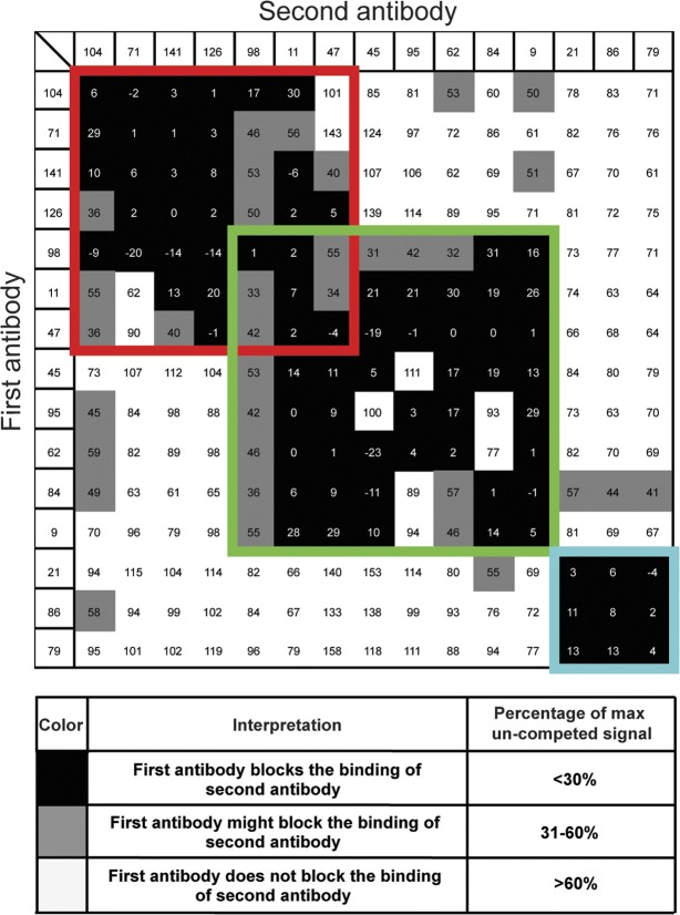
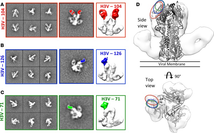
Since 2011, over 300 human cases of infection, especially in exposed children, with the influenza A H3N2 variant (H3N2v) virus that circulates in swine in the US have been reported. The structural and genetic basis for the lack of protection against H3N2v induced by vaccines containing seasonal H3N2 antigens is poorly understood. We isolated 17 human monoclonal antibodies (mAbs) that neutralized H3N2v virus from subjects experimentally immunized with an H3N2v candidate vaccine. Six mAbs exhibited very potent neutralizing activity (IC50 < 200 ng/ml) against the H3N2v virus but not against current human H3N2 circulating strains. Fine epitope mapping and structural characterization of antigen-antibody complexes revealed that H3N2v specificity was attributable to amino acid polymorphisms in the 150-loop and the 190-helix antigenic sites on the hemagglutinin protein. H3N2v-specific antibodies also neutralized human H3N2 influenza strains naturally circulating between 1995 and 2005. These results reveal a high level of antigenic relatedness between the swine H3N2v virus and previously circulating human strains, consistent with the fact that early human H3 seasonal strains entered the porcine population in the 1990s and reentered the human population, where they had not been circulating, as H3N2v about a decade later. The data also explain the increased susceptibility to H3N2v viruses in young children, who lack prior exposure to human seasonal strains from the 1990s.


Figures

Figure 1
Figure 1. Characterization of 17 neutralizing monoclonal antibodies (mAbs).
The antibodies are arranged in the order of neutralization potency (column 9) with the most potent antibodies at the top. Seventeen mAbs isolated by human B cell hybridoma generation exhibited neutralization potential (shown as half-maximal inhibitory concentration [IC50]) at <5 μg/ml against the H3N2v virus by microneutralization assay. Nine antibodies exhibited hemagglutinin inhibition (HAI) activity, indicating that they disrupt receptor-binding function of the virus. The mAbs were tested for binding against HA from H3N2v or 3 seasonal strains (shown as half-maximal effective concentration [EC50]). The > symbol indicates that binding was not detected at the maximum concentration tested (2 μg/ml). The experiments for determining EC50 (n = 4), IC50 (n = 3), and HAI (n = 3) were conducted twice independently.
Figure 2
Figure 2. Prophylactic efficacy of H3v-104, H3v-126, and H3v-71 in mice.
Groups of mice (n = 5) were treated with 100 μg of individual mAbs 24 hours before challenge with mouse-adapted A/Minnesota/11/2010 X203 virus. Controls (n = 10) were injected with PBS. The weight loss of mice was measured daily for 14 days after inoculation (day 0).
Figure 3
Figure 3. Competition binding of neutralizing antibodies to H3N2v A/Minnesota/11/2010 hemagglutinin (HA) protein.
Biolayer interferometry was used to perform competition-binding assays. The HA was loaded onto Ni-NTA tips, and binding to 2 successive antibodies was tested. The binding signal for each antibody was obtained from a single association step of the mAb onto HA. If binding of the first antibody blocked the binding of the second antibody by reducing its binding signal by more than 70%, it was defined as a competitor, indicated in black. The values in the table indicate the percentage of the maximum uncompeted binding signal. The red box indicates group 1, the binding group comprising the potently neutralizing mAbs. The green box and the blue box represent group 2 (partially overlaps with group 1) and group 3, respectively. The experiment was conducted twice independently.
Figure 4
Figure 4. Binding of H3 variant-specific antibodies to mutated Minnesota hemagglutinin (HA) proteins.
Mutagenesis of MNv HA was performed to determine antigenic residues important for recognition by variant-specific mAbs. Mutants of H3N2v A/Minnesota/11/2010 (MN) HA were generated by site-directed mutagenesis, and the half-maximal effective concentration (EC50) values were determined by performing ELISA with serial dilutions of each antibody against the mutant HAs. The table (A) shows EC50 values and the graph (B) shows binding curves. The mutants that disrupted binding completely or decreased the EC50 by greater than 5-fold are represented as dashed or dotted lines, respectively, and are indicated by red EC50 values in the table. The > symbol indicates that binding was not detected at the maximum concentration tested (10 μg/ml). The experiments for determining the EC50 (n = 4) values were performed twice independently.
Figure 5
Figure 5. Negative stain EM images of hemagglutinin-Fab complexes.
In each case the stem-binding antibody CR9114 was added to the complex in order to improve 3D reconstructions. (A) Reference-free 2D class averages of complex containing Fab 104 (left), single class average with Fab colored in red (middle), and 3D reconstruction (right). (B) Reference-free 2D class averages of complex containing Fab 126 (left), single class average with Fab colored in blue (middle), and 3D reconstruction (right). (C) Reference-free 2D class averages of complex containing Fab 71 (left), single class average with Fab colored in green (middle), and 3D reconstruction (right). (D) Side and top views of HA-Fab 126-CR9114 with Fab 126 removed and crystal structure of H3V (4FNK) fitted. Binding sites of the 3 antibodies described in AC is highlighted using colors corresponding to Fabs.
Figure 6
Figure 6. Neutralization of H3N2 seasonal strains.
The monoclonal antibodies were tested for neutralization activity by microneutralization assay against 11 seasonal strains isolated between 1968 and 2012. The half-maximal inhibitory concentration (IC50) values for H3v-98 (pink), H3v-104 (orange), or H3v-71 (green) are indicated on the y axis, and the H3N2 strains used for neutralization are indicated on the x axis. The IC50 values are represented as baseline if neutralization was not detected at any concentration less than 5 μg/ml. The microneutralization assay for determining the IC50 (n = 3) values was performed twice independently.

References

    1. World Health Organization Influenza (Seasonal) [June 13, 2016];WHO Web site. http://www.who.int/mediacentre/factsheets/fs211/en/
    1. Dreyfus C, et al. Structure of a classical broadly neutralizing stem antibody in complex with a pandemic H2 influenza virus hemagglutinin. J Virol. 2013;87(12):7149–7154. doi: 10.1128/JVI.02975-12. - DOI - PMC - PubMed
    1. Tong S, et al. New world bats harbor diverse influenza A viruses. PLoS Pathog. 2013;9(10):e86673. doi: 10.1371/journal.ppat.1003657. - DOI - PMC - PubMed
    1. Suzuki Y, et al. Sialic acid species as a determinant of the host range of influenza A viruses. J Virol. 2000;74(24):11825–11831. doi: 10.1128/JVI.74.24.11825-11831.2000. - DOI - PMC - PubMed
    1. Rogers GN, Paulson JC. Receptor determinants of human and animal influenza virus isolates: Differences in receptor specificity of the H3 hemagglutinin based on species of origin. Virology. 1983;127(2):361–373. doi: 10.1016/0042-6822(83)90150-2. - DOI - PubMed