Randomized Controlled Trial
. 2016 Sep 1;126(9):3207-18.
doi: 10.1172/JCI86249. Epub 2016 Aug 2.

Dasatinib induces lung vascular toxicity and predisposes to pulmonary hypertension

Randomized Controlled Trial

Dasatinib induces lung vascular toxicity and predisposes to pulmonary hypertension

Christophe Guignabert et al. J Clin Invest. .

Abstract

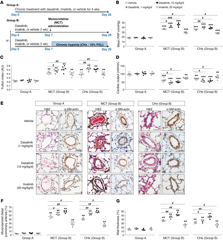
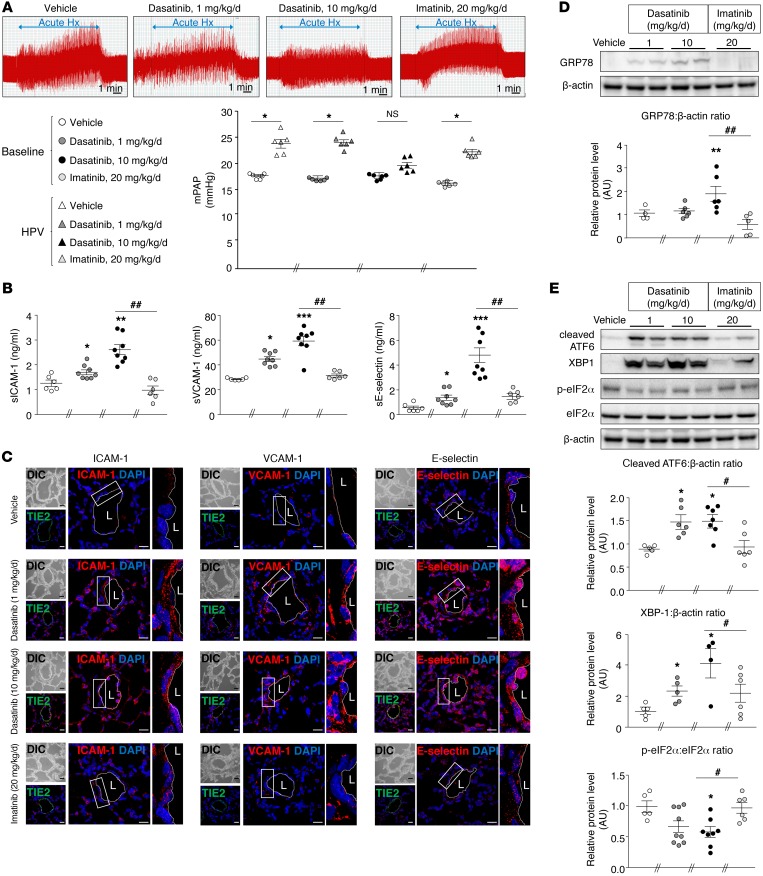
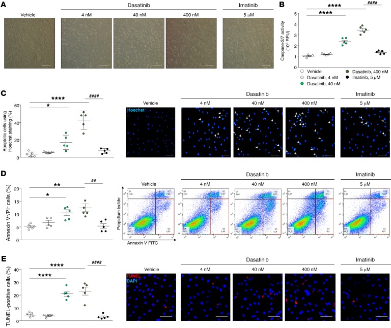
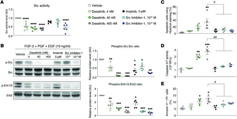
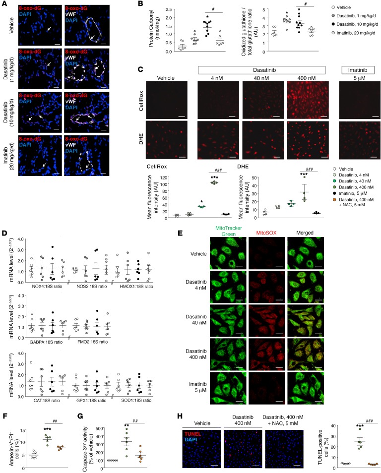
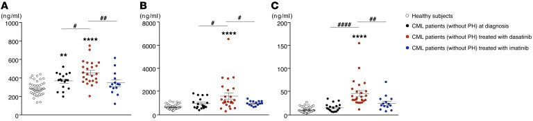
Pulmonary arterial hypertension (PAH) is a life-threatening disease that can be induced by dasatinib, a dual Src and BCR-ABL tyrosine kinase inhibitor that is used to treat chronic myelogenous leukemia (CML). Today, key questions remain regarding the mechanisms involved in the long-term development of dasatinib-induced PAH. Here, we demonstrated that chronic dasatinib therapy causes pulmonary endothelial damage in humans and rodents. We found that dasatinib treatment attenuated hypoxic pulmonary vasoconstriction responses and increased susceptibility to experimental pulmonary hypertension (PH) in rats, but these effects were absent in rats treated with imatinib, another BCR-ABL tyrosine kinase inhibitor. Furthermore, dasatinib treatment induced pulmonary endothelial cell apoptosis in a dose-dependent manner, while imatinib did not. Dasatinib treatment mediated endothelial cell dysfunction via increased production of ROS that was independent of Src family kinases. Consistent with these findings, we observed elevations in markers of endothelial dysfunction and vascular damage in the serum of CML patients who were treated with dasatinib, compared with CML patients treated with imatinib. Taken together, our findings indicate that dasatinib causes pulmonary vascular damage, induction of ER stress, and mitochondrial ROS production, which leads to increased susceptibility to PH development.


Figures

Figure 1
Figure 1. Pretreatment of rats with dasatinib confers an exaggerated response to monocrotaline (MCT) and chronic hypoxia (CHx), 2 inducers of experimental pulmonary hypertension in vivo.
(A) Rodent models used to study the in vivo effects of dasatinib on pulmonary hemodynamic parameters and vascular remodeling: chronic treatment group (group A) and pretreatment group (group B). (BD) Values of mean pulmonary arterial pressure (PAP) (B), Fulton index (C), and cardiac output (D) in vehicle-, dasatinib-, and imatinib-treated rats. (E) Representative images of H&E- and α-smooth muscle (SM) actin–stained sections of distal pulmonary arteries in vehicle-, dasatinib-, and imatinib-treated rats. (F and G) Quantification of the percentage of muscularized pulmonary arteries (F) and wall thickness (G) in lungs of vehicle-, dasatinib-, and imatinib-treated rats. Horizontal lines display the mean ± SEM (n = 5–6). **P < 0.01, ***P < 0.001 vs. vehicle-treated rats; #P < 0.05, ##P < 0.01, ###P < 0.001 vs. vehicle-treated rats exposed to MCT or to CHx. Scale bars: 20 μm.
Figure 2
Figure 2. Dasatinib treatment causes pulmonary EC dysfunction in vivo.
(A) Values of mean pulmonary arterial pressure (mPAP) in vehicle-, dasatinib-, and imatinib-treated rats while ventilated with room air (baseline) and after 10 minutes of ventilation with a hypoxic gas mixture (acute Hypoxia [Hx]). mPAP increased in rats treated with vehicle, dasatinib (1×), and imatinib, but not in rats treated with dasatinib (10×). (B) Quantification of the circulating levels of soluble forms of ICAM-1 (sICAM-1), VCAM-1 (sVCAM-1), and E-selectin (sE-selectin) in vehicle-, dasatinib-, and imatinib-treated rats. (C) Confocal microscopic analyses and double labeling with ICAM-1, VCAM-1, and E-selectin with the endothelial-specific marker Tie2 in lungs of vehicle-, dasatinib-, and imatinib-treated rats. (D and E) Representative Western blots and quantification of the glucose-regulated protein 78 (GRP78)/β-actin ratio (D) and of the cleaved activating transcription factor 6 (ATF6)/β-actin ratio, the X-box–binding protein 1 (XBP1)/β-actin ratio, and the phospho–eukaryotic initiation factor 2α (eIF2α)/eIF2α ratio (E) in lungs of vehicle-, dasatinib-, and imatinib-treated rats. Horizontal lines display the mean ± SEM (n = 4–9). *P < 0.05, **P < 0.01, ***P < 0.001 vs. vehicle-treated rats or baseline; #P < 0.05, ##P < 0.01 vs. rats treated with dasatinib (10×). Scale bars: 20 μm. HPV, hypoxic pulmonary vasoconstriction; L, lumen.
Figure 3
Figure 3. Dasatinib-induced apoptosis of human pulmonary ECs in vitro.
(A) Representative images of human pulmonary EC monolayers (passage <5) 2 hours after treatment with vehicle, dasatinib, or imatinib. (B) Determination of caspase-3/7 activity in human pulmonary ECs treated with vehicle, dasatinib, or imatinib. (C) Quantification of the percentage of apoptotic cells using Hoechst 33342 staining and representative images. (D) Quantification of annexin V and propidium iodide (PI) dual labeling in human pulmonary ECs treated with vehicle, dasatinib, or imatinib and representative FACS dot plots. (E) Quantification of TUNEL-positive cells in human pulmonary ECs treated with vehicle, dasatinib, or imatinib and representative images. Horizontal lines display the mean ± SEM (n = 5). *P < 0.05, **P < 0.01, ****P < 0.0001 vs. vehicle-treated cells; ##P < 0.01, ####P < 0.0001 vs. human pulmonary ECs treated with 400 nM dasatinib. Scale bars: 20 μm.
Figure 4
Figure 4. Dasatinib-induced apoptosis in cultured human pulmonary ECs is independent of its action on Src family kinases.
(A) Quantification of the activity of Src directly in human pulmonary ECs treated with vehicle, dasatinib, imatinib, or Src inhibitor-1. (B) Representative Western blots and quantification of the phospho-Src/Src ratio and phospho-ERK1/2/ERK2 ratio in human pulmonary ECs exposed to a growth factor cocktail and treated with vehicle, dasatinib, imatinib, or Src inhibitor-1. (CE) Quantification of pulmonary EC apoptosis using Hoechst 33342 (C), caspase-3/7 activity (D), and double staining with annexin V and propidium iodide (PI) by flow cytometry (E) in pulmonary ECs treated with vehicle, dasatinib, imatinib, or Src inhibitor-1. Horizontal lines display the mean ± SEM (n = 4–6). *P < 0.05, **P < 0.01, ***P < 0.001, ****P < 0.0001 vs. vehicle-treated cells; #P < 0.05, ##P < 0.01 vs. human pulmonary ECs treated with 400 nM dasatinib. FGF-2, fibroblast growth factor 2 (basic); PDGF, platelet-derived growth factor; EGF, epidermal growth factor.
Figure 5
Figure 5. Dasatinib stimulates mitochondrial ROS production in vivo and in vitro.
(A) Representative images obtained by confocal microscopic analyses and double labeling with 8-oxo-2′-deoxyguanosine (8-oxo-dG) and the endothelial-specific marker vWF in lungs from vehicle-, dasatinib-, and imatinib-treated rats. (B) Quantifications of the oxidized glutathione/total glutathione ratio and of the protein carbonyl products in lungs and serum of vehicle-, dasatinib-, and imatinib-treated rats, respectively. (C) Representative images and quantification of intracellular ROS generation using fluorogenic probes (CellRox) and dihydroethidium (DHE) in pulmonary ECs treated 30 minutes with vehicle, dasatinib, or imatinib. (D) Gene expression analysis of pro-oxidative enzymes (NADPH oxidase 4 [NOX4], NO synthase 2 [NOS2], heme oxygenase 1 [HMOX1], GA-binding protein α chain [GABPA], flavin-containing monooxygenase 2 [FMO2]) and antioxidative enzymes (catalase [CAT], glutathione peroxidase 1 [GPX1], superoxide dismutase 1 [SOD1]) in lungs of rats treated with vehicle, dasatinib, and imatinib. (E) Mitochondria were labeled with MitoTracker Green (green) and mitochondrial ROS with MitoSOX Red (red). (FG) Quantitative FACS analysis of the annexin V and propidium iodide (PI) dual labeling and quantification of caspase-3/7 activity in pulmonary ECs pretreated or not for 16 hours with N-acetylcysteine (NAC) at 5 mM and exposed or not to 400 nM dasatinib. (H) Representative images and quantification of the TUNEL staining in pulmonary ECs pretreated or not for 1 hour with NAC at 5 mM and exposed or not to 400 nM dasatinib. Horizontal lines display the mean ± SEM (n = 4–9). *P < 0.05, **P < 0.01, ***P < 0.001 vs. vehicle-treated cells or -treated rats; #P < 0.05, ##P < 0.01, ###P < 0.001 vs. pulmonary ECs treated with 400 nM dasatinib or rats treated with dasatinib (10×). Scale bars: 20 μm.
Figure 6
Figure 6. Patients with CML treated with dasatinib display increased serum concentrations of sICAM-1, sVCAM-1, and sE-selectin.
Quantifications of sICAM-1 (A), sVCAM-1 (B), and sE-selectin (C) in CML patients at diagnosis (n = 17), in CML patients treated with dasatinib or imatinib after less than 3 months of therapy (n = 24 and 14, respectively), and in healthy subjects (n = 39). Horizontal lines display the mean ± SEM. **P < 0.01, ****P < 0.0001 vs. healthy subjects; #P < 0.05, ##P < 0.01, ####P < 0.0001 vs. CML patients treated with dasatinib.

Similar articles

Cited by

References

    1. Galiè N, et al. 2015 ESC/ERS Guidelines for the diagnosis and treatment of pulmonary hypertension: The Joint Task Force for the Diagnosis and Treatment of Pulmonary Hypertension of the European Society of Cardiology (ESC) and the European Respiratory Society (ERS). Endorsed by: Association for European Paediatric and Congenital Cardiology (AEPC), International Society for Heart and Lung Transplantation (ISHLT) Eur Respir J. 2015;46(4):903–975. doi: 10.1183/13993003.01032-2015. - DOI - PubMed
    1. Guignabert C, Dorfmuller P. Pathology and pathobiology of pulmonary hypertension. Semin Respir Crit Care Med. 2013;34(5):551–559. doi: 10.1055/s-0033-1356496. - DOI - PubMed
    1. Guignabert C, et al. New molecular targets of pulmonary vascular remodeling in pulmonary arterial hypertension: importance of endothelial communication. Chest. 2015;147(2):529–537. doi: 10.1378/chest.14-0862. - DOI - PubMed
    1. Guignabert C, et al. Pathogenesis of pulmonary arterial hypertension: lessons from cancer. Eur Respir Rev. 2013;22(130):543–551. doi: 10.1183/09059180.00007513. - DOI - PMC - PubMed
    1. Huertas A, et al. Immune dysregulation and endothelial dysfunction in pulmonary arterial hypertension: a complex interplay. Circulation. 2014;129(12):1332–1340. doi: 10.1161/CIRCULATIONAHA.113.004555. - DOI - PubMed

Publication types

MeSH terms