. 2016 Aug 2;5(8):e341.
doi: 10.1038/mtna.2016.51.

Poly(ester amine) Composed of Polyethylenimine and Pluronic Enhance Delivery of Antisense Oligonucleotides In Vitro and in Dystrophic mdx Mice

Affiliations

Poly(ester amine) Composed of Polyethylenimine and Pluronic Enhance Delivery of Antisense Oligonucleotides In Vitro and in Dystrophic mdx Mice

Mingxing Wang et al. Mol Ther Nucleic Acids. .

Abstract

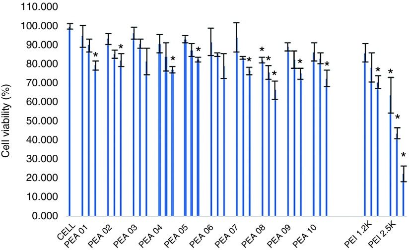
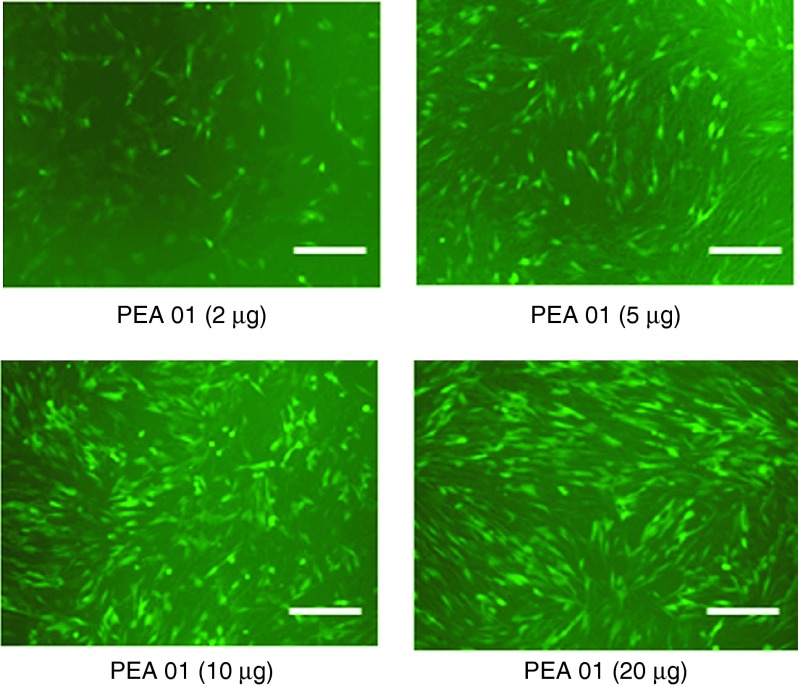
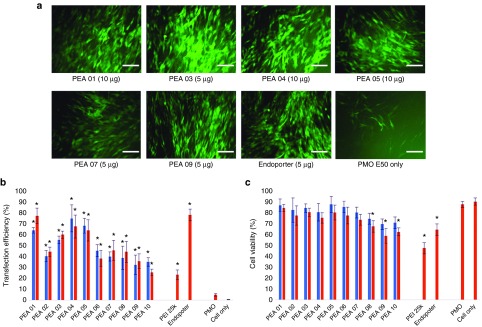
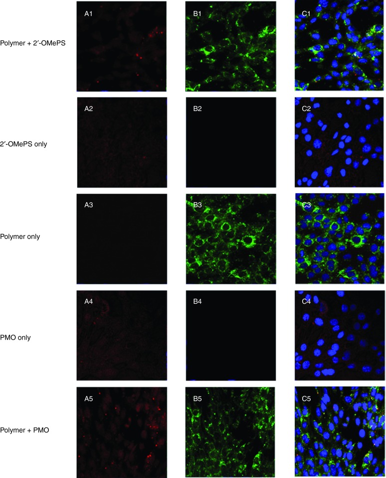
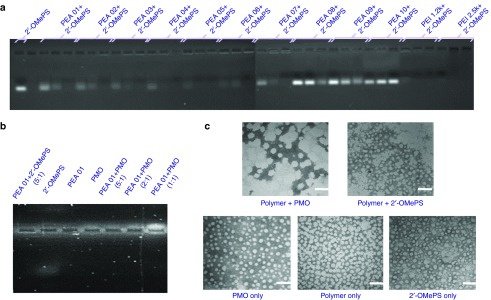
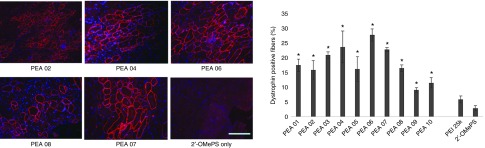
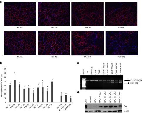
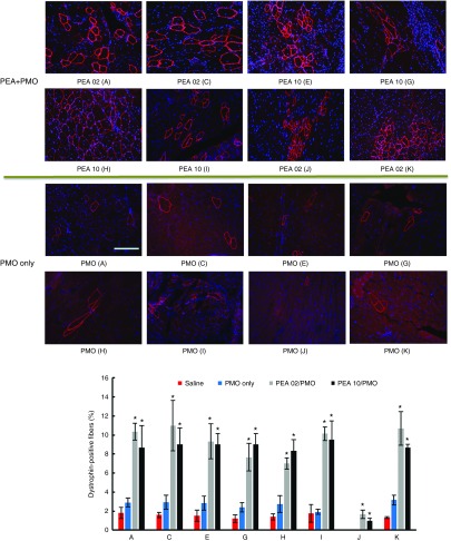
A series of poly(esteramine)s (PEAs) constructed from low molecular weight polyethyleneimine (LPEI) and Pluronic were evaluated for the delivery of antisense oligonuclotides (AOs), 2'-O-methyl phosphorothioate RNA (2'-OMePS) and phosphorodiamidate morpholino oligomer (PMO) in cell culture and dystrophic mdx mice. Improved exon-skipping efficiency of both 2'-OMePS and PMO was observed in the C2C12E50 cell line with all PEA polymers compared with PEI 25k or LF-2k. The degree of efficiency was found in the order of PEA 01, PEA 04 > PEA 05 > others. The in vivo study in mdx mice demonstrated enhanced exon-skipping of 2'-OMePS with the order of PEA 06 > PEA 04, PEA 07 > PEA 03 > PEA 01 > others, and much higher than PEI 25k formulated 2'-OMePS. Exon-skipping efficiency of PMO in formulation with the PEAs were significantly enhanced in the order of PEA 02 > PEA 10 > PEA 01, PEA 03 > PEA 05, PEA 07, PEA 08 > others, with PEA 02 reaching fourfold of Endo-porter formulated PMO. PEAs improve PMO delivery more effectively than 2'-OMePS delivery in vivo, and the systemic delivery evaluation further highlight the efficiency of PEA for PMO delivery in all skeletal muscle. The results suggest that the flexibility of PEA polymers could be explored for delivery of different AO chemistries, especially for antisense therapy.


Figures

Figure 1
Figure 1
The cytotoxicity of the PEAs polymers in C2C12E50 cell lines via MTS-based cell viability assay. The polymer dosages are 4, 10, 20 µg/ml from left to right for each sample. Cells were seeded in 96-well plates at an initial density of 1 × 104 cells/well in 200 µl growth media. The results are presented as the mean ± SD in triplicate (Student's t-test, *P ≤ 0.05 compared with untreated cell as a control).
Figure 2
Figure 2
Dose–response GFP expression of polymer PEA 01 formulated 2′-OMePSE50. C2C12E50 cells were treated with 2 µg 2′-OMePS and the polymer in 500 µl 10% FBS-DMEM and incubated under 37 °C, 10% CO2 environment. The images were taken 48 hours after transfection (scale bar = 500 µm).
Figure 3
Figure 3
Delivery efficiency and toxicity of PEAs mediated 2′-OMePS in C2C12 cell line determined by fluorescence microscopy and flow cytometer. (a) Representative fluorescence images of 2′-OMePSE50-induced exon-skipping in C2C12E50 cell line. The images were taken 48 hours after treatment (scale bar = 500 µm). (b) Transfection efficiency of 2′-OMePSE50 formulated with the polymers (Student's t-test, *P ≤ 0.05 compared with 2′-OMePS only). (c) Cell viability (Student's t-test, *P ≤ 0.05 compared with untreated cells as a control). Two micrograms of 2′-OMePSE50 were formulated with PEAs (5,10 µg), PEI-25k (2 µg) and LF-2k (4 µg) in 500 µl 10% FBS-DMEM medium. The results are presented as the mean ± SD in triplicate.
Figure 4
Figure 4
Delivery efficiency and toxicity of PEAs mediated PMO in C2C12 cell line determined by fluorescence microscopy and flow cytometer. (a) Representative fluorescent microscopy images of PMOE50 -induced exon-skipping in C2C12E50 cell line. The images were taken 48 hours after treatment (scale bar = 500 µm). (b) Transfection efficiency of PMOE50 formulated with polymers (Student's t-test, *P ≤ 0.05 compared with PMO only). (c) Cell viability (Student's t-test, *P ≤ 0.05 compared with untreated cells as a control). Five micrograms of PMOE50 were formulated with PEAs (5,10 µg), and PEI-25k (2 µg), Endo-porter (5 µg) formulated as controls in 500 µl 10% FBS-DMEM medium, respectively. The results are presented as the mean ± SD in triplicate.
Figure 5
Figure 5
Intracellular interaction of FITC-labeled PEA formulated with Cy3-labeled-oligonucleotide (Cy3-Oligo) in C2C12 cell line (5 µg polymer in 500 µl 10% FBS-DMEM with Cy3-Oligo 1 µg). (A1-A5) Red fluorescence from Cy3-Oligo; (B1-B5) Green fluorescence arising from FITC-polymer; (C1-C5) Merged image. The yellow fluorescence indicates that FITC-polymer colocalized with Cy3-Oligo. The images were obtained using a Zeiss LSM-710 inverted confocal microscope with 63× magnification.
Figure 6
Figure 6
Affinity between polymer and oligonucleotide. (a) Electrophoretic mobility of Polymer/2′-OMePS complexes at three weight ratio of Rw = 1, 2, and 5 (from left to right for each polymer, 1 µg oligonucleotide in a total of 24 µl medium was used. The first lane on the left is loaded with 1 µg oligonucleotide only). (b) PEA 01 with 2′-OMePS/PMO at different weight ratios (5 µg PEA 01 used in this experiment in total of 24 µl). (c) Negative staining TEM images of PEA 01/oligonucleotide condensates: PEI 01/PMO (10 µg, 5 µg) and PEI 01/2′-OMePS (10 µg, 2 µg) in 500 µl 0.9% Saline (scale bar = 100 nm).
Figure 7
Figure 7
Dystrophin exon-skipping and protein expression following i.m. administration of 2′-OMePSE23 without and with polymers in TA muscle of mdx mice (aged 4–5 weeks) 2 weeks after treatment. Muscles were treated with 5 µg 2′-OMePSE23 and 10 µg PEAs (5 µg PEI 25k as comparison) in 40 µl saline. 2′-OMePSE23 only was used as control: (Left) Restoration of dystrophin in TA muscle was detected by immunohistochemistry with rabbit polyclonal antibody P7 against dystrophin. Blue nuclear staining with DAPI (4,6-diamidino-2-phenylindole). Scale bar = 200 µm. (Right) The numbers of dystrophin-positive fiber in a single cross-section induced by 2′-OMePSE23 with/without polymer formulation (mean ± SD, n = 5, Student's t-test, *P ≤ 0.05 compared with 2′-OMePSE23).
Figure 8
Figure 8
Dystrophin exon-skipping and protein expression following i.m. administration of PMOE23 with polymers in TA muscle of mdx mice (aged 4–5 weeks) 2 weeks after treatment. Muscles were treated with PMOE23 (2 µg) and PEAs (10 µg) or PEI 25k (2 µg)/Endo-porter (5 µg) in 40 µl saline. PMOE23 (2 µg) only was used as control: (a) Restoration of dystrophin in TA muscle was detected by immunohistochemistry with rabbit polyclonal antibody P7. Blue nuclear staining with DAPI (4,6-diamidino-2-phenylindole). (scale bar = 200 µm). (b) The percentage of dystrophin-positive fibers in a single cross-section induced by PMOE23 with/without polymer formulation (mean ± SD, n = 5, Student's t-test, *P ≤ 0.05 compared with PMOE23). (c) Detection of exon 23 skipping by RT-PCR. Total RNA (100 ng) of from each sample was used for amplification of dystrophin mRNA from exon 20 to exon 26. The upper bands (indicated by E22+E23+E24) correspond to the normal mRNA and the lower bands (indicated by E22+E24) correspond to the mRNA with exon E23 skipped. (d) Western blot demonstrates the expression of dystrophin protein from treated mdx mice compared with C57BL/6 and untreated mdx mice. Dys: dystrophin detected with a monoclonal antibody ManDys 1. α-Actin was used as the loading control.
Figure 9
Figure 9
Dystrophin expression in different muscles of mdx mice (aged 4–5 weeks) 2 weeks after systemic administration of PMO with polymers. Each mouse was injected with 1 mg PMOE23 with and without polymer (0.5 mg). Upper panel, immunohistochemistry with antibody P7 for the detection of dystrophin (scale bar = 200 µm). Down panel, percentage of dysrophin-positive fibers in different muscle tissues (mean ± SD, n = 3, Student's t-test, *P ≤ 0.05 compared with 1 mg PMO. A: Tibialis anterior; C: Quadriceps; E: Gastronemieus; G: Abdomen; H: Intercostals; I: Diaphragm; J: Heart; K: Bicep).

References

    1. Hoffman, EP, Brown, RH Jr and Kunkel, LM (1987). Dystrophin: the protein product of the Duchenne muscular dystrophy locus. Cell 51: 919–928. - PubMed
    1. Koenig, M, Beggs, AH, Moyer, M, Scherpf, S, Heindrich, K, Bettecken, T et al. (1989). The molecular basis for Duchenne versus Becker muscular dystrophy: correlation of severity with type of deletion. Am J Hum Genet 45: 498–506. - PMC - PubMed
    1. Wagner, KR, Lechtzin, N and Judge, DP (2007). Current treatment of adult Duchenne muscular dystrophy. Biochim Biophys Acta 1772: 229–237. - PubMed
    1. McNeil, DE, Davis, C, Jillapalli, D, Targum, S, Durmowicz, A and Coté, TR (2010). Duchenne muscular dystrophy: Drug development and regulatory considerations. Muscle Nerve 41: 740–745. - PubMed
    1. Kole, R and Krieg, AM (2015). Exon skipping therapy for Duchenne muscular dystrophy. Advanced Drug Delivery Reviews 87: 104–107. - PubMed

LinkOut - more resources