. 2016 Jul 28;17(8):1206.
doi: 10.3390/ijms17081206.

Full-Length cDNA Cloning, Molecular Characterization and Differential Expression Analysis of Lysophospholipase I from Ovis aries

Affiliations

Full-Length cDNA Cloning, Molecular Characterization and Differential Expression Analysis of Lysophospholipase I from Ovis aries

Nan-Nan Liu et al. Int J Mol Sci. .

Abstract

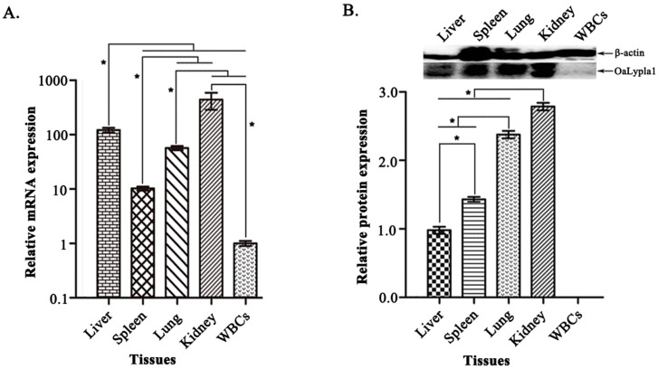
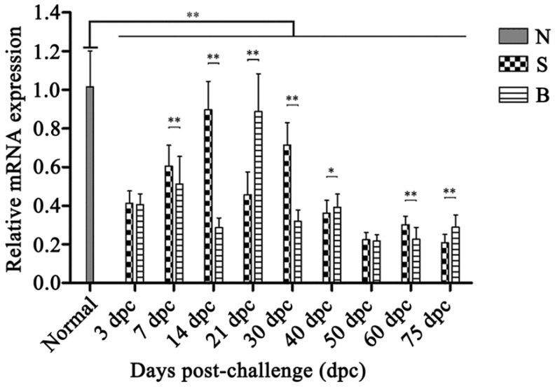
Lysophospholipase I (LYPLA1) is an important protein with multiple functions. In this study, the full-length cDNA of the LYPLA1 gene from Ovis aries (OaLypla1) was cloned using primers and rapid amplification of cDNA ends (RACE) technology. The full-length OaLypla1 was 2457 bp with a 5'-untranslated region (UTR) of 24 bp, a 3'-UTR of 1740 bp with a poly (A) tail, and an open reading frame (ORF) of 693 bp encoding a protein of 230 amino acid residues with a predicted molecular weight of 24,625.78 Da. Phylogenetic analysis showed that the OaLypla1 protein shared a high amino acid identity with LYPLA1 of Bos taurus. The recombinant OaLypla1 protein was expressed and purified, and its phospholipase activity was identified. Monoclonal antibodies (mAb) against OaLypla1 that bound native OaLypla1 were generated. Real-time PCR analysis revealed that OaLypla1 was constitutively expressed in the liver, spleen, lung, kidney, and white blood cells of sheep, with the highest level in the kidney. Additionally, the mRNA levels of OaLypla1 in the buffy coats of sheep challenged with virulent or avirulent Brucella strains were down-regulated compared to untreated sheep. The results suggest that OaLypla1 may have an important physiological role in the host response to bacteria. The function of OaLypla1 in the host response to bacterial infection requires further study in the future.

Keywords: Brucella; Lysophospholipase I; Ovis aries; differential expression; tissue distribution.


Figures

Figure 1
Figure 1
The full-length cDNA and the corresponding amino acid sequence of OaLypla1. The polyadenylation signal sequence (AATAAA) is shown in bold and underlined. The lipase motif GXSXG is boxed, and the catalytic triad is marked by asterisks (*). The arrows represent the places and lengths of the primers (5’-3’) used in this study.
Figure 2
Figure 2
Multiple alignment analysis of the amino acid sequences of LYPLA1s from different vertebrates. The conserved amino acid residues of LYPLA1s are indicated by asterisks (*) above the column. Conserved substitutions are shown by colons (:), and dots (.) indicate semi-conserved amino acids. GenBank GI numbers of LYPLA1 protein sequences are given as follows: Ovis aries LYPLA1: gi (592882195); Bos taurus LYPLA1: gi (77736321); Cricetulus griseus LYPLA1: gi (537237418); Homo sapiens LYPLA1: gi (5453722); Macaca mulatta LYPLA1: gi(388453011); Mus musculus LYPLA1: gi (6678760); Oryctolagus cuniculus LYPLA1: gi (157954426); Pongo abelii LYPLA1: gi (197099340); Rattus norvegicus LYPLA1: gi (6981362); and Xenopus (Silurana) tropicalis LYPLA1: gi(54020910). The different colors are used to differ the proteinogenic 20 amino acids. Red: R and K; Brick red: G; Orange: C; Yellow: P; Green: N, S, T, and Q; Indigo: H and Y; Blue: M, A, L, I, V, F and W; Purple: D and E.
Figure 3
Figure 3
Phylogenetic relationship of LYPLA1 proteins from different species. The phylogenetic tree was constructed using MEGA 4.1 with the ClustalW algorithm. The number on the nodes indicates bootstrap values from 1000 replications.
Figure 4
Figure 4
Expression and purification analysis of the recombinant OaLypla1 protein. (A) Expression analysis of the recombinant OaLypla1 by SDS-PAGE. M: protein marker (Thermo, Waltham, MA, USA); lane 1: total proteins from the uninduced pET-30a-LYPLA1-W cells; lane 2: total proteins from the induced pET-30a-LYPLA1-W cells (expressing OaLypla1 with no His6-tag); lane 3: total proteins from the induced pET-30a-LYPLA1-H cells (expressing OaLypla1 with a His6-tag) showing the shifted band due to the His6-tag; (B) Purification analysis of the recombinant OaLypla1H by SDS-PAGE. M: protein marker (Thermo, Waltham, MA, USA); lane 1: total proteins from the uninduced pET-30a-LYPLA1-H cells; lane 2: total proteins from the induced pET-30a-LYPLA1-H cells; lane 3: the purified recombinant protein OaLypla1H; (C) Expression and purification analysis of the recombinant OaLypla1H by Western blotting. M: protein marker (Thermo, Waltham, MA, USA); lane 1: total proteins from the uninduced pET-30a-LYPLA1-H cells; lane 2: total proteins from the induced pET-30a-LYPLA1-H cells; lane 3: the purified recombinant protein OaLypla1H.
Figure 5
Figure 5
Phospholipase activity assay of the OaLypla1 protein. (A) Egg yolk/agarose diffusion test: a, 0.8 mg/mL OaPDCD10 with a His6-tag; b, 1.6 mg/mL BSA; and c–h, 0.05, 0.1, 0.2, 0.4, 0.8 and 1.2 mg/mL of OaLypla1H; (B) Relative area ratio. The results were calculated as a measure of the relative area ratio with the area of the hole in the center as 100%.
Figure 6
Figure 6
Immunoassay specificity of the monoclonal antibody against OaLypla1. (A) Immunoassay specificity against the recombinant OaLypla1. M: protein marker (Thermo, Waltham, MA, USA); lane 1: total proteins from the uninduced pET-30a-LYPLA1-W cells; lane 2: total proteins from the induced pET-30a-LYPLA1-W cells; lane 3: total proteins from the induced pET-30a-LYPLA1-H cells; (B) Immunoassay specificity against the native OaLypla1. M: protein marker (Thermo, Waltham, MA, USA); lane 1: total proteins from the induced pET-30a-LYPLA1-H cells as a positive control; lane 2: total proteins extracted from the kidney of O. aries. The pET-30a-LYPLA1-W cells were induced to express the recombinant OaLypla1 with no His6-tag, and the pET-30a-LYPLA1-H cells expressed OaLypla1 fused with a His6-tag.
Figure 7
Figure 7
Tissue distribution of OaLypla1. (A) The tissue distribution of OaLypla1 determined by quantitative real-time PCR. The mRNA levels in tissues were normalized with β-actin; (B) OaLypla1 protein detected in the tissues using Western blotting. Statistical differences among liver, spleen, lung, kidney, and white blood cells were determined by one-way analysis of variance (ANOVA) using SPSS 13.0 software (IBM, Armonk, NY, USA). Data are presented as the mean relative expression ± SD (n = 3). An asterisk indicates a statistically significant difference (* p < 0.05). WBCs: white blood cells.
Figure 8
Figure 8
Differential expression of OaLypla1 from buffy coats of O. aries challenged with different virulent Brucella strains. N: the normal sheep group; S: the group inoculated with the avirulent Brucella suis S2 strain; and B: the group challenged with the virulent field strain Brucella melitensis. Relative expression was calculated by the 2−∆∆Ct method using O. aries β-actin as an endogenous control. Statistical differences among the groups were determined by a one-way analysis of variance (ANOVA) using SPSS 13.0 software. Data are presented as the mean relative expression ± SD (n = 3) (* p < 0.05, ** p < 0.01).

Similar articles

Cited by

References

    1. Portilla D., Crew M.D., Grant D., Serrero G., Bates L.M., Dai G., Sasner M., Cheng J., Buonanno A. cDNA cloning and expression of a novel family of enzymes with calcium-independent phospholipase A2 and lysophospholipase activities. J. Am. Soc. Nephrol. 1998;9:1178–1186. - PubMed
    1. Wang A., Yang H.C., Friedman P., Johnson C.A., Dennis E.A. A specific human lysophospholipase: cDNA cloning, tissue distribution and kinetic characterization. Biochim. Biophys. Acta. 1999;1437:157–169. doi: 10.1016/S1388-1981(99)00012-8. - DOI - PubMed
    1. Duncan J.A., Gilman A.G. A cytoplasmic acyl-protein thioesterase that removes palmitate from G protein alpha subunits and p21RAS. J. Biol. Chem. 1998;273:15830–15837. doi: 10.1074/jbc.273.25.15830. - DOI - PubMed
    1. Sugimoto H., Hayashi H., Yamashita S. Purification, cDNA cloning, and regulation of lysophospholipase from rat liver. J. Biol. Chem. 1996;271:7705–7711. - PubMed
    1. Blaskovic S., Adibekian A., Blanc M., van der Goot G.F. Mechanistic effects of protein palmitoylation and the cellular consequences thereof. Chem. Phys. Lipids. 2014;180:44–52. doi: 10.1016/j.chemphyslip.2014.02.001. - DOI - PubMed

LinkOut - more resources