Review
. 2016 Jul 28;8(8):210.
doi: 10.3390/v8080210.

Measles Virus Host Invasion and Pathogenesis

Affiliations
Review

Measles Virus Host Invasion and Pathogenesis

Brigitta M Laksono et al. Viruses. .

Abstract

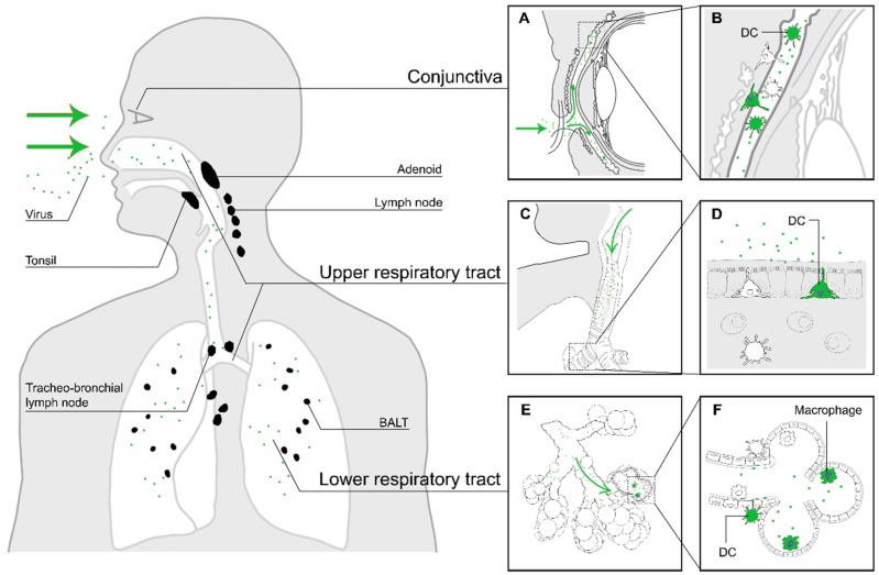
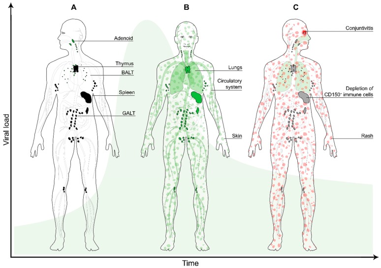
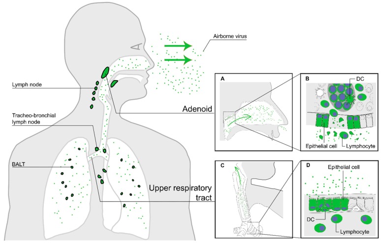
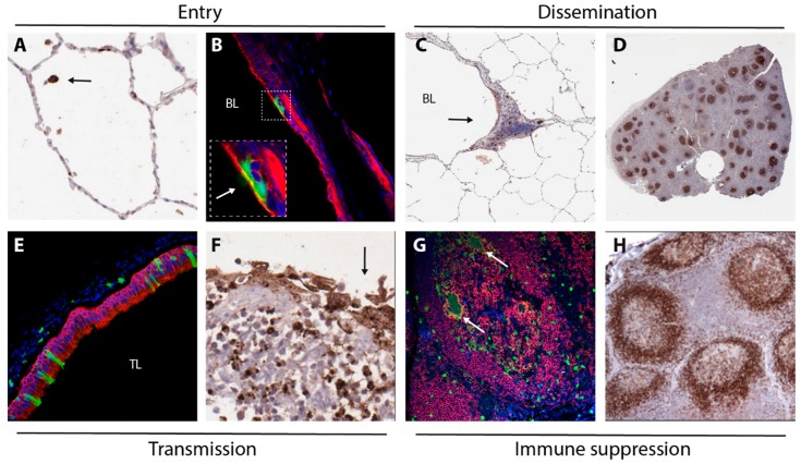
Measles virus is a highly contagious negative strand RNA virus that is transmitted via the respiratory route and causes systemic disease in previously unexposed humans and non-human primates. Measles is characterised by fever and skin rash and usually associated with cough, coryza and conjunctivitis. A hallmark of measles is the transient immune suppression, leading to increased susceptibility to opportunistic infections. At the same time, the disease is paradoxically associated with induction of a robust virus-specific immune response, resulting in lifelong immunity to measles. Identification of CD150 and nectin-4 as cellular receptors for measles virus has led to new perspectives on tropism and pathogenesis. In vivo studies in non-human primates have shown that the virus initially infects CD150⁺ lymphocytes and dendritic cells, both in circulation and in lymphoid tissues, followed by virus transmission to nectin-4 expressing epithelial cells. The abilities of the virus to cause systemic infection, to transmit to numerous new hosts via droplets or aerosols and to suppress the host immune response for several months or even years after infection make measles a remarkable disease. This review briefly highlights current topics in studies of measles virus host invasion and pathogenesis.

Keywords: immune suppression; measles virus; pathogenesis; transmission; tropism.


Figures

Figure 1
Figure 1
The first stage of MV infection: entry of MV into a susceptible host. The virus enters the respiratory tract (green arrows in panels (C) and (E)), where it binds to DC-SIGN+ DCs or infects CD150+ myeloid or lymphoid cells in the mucocilliary epithelium or the alveolar spaces. Another potential site of entry is through the conjunctiva, which is rich in DCs and CD150+ lymphocytes (A). Panels on the right show an enlarged illustration of potential entry events. MV particles deposited on the conjunctiva will enter the space between cornea and eyelids ((A), green arrows), where they can infect myeloid or lymphoid cells (B). MV particles inhaled into the respiratory tract ((C) and (E), green arrows) can either infect DC-SIGN+ dendritic cells in the upper respiratory tract, with dendrites protruding into the respiratory mucosa (D), or dendritic cells or macrophages in the alveolar lumina of the lower respiratory tract (F). The infected immune cells subsequently migrate to nearby tertiary lymphoid tissues and draining lymph nodes (black).
Figure 2
Figure 2
The second stage of MV infection: systemic dissemination. (A) The MV-infected myeloid cells migrate to the draining lymph nodes (black), where they transmit the virus to CD150+ lymphocytes (predominantly B-cells and memory CD4+ and CD8+ T-cells); (B) during viremia infected cells enter the circulation and migrate systemically to various organs and tissues (green), where the infection is further amplified. Infection of skin-resident immune cells results in virus transmission to nectin-4+ epithelial cells (green patches); (C) a few days later, depletion of immune cells in lymphoid organs and tissues results in transient immune suppression (grey). MV-specific T-cells infiltrate the skin where they clear the infected cells, which results in the typical measles skin rash (red patches). The green bell-shaped curve in the background represents the viral load over time.
Figure 3
Figure 3
The third stage of MV infection: transmission of new MV particles via the air. Nectin-4+ epithelial cells in the upper and lower respiratory tract epithelium produce new virus particles and release them into the mucus lining the lumen of the respiratory tract (green arrows in panels (A) and (C)). Epithelial damage in infected lymphoid tissues, such as the tonsils (A), releases virus particles produced by lymphocytes into the upper respiratory tract (B). Epithelial damage in the lower respiratory tract induces cough (panels (C) and (D)), enhancing the discharge of aerosols containing MV particles.
Figure 4
Figure 4
Images collected from experimentally infected NHPs, illustration mechanisms underlying MV entry (A,B), dissemination (C,D), transmission (E,F) and immune suppression (G,H). MV-infected cells were detected by immunohistochemical staining (A,C,D,F) or by immunofluorescent double-staining (B,E,G). (A) infection of a single cell (arrow, likely an alveolar macrophage) in the alveolar lumen 3 DPI; (B) infection of epithelial cells in the trachea 5 DPI (arrow in insert points at green cilia), green = GFP, red = cytokeratin, blue = DAPI; (C) infection of myeloid and lymphoid cells in BALT (arrow) 4 DPI, BL = bronchial lumen; (D) low-magnification image of a lymph node 9 DPI, with many B-cell follicles containing large concentrations of MV-infected lymphocytes; (E) MV-infected epithelial cells in the trachea 9 DPI (green = GFP, red = cytokeratin, blue = DAPI, TL = tracheal lumen); (F) Disruption of the epithelium (arrow) of an adenoid containing many MV-infected lymphocytes 9 DPI; (G) MV-infected B-lymphocytes (including Warthin-Finkeldey syncytia, arrows) in a B-cell follicle 9 DPI (green = GFP, red = CD20, blue = DAPI); (H) follicular exhaustion of B-cell follicles 11 DPI (brown = CD20).

Similar articles

Cited by

References

    1. De Vries R.D., Duprex W.P., de Swart R.L. Morbillivirus infections: An introduction. Viruses. 2015;7:699–706. doi: 10.3390/v7020699. - DOI - PMC - PubMed
    1. Yanagi Y., Takeda M., Ohno S. Measles virus: Cellular receptors, tropism and pathogenesis. J. Gen. Virol. 2006;87:2767–2779. doi: 10.1099/vir.0.82221-0. - DOI - PubMed
    1. Griffin D.E. Fields Virology. 6th ed. Lippincott Williams & Wilkins; Philadelphia, PA, USA: 2013. Measles virus.
    1. Mina M.J., Metcalf C.J., de Swart R.L., Osterhaus A.D., Grenfell B.T. Long-term measles-induced immunomodulation increases overall childhood infectious disease mortality. Science. 2015;348:694–699. doi: 10.1126/science.aaa3662. - DOI - PMC - PubMed
    1. Perry R.T., Murray J.S., Gacic-Dobo M., Dabbagh A., Mulders M.N., Strebel P.M., Okwo-Bele J.M., Rota P.A., Goodson J.L. Progress towards regional measles elimination, worldwide, 2000–2014. Wkly. Epidemiol. Rec. 2015;90:623–631. doi: 10.15585/mmwr.6444a4. - DOI - PubMed

Publication types

LinkOut - more resources