. 2016 Aug 1;9(8):885-97.
doi: 10.1242/dmm.024851. Epub 2016 Jun 9.

Alterations in nuclear structure promote lupus autoimmunity in a mouse model

Affiliations

Alterations in nuclear structure promote lupus autoimmunity in a mouse model

Namrata Singh et al. Dis Model Mech. .

Abstract

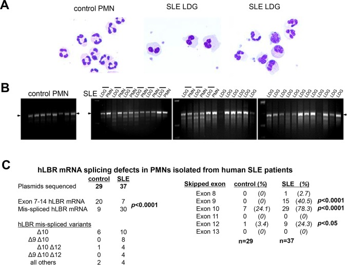
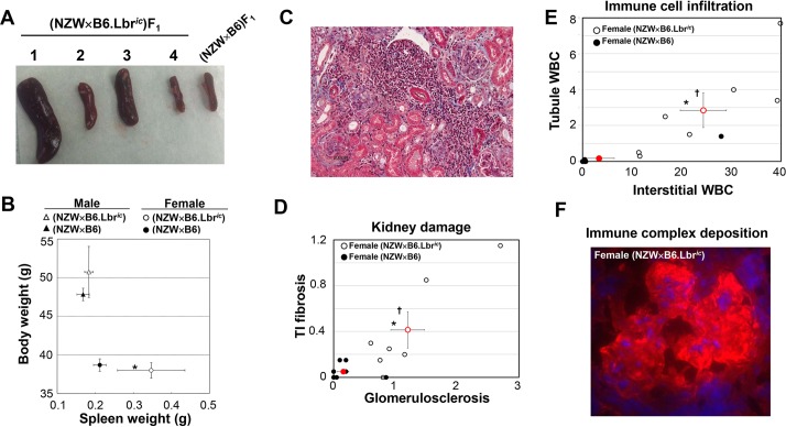
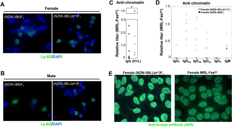
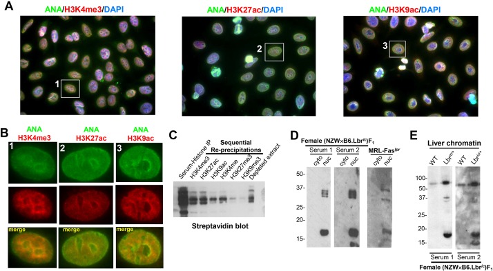
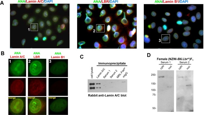
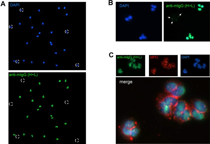
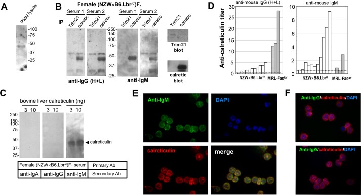
Systemic lupus erythematosus (SLE) is an autoimmune disorder characterized by the development of autoantibodies that recognize components of the cell nucleus. The vast majority of lupus research has focused on either the contributions of immune cell dysfunction or the genetics of the disease. Because granulocytes isolated from human SLE patients had alterations in neutrophil nuclear morphology that resembled the Pelger-Huet anomaly, and had prominent mis-splicing of mRNA encoding the nuclear membrane protein lamin B receptor (LBR), consistent with their Pelger-Huet-like nuclear morphology, we used a novel mouse model system to test the hypothesis that a disruption in the structure of the nucleus itself also contributes to the development of lupus autoimmunity. The lupus-prone mouse strain New Zealand White (NZW) was crossed with c57Bl/6 mice harboring a heterozygous autosomal dominant mutation in Lbr (B6.Lbr(ic/+)), and the (NZW×B6.Lbr(ic))F1 offspring were evaluated for induction of lupus autoimmunity. Only female (NZW×B6.Lbr(ic))F1 mice developed lupus autoimmunity, which included splenomegaly, kidney damage and autoantibodies. Kidney damage was accompanied by immune complex deposition, and perivascular and tubule infiltration of mononuclear cells. The titers of anti-chromatin antibodies exceeded those of aged female MRL-Fas(lpr) mice, and were predominantly of the IgG2 subclasses. The anti-nuclear antibody staining profile of female (NZW×B6.Lbr(ic))F1 sera was complex, and consisted of an anti-nuclear membrane reactivity that colocalized with the A-type lamina, in combination with a homogeneous pattern that was related to the recognition of histones with covalent modifications that are associated with gene activation. An anti-neutrophil IgM recognizing calreticulin, but not myeloperoxidase (MPO) or proteinase 3 (PR3), was also identified. Thus, alterations in nuclear structure contribute to lupus autoimmunity when expressed in the context of a lupus-prone genetic background, suggesting a mechanism for the development of lupus autoimmunity in genetically predisposed individuals that is induced by the disruption of nuclear architecture.

Keywords: Autoantibody; Calreticulin; Chromatin; Histone modifications; Lamina; Nucleus.


Conflict of interest statement

The authors declare no competing or financial interests.

Figures

Fig. 1.
Fig. 1.
LBR splicing defects in PMNs isolated from SLE patients. (A) Differential staining of normal density neutrophils isolated from a healthy individual, and the LDGs from two SLE patients, demonstrates the Pelger–Huet-like nuclear morphology of SLE neutrophils. Image magnification, 400×. (B) PCR amplification of exons 7 to 14 of human LBR cDNA prepared from control PMNs, SLE normal density PMNs and LDGs. The horizontal lines above the sample lanes reflect autologous pairs of LDGs and normal density PMNs isolated in parallel. The arrow indicates the position of properly spliced human LBR. (C) Subcloning and sequencing of human LBR cDNA amplicons verified the mis-splicing of mRNA, and identified extensive skipping of LBR exon 10, both alone and in combination with exons 9 and/or 12. Statistical significance assessed by Fisher's exact test, one-tailed.
Fig. 2.
Fig. 2.
Development of splenomegaly and kidney damage in female (NZW×B6.Lbric)F1 mice. (A) Intact spleens isolated from four female (NZW×B6.Lbric)F1 mice and one female (NZW×B6)F1 control. The spleens of three female (NZW×B6.Lbric)F1 mice are enlarged, whereas the size of the fourth is similar to that of the control. (B) Mean body weight (y-axis) and splenic mass (x-axis) in male (triangles) and female (circles) mice of the NZW×B6 (filled symbols) and NZW×B6.Lbric (open symbols) genotype. Error bars represent s.e.m. Asterisk (*) indicates significant difference spleen weight in female (NZW×B6.Lbric)F1 mice relative to female (NZW×B6)F1 littermates (Student's t-test, two-tailed, P<0.05). (C) Masson's Trichrome-stained kidney section representative of the female (NZW×B6.Lbric)F1 mice with the most severe kidney damage and perivascular cellular infiltration. Image magnification, 200×. (D) Histological evaluation of Masson's Trichrome-stained sections for kidney damage. Female (NZW×B6)F1 (solid circles) and (NZW×B6.Lbric)F1 (open circles) were assessed for tubulointerstitial fibrosis (y-axis) and glomerulosclerosis (x-axis). Red symbols represent the mean and s.e.m. of the corresponding group. Asterisk (*) indicates significant difference in glomerulosclerosis, dagger (†) indicates significant difference in tubulointerstitial fibrosis, Mann–Whitney test, one-tailed. (E) Histological evaluation of Masson's Trichrome-stained sections for cellular infiltration. Female (NZW×B6)F1 (solid circles) and (NZW×B6.Lbric)F1 (open circles) were assessed for tubule white blood cell infiltration (y-axis) and interstitial infiltration (x-axis). Red symbols represent the mean and s.e.m. of corresponding group. Asterisk (*) indicates significant difference in interstitial white blood cell number, dagger (†) indicates significant difference in tubule white blood cell number, Student's t-test, one-tailed. (F) Immune complex deposition in the glomerulus of female (NZW×B6.Lbric)F1 mice. Frozen kidney sections were stained for the presence of glomerular IgG with Cy3-conjugated sheep anti-mouse IgG, and nuclei counter-stained with DAPI. The corresponding staining of a kidney section from a female (NZW×B6)F1 mouse is presented in Fig. S1. Image magnification, 400×.
Fig. 3.
Fig. 3.
Development of lupus autoantibodies in female (NZW×B6.Lbric)F1 mice. (A,B) Disruption of neutrophil nuclear morphology in (NZW×B6.Lbric)F1 mice. White blood cells isolated from female (A) and male (B) (NZW×B6.Lbric)F1 and (NZW×B6)F1 mice were stained with FITC-conjugated anti-Ly-6G to identify neutrophils, and the nuclei counter-stained with DAPI. The ring-shaped nuclear morphology of (NZW×B6)F1 mice is characteristic of mouse neutrophils, whereas the disorganized nuclear morphology of (NZW×B6.Lbric)F1 neutrophils is consistent with the autosomal dominant disruption in Lbr. (C) Anti-chromatin ELISA assays of serum from female (NZW×B6)F1 (filled circles) and (NZW×B6.Lbric)F1 mice (open circles). Serum was screened initially for anti-chromatin IgG titers relative to female MRL-Faslpr. (D) Sera from six anti-chromatin IgG-positive (NZW×B6.Lbric)F1 mice and one anti-chromatin-negative (NZW×B6)F1 mouse were subsequently screened for anti-chromatin titers of IgG1, IgG2a, IgG2b, IgG2c, and IgM relative to female MRL-Faslpr. (E) Distinct anti-nuclear antibody (ANA) staining patterns of the HEp-2 cell line with sera from female (NZW×B6.Lbric)F1 (left panel) and MRL-Faslpr mice (right panel). Additional images for female (NZW×B6.Lbric)F1 mice, and female (NZW×B6)F1 are presented in Fig. S2. Anti-nuclear antibody staining for male mice is presented in Fig. S3. Image magnification, 1000×.
Fig. 4.
Fig. 4.
Female (NZW×B6.Lbric)F1 mice develop anti-nuclear autoantibodies recognizing histone H3 modifications associated with gene activation. (A) Co-localization of anti-nuclear reactivity and the activation-associated histone H3 modifications H3K4me3 (left panel), H3K27ac (middle panel), and H3K9ac (right panel). Anti-nuclear antibody (ANA) staining in the female (NZW×B6.Lbric)F1 serum was detected with an Alexa Fluor 488-conjugated goat anti-mouse antibody, anti-modified H3 histones were detected by an Alexa Fluor 594-conjugated goat anti-rabbit secondary antibody. Nuclei were counterstained with DAPI. Image magnification, 1000×. The individual red, green and blue color channels demonstrating the co-localization of the homogenous component of anti-nuclear autoantibody reactivity with modified histones are presented in Fig. S4. (B) Digital enlargement of the individual cells indicated in A. Anti-nuclear staining mediated by the (NZW×B6.Lbric)F1 serum is shown in the green channel, and the indicated modified histone is counterstained in the red channel. The yellow overlap signal represents nuclear co-localization of mouse serum immunoreactivity with anti-modified H3 histones. (C) Histones purified from HeLa cells were biotinylated in vitro, immunoprecipitated with (NZW×B6.Lbric)F1 mouse sera, collected on Protein A/G sepharose beads, washed, and eluted. The contents of the recovered histone extracts, consisting of histones that were specifically recognized by (NZW×B6.Lbric)F1 mouse sera, were sequentially re-precipitated with rabbit sera recognizing H3K4me3, H3K27ac, H3K9ac, H3K4me, H3K27me3, H3K9me3. Samples prepared from histone extracts collected from each step before, during, and after the re-precipitation process were blotted using peroxidase-conjugated streptavidin and enhanced chemiluminescence. (D) Cytoplasmic and nuclear extracts prepared from 1×10⁶ B6 mouse embryonic fibroblasts (MEFs) were resolved using a 12% SDS-PAGE gel, and immunoblotted with sera from two female (NZW×B6.Lbric)F1 mice, and pooled sera from aged female MRL-Faslpr mice. Serum staining from a representative female (NZW×B6)F1 mouse, and verification of cytoplasmic and nuclear fractionation, is presented in Fig. S5. (E) Chromatin (5 µg) was isolated from the liver of female wild-type B6 and B6.Lbric/+ mice, and immunoblotted with serum from two female (NZW×B6.Lbric)F1 mice.
Fig. 5.
Fig. 5.
Female (NZW×B6.Lbric)F1 mice develop anti-nuclear autoantibodies recognizing the A-type lamina. (A) Co-staining of HEp-2 cells with female (NZW×B6.Lbric)F1 serum and lamin A/C (left panel), LBR (middle panel), and lamin B1 (right panel). Anti-nuclear antibody (ANA) staining in the serum was detected with an Alexa Fluor 488-conjugated goat anti-mouse antibody, lamina proteins were labeled with the indicated rabbit anti-serum and an Alexa Fluor 594-conjugated goat anti-rabbit secondary antibody. Nuclei were counterstained with DAPI. Image magnification, 1000×. The individual red, green and blue color channels demonstrating the co-localization of the nuclear membrane component of anti-nuclear autoantibody reactivity with the A-type lamina are presented in Fig. S6. (B) Digital enlargement of the individual cells indicated in A. Anti-nuclear antibody staining mediated by the (NZW×B6.Lbric)F1 serum is shown in the green channel, and the indicated nuclear envelope protein is counterstained in the red channel. The nuclear membrane colocalization of mouse serum with the anti-lamin A/C is represented by the yellow overlap signal. (C) Lamin A/C immunoblots of 5×10⁶ MEFs immunoprecipitated with a mouse lamin A/C monoclonal antibody, the sera from two female (NZW×B6.Lbric)F1 mice (Serum 1 and 2), or pooled aged female MRL-Faslpr sera. Non-specific mouse IgG (mIgG) served as a serum control. (D) Cytoplasmic and nuclear extracts prepared from 1×10⁶ MEFs were resolved on a 7.5% SDS-PAGE gel, and immunoblotted with sera from two female (NZW×B6.Lbric)F1 mice.
Fig. 6.
Fig. 6.
Anti-neutrophil serum reactivity develops in (NZW×B6.Lbric)F1 mice. (A) Fluorescence microscopy of ethanol-fixed human PMNs stained with DAPI (top panel), and female (NZW×B6.Lbric)F1 mouse serum and an Alexa Fluor 488-conjugated goat anti-mouse secondary antibody (bottom panel). Broken circles indicate DAPI-positive bilobed eosinophils that were not readily detected by the female (NZW×B6.Lbric)F1 mouse serum. Image magnification, 100×. (B) High-power magnification of ethanol-fixed PMN staining. Neutrophils displayed strong nuclear staining, but eosinophils had comparatively weak and patchy perinuclear staining. Image magnification, 400×. The preferential staining of isolated neutrophils relative to peripheral blood mononuclear cells and isolated monocytes is demonstrated in greater detail in Fig. S7. (C) Ethanol-fixed PMNs were co-stained with female (NZW×B6.Lbric)F1 mouse serum (green) and anti-MPO (red), nuclei were counterstained with DAPI (blue). The mouse serum staining did not colocalize with MPO. Image magnification, 1000×.
Fig. 7.
Fig. 7.
Anti-calreticulin IgM mediates the anti-neutrophil reactivity in (NZW×B6.Lbric)F1 mice. (A) Triton X-100-soluble extracts of human PMNs were resolved on a 10% SDS-PAGE gel and immunoblotted with female (NZW×B6.Lbric)F1 mouse serum. (B) Immunoprecipitates of Trim21 and calreticulin were prepared from 20×106 human PMNs, resolved on a 10% SDS-PAGE gel, serum immunoblotted, and detected with peroxidase-conjugated goat anti-mouse secondary antibodies recognizing IgG (H+L) (left panel) or IgM (right panel). Exposure time for development of the IgM signal was ∼1/10 that of IgG (H+L). Control blots (far right panels) verified recovery of Trim21 and calreticulin in immunoprecipitates. (C) Calreticulin purified from bovine liver was serum immunoblotted and detected with peroxidase-conjugated goat anti-mouse secondary antibodies recognizing IgA (left panel), IgG (middle panel), or IgM (right panel). The location of the calreticulin standard immunoblot using a calreticulin monoclonal antibody is indicated by the arrowhead. (D) IgG (H+L) (left panel) and IgM (right panel) anti-calreticulin ELISA assay of individual female (NZW×B6.Lbric)F1 or MRL-Faslpr mouse sera. Dashed line represents the mean plus 2 standard deviations from n=7 wild-type B6 mice. (E) Paraformaldehyde-fixed PMNs were co-stained with female (NZW×B6.Lbric)F1 mouse serum (green) and rabbit anti-calreticulin (red), nuclei were counterstained with DAPI (blue). The co-localization of mouse serum staining and calreticulin was detected using an anti-IgM secondary antibody (merge: yellow signal). Image magnification, 400×. (F) Paraformaldehyde-fixed PMNs were co-stained with female (NZW×B6.Lbric)F1 mouse serum (green) and rabbit anti-calreticulin (red), nuclei were counterstained with DAPI (blue). Neither the anti-IgG (top panel), nor anti-IgA (bottom panel) secondary antibodies colocalized the mouse serum staining with calreticulin. Image magnification, 400×.

References

    1. Armstrong D. L., Zidovetzki R., Alarcón-Riquelme M. E., Tsao B. P., Criswell L. A., Kimberly R. P., Harley J. B., Sivils K. L., Vyse T. J., Gaffney P. M. et al. (2014). GWAS identifies novel SLE susceptibility genes and explains the association of the HLA region. Genes Immun. 15, 347-354. 10.1038/gene.2014.23 - DOI - PMC - PubMed
    1. Bennett L., Palucka A. K., Arce E., Cantrell V., Borvak J., Banchereau J. and Pascual V. (2003). Interferon and granulopoiesis signatures in systemic lupus erythematosus blood. J. Exp. Med. 197, 711-723. 10.1084/jem.20021553 - DOI - PMC - PubMed
    1. Binda O., LeRoy G., Bua D. J., Garcia B. A., Gozani O. and Richard S. (2010). Trimethylation of histone H3 lysine 4 impairs methylation of histone H3 lysine 9: regulation of lysine methyltransferases by physical interaction with their substrates. Epigenetics 5, 767-775. 10.4161/epi.5.8.13278 - DOI - PMC - PubMed
    1. Bleck T. P. and Smith M. C. (1990). Possible induction of systemic lupus erythematosus by valproate. Epilepsia 31, 343-345. 10.1111/j.1528-1157.1990.tb05386.x - DOI - PubMed
    1. Broers J. L. V., Machiels B. M., Kuijpers H. J. H., Smedts F., van den Kieboom R., Raymond Y. and Ramaekers F. C. S. (1997). A- and B-type lamins are differentially expressed in normal human tissues. Histochem. Cell Biol. 107, 505-517. 10.1007/s004180050138 - DOI - PubMed

Publication types

MeSH terms