Review
. 2016 Aug 2;16(1):19.
doi: 10.1186/s40644-016-0076-2.

CT in ovarian cancer staging: how to review and report with emphasis on abdominal and pelvic disease for surgical planning

Affiliations
Review

CT in ovarian cancer staging: how to review and report with emphasis on abdominal and pelvic disease for surgical planning

Anju Sahdev. Cancer Imaging. .

Abstract

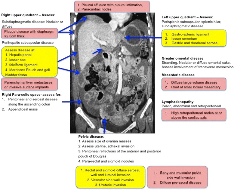
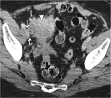
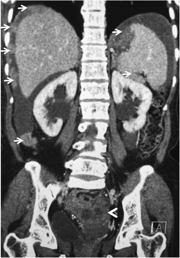
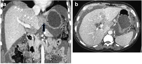
CT of the abdomen and pelvis is the first line imaging modality for staging, selecting treatment options and assessing disease response in ovarian cancer. The staging CT provides disease distribution, disease burden and is the imaging surrogate for surgico-pathological FIGO staging. Optimal cyto-reductive surgery offers patients' the best chance for disease control or cure, but sub-optimal resection confers no advantage over chemotherapy and adversely increases the risk of post surgical complications. Although there is extensive literature comparing performance of CT against laparoscopy and surgery, for the staging abdominal and pelvic CT, there are currently no accepted guidelines for interpretation or routinely used minimum data set templates for reporting these complex CT scans often with extensive radiological findings. This review provides a systematic approach for identifying the important radiological findings and highlighting important sites of disease within the abdomen and pelvis, which may alter or preclude surgery at presentation or after adjuvant chemotherapy. The distribution of sites and volume of disease can be used to categorize patients as suitable, probably suitable or not suitable for optimal cyto-reductive surgery. This categorization can potentially assist oncological surgeons and oncologists as a semi objective assessment tool useful for selecting patient treatment, streamlining multi disciplinary discussion and improving the reproducibility and correlation of CT with surgical findings. The review also highlights sites of disease and complications of ovarian cancer which should be included as part of the radiological report as these may require additional surgical input from non gynaecological surgeons or influence treatment selection.

Keywords: CT; Disease distribution; Ovarian cancer; Surgical resection; Template reporting.


Figures

Fig. 1
Fig. 1
Schematic of the common sites of disease in ovarian cancer. The features in yellow are sites of potentially non resectable disease. The features in red are sites of non resectable disease
Fig. 2
Fig. 2
Seventy-six year old woman with a right sided malignant ovarian mass (arrow head). The mass extends to the right pelvic side wall and abuts the right external iliac vein (arrows). A distance of less than 3 mm from the pelvic side wall structures is highly suggestive of invasion
Fig. 3
Fig. 3
Eighty year old woman with stage IIIC papillary serous adenocarcinoma of the ovary (arrowhead). Extensive resectable metastatic peritoneal nodules are arrowed in the abdomen. The pelvic disease causes bilateral ureteric obstruction with resultant bilateral hydronephrosis
Fig. 4
Fig. 4
a and b 64 year old woman with stage IIIC clear cell carcinoma of the ovary. Axial and Coronal reconstructed CT demonstrating non resectable dense confluent right subdiaphragmatic disease (arrows), diffuse thick plaque disease in the gastro-hepatic ligament (block arrow) and an deposit >2 cm in the hepato-duodenal ligament and porta hepatis (dashed arrows)
Fig. 5
Fig. 5
a and b 76 year old woman with non resectable stage IV undifferentiated adenocarcinoma of the ovary. The dashed arrows show extensive diffuse meso colic disease posterior to the transverse colon confluent with diffuse small bowel serosal disease. The disease results in small bowel obstruction. Solid arrows show a typical thick omental cake in the lower abdomen and a large peritoneal deposit (block arrow)
Fig. 6
Fig. 6
Large volume >2 cm non-resectable disease in the lesser sac (arrows) with confluent disease between the pancreas and stomach and the gastro-splenic ligament. Further disease is present in the spleno-renal ligament (dashed arrows) and ascites around the liver
Fig. 7
Fig. 7
Sixty-six year old woman with Stage IV serous cystadenocarcinoma of the ovary. Dense infiltrative non-resectable disease in the mid abdominal mesentery (block arrow) with resultant cicatrization of the surrounding bowel loops. Innumerable additional nodular deposits (arrows) are scattered in the remaining small bowel mesentery which would also constitute non resectable disease
Fig. 8
Fig. 8
Seventy-three year old woman with serous cystadenocarcinoma. Extensive diffuse non resectable retroperitoneal nodal metastases encasing the aorta, root of coeliac artery and invading the diaphragm (arrows). Enlarged right para-cardiac node (dashed arrow) indicating stage IV disease
Fig. 9
Fig. 9
Fifty-five year old woman with endometriod carcinoma of the ovary. Hepatic subcapsular deposits (arrows) with invasion of underlying liver parenchyma (dashed arrows). The invasion requires hepato-biliary surgical assistance as segmentectomy or lobectomy may be required in patients being considered for optimal resection
Fig. 10
Fig. 10
58 year old woman with stage IIIC undifferentiated carcinoma of the ovary. Axial (a) and coronal CT (b) shows extensive small bowel mesenteric disease (arrows) invading small bowel serosa and wall with resultant small bowel obstruction. Resection of this degree of mesenteric invasion would require sacrificing the entire small bowel

References

    1. Chang SJ, Bristow RE, Chi DS, Cliby WA. Role of aggressive surgical cytoreduction in advanced ovarian cancer. J Gynecol Oncol. 2015;26(4):336–42. doi: 10.3802/jgo.2015.26.4.336. - DOI - PMC - PubMed
    1. Suidan RS, Ramirez PT, Sarasohn DM, Teitcher JB, Mironov S, Iyer RB, et al. A multicenter prospective trial evaluating the ability of preoperative computed tomography scan and serum CA-125 to predict suboptimal cytoreduction at primary debulking surgery for advanced ovarian, fallopian tube, and peritoneal cancer. Gynecol Oncol. 2014;134(3):455–61. doi: 10.1016/j.ygyno.2014.07.002. - DOI - PMC - PubMed
    1. Borley J, Wilhelm-Benartzi C, Yazbek J, Williamson R, Bharwani N, Stewart V, et al. Radiological predictors of cytoreductive outcomes in patients with advanced ovarian cancer. BJOG. 2015;122(6):843–9. doi: 10.1111/1471-0528.12992. - DOI - PubMed
    1. Hoskins WJ, McGuire WP, Brady MF, Homesley HD, Creasman WT, Berman M, et al. The effect of diameter of largest residual disease on survival after primary cytoreductive surgery in patients with suboptimal residual epithelial ovarian carcinoma. Am J Obstet Gynecol. 1994;170(4):974–9. doi: 10.1016/S0002-9378(94)70090-7. - DOI - PubMed
    1. Holloway BJ, Gore ME, A'Hern RP, Parsons C. The significance of paracardiac lymph node enlargement in ovarian cancer. Clin Radiol. 1997;52(9):692–7. doi: 10.1016/S0009-9260(97)80034-7. - DOI - PubMed

MeSH terms