Review
. 2017 Feb 5:441:7-16.
doi: 10.1016/j.mce.2016.07.036. Epub 2016 Jul 30.

A brief history of the search for the protein(s) involved in the acute regulation of steroidogenesis

Affiliations
Review

A brief history of the search for the protein(s) involved in the acute regulation of steroidogenesis

Douglas M Stocco et al. Mol Cell Endocrinol. .

Abstract

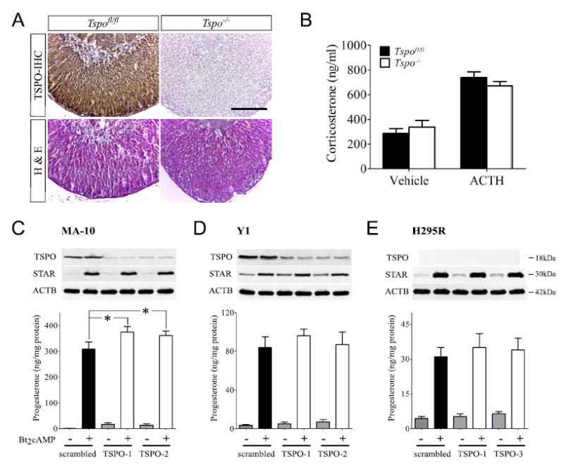
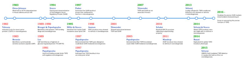
The synthesis of steroid hormones occurs in specific cells and tissues in the body in response to trophic hormones and other signals. In order to synthesize steroids de novo, cholesterol, the precursor of all steroid hormones, must be mobilized from cellular stores to the inner mitochondrial membrane (IMM) to be converted into the first steroid formed, pregnenolone. This delivery of cholesterol to the IMM is the rate-limiting step in this process, and has long been known to require the rapid synthesis of a new protein(s) in response to stimulation. Although several possibilities for this protein have arisen over the past few decades, most of the recent attention to fill this role has centered on the candidacies of the proteins the Translocator Protein (TSPO) and the Steroidogenic Acute Regulatory Protein (StAR). In this review, the process of regulating steroidogenesis is briefly described, the characteristics of the candidate proteins and the data supporting their candidacies summarized, and some recent findings that propose a serious challenge for the role of TSPO in this process are discussed.

Keywords: Conditional knockout; Global knockout; Mitochondria; Steroidogenesis; Steroidogenic Acute Regulatory Protein (StAR); Translocator Protein (TSPO).


Figures

Fig. 1
Fig. 1
Illustration of Newly Synthesized StAR Proteins. Representative fluorograms of two-dimensional polyacrylamide gel electrophoresis (2-D PAGE) of 35S-methionine labeled mitochondrial proteins isolated from control and stimulated MA-10 mouse Leydig tumor cells. Cells were incubated in the presence of 1 mCi/ml 35S-methionine and either in the presence or absence of 1 mM dibutyryl cAMP (dbcAMP) for 6 h. Mitochondria were isolated and the proteins were prepared for 2-D PAGE. Following electrophoresis the gels were dried and placed in cassettes with X-ray film. After suitable periods of time, the films were developed and the radioactive proteins could be seen. Shown are selected areas of fluorograms from control (A) and stimulated (B) cells. The arrows (1–4), illustrate the positions of the 30 kDa StAR protein forms. Proteins 3 and 4 represent the phosphorylated forms of proteins 1 and 2 respectively. Adapted from Stocco and Clark (1993).
Fig. 2
Fig. 2
In vitro Expression of StAR Protein and Steroid Production in MA-10 and COS-1 Cells. A, MA-10 cells were stimulated with dbcAMP and subjected to Western blot analysis as were unstimulated MA-10 cells that had been transfected with pCMV vector alone or pCMV containing the 37 kDa cDNA for StAR. C, Progesterone production in control and StAR transfected MA-10 cells were measured by RIA 16 h post transfection. B, COS-1 cells rendered steroidogenic through transfection with the proteins for the cholesterol side chain cleavage enzyme system (SCC) were transfected with control pMCV and pCMV containing 37 kDa StAR cDNA and western blot analysis was performed. D, pregnenolone synthesis was measured in cells transfected with empty vector, StAR alone, SCC proteins alone and cells containing both StAR and the SCC system. It can clearly be seen that in both MA-10 and COS-1 cells, expression of the StAR protein is required for the cells to increase steroid synthesis.
Fig. 3
Fig. 3
Adrenal Glands and Testes in StAR Knockout Mice. Adrenal glands and testes were isolated from wild type and StAR knockout mice at six weeks after birth. Frozen sections from each tissue were stained with oil red O for the detection of neutral lipids. A, the adrenal glands from StAR null mice contained significantly higher levels of lipid than did wild type animals. In the upper panels of A, the whole gland is shown, and a higher magnification of the gland is shown in the lower panels. B, similar procedures were performed in the testis and in the interstitial areas, the location of the steroidogenic Leydig cells, significantly higher deposits of lipid can be seen at both the lower and higher magnifications.
Fig. 4
Fig. 4
Effects of Conditional Knockout of TSPO in Mouse Testis. A, Immunohistochemical (IHC) localization showing complete absence of TSPO in Leydig and Sertoli cells of TSPOc–/– testes. Hematoxylin and eosin (H&E) staining showing unaltered seminiferous tubule morphology and spermatogenesis in TSPOc–/– testes (n = 5). Scale bars, 50 μm. B, Plasma testosterone levels were not significantly different between TSPOfl/fl and TSPOc–/– mice (n = 19-22/group), before, or C, when sampled 1 h after hCG stimulation (n 7/group). D, a modest but significant increase in testis weights was observed in TSPOc–/– mice compared with TSPOfl/fl mice (P < 0.05; n = 18/group). E, seminal vesicle weights were not significantly different between TSPOfl/fl and TSPOc–/– mice (mean SEM; n = 18/group). Panels from Morohaku et al., 2014, Endocrinology, 155 (1): 89–97.
Fig. 5
Fig. 5
A-B: Effect of TSPO Global Knockout on Adrenal Gland Morphology and Steroid Synthesis. A, Adrenal sections from Tspofl/fl and Tspo–/– mice showing TSPO localization in adrenocortical cells with a higher density in the zona glomerulosa; no staining was observed in Tspo–/– adrenal. No difference in adrenocortical morphology was apparent between the two genotypes. B, both basal and ACTH stimulated plasma corticosterone levels were similar in Tspofl/fl and Tspo–/– mice (n = 10–14/group). C-E: TSPO Knockdown in Steroidogenic Cells Does Not Affect Steroid Hormone Biosynthesis. C and D, TSPO knockdown resulted in substantial decreases in TSPO protein in MA-10 and Y1 cells compared to controls. E, no base-line TSPO was detected in H295R cells. Bt2cAMP treatment resulted in similar increases in StAR protein expression and higher progesterone production in MA-10, Y1 and H295R cells. TSPO knockdown in MA-10 cells showed significantly higher progesterone production (p < 0.05) while progesterone production in TSPO knockdown Y1 cells was similar to the scrambled controls. H295R cells showed no TSPO expression but still produced progesterone upon Bt2cAMP treatment. Panels from Tu et al., 2014, Journal of Biological Chemistry, 289 (40): 27444-54.
Fig 6
Fig 6
Timeline representing key studies focused on the mechanism of mitochondrial cholesterol import for steroidogenesis and their outcomes. References in chronology: (Chanderbhan et al., 1982), (Krueger and Orme-Johnson, 1983), (Pedersen and Brownie, 1983), (Mukhin et al., 1989), (Krueger and Papadopoulos, 1990), (Li et al., 1989), (Papadopoulos et al., 1991a, b, c), (Clark et al., 1994), (Lin et al., 1995), (Caron et al., 1997), (Papadopoulos et al., 1997a, b), (Seedorf et al., 1998), (West et al., 2001), (Hauet et al., 2005), (Bogan et al., 2007), (Wisniewska et al., 2010), (Neess et al., 2011), (Morohaku et al., 2014), (Tu et al., 2014), (Banati et al., 2014), (Tu et al., 2015).

References

    1. Aghazadeh Y, Zirkin BR, Papadopoulos V. Pharmacological regulation of the cholesterol transport machinery in steroidogenic cells of the testis. Vitam Horm. 2015;98:189–227. - PubMed
    1. Alberta JA, Epstein LF, Pon LA, Orme-Johnson NR. Mitochondrial localization of a phosphoprotein that rapidly accumulates in adrenal cortex cells exposed to adrenocorticotropic hormone or to cAMP. J Biol Chem. 1989;264:2368–2372. - PubMed
    1. Anuka E, Gal M, Stocco DM, Orly J. Expression and roles of steroidogenic acute regulatory (StAR) protein in ‘non-classical’, extra-adrenal and extra-gonadal cells and tissues. Mol Cell Endocrinol. 2013;371:47–61. - PubMed
    1. Arakane F, Sugawara T, Nishino H, Liu Z, Holt JA, Pain D, Stocco DM, Miller WL, Strauss JF., 3rd Steroidogenic acute regulatory protein (StAR) retains activity in the absence of its mitochondrial import sequence: implications for the mechanism of StAR action. Proc Natl Acad Sci U S A. 1996;93:13731–13736. - PMC - PubMed
    1. Arensburg J, Payne AH, Orly J. Expression of steroidogenic genes in maternal and extraembryonic cells during early pregnancy in mice. Endocrinology. 1999;140:5220–5232. - PubMed

Publication types

LinkOut - more resources