Review
. 2016 Sep 30;44(17):8041-51.
doi: 10.1093/nar/gkw693. Epub 2016 Aug 2.

Origins of tmRNA: the missing link in the birth of protein synthesis?

Affiliations
Review

Origins of tmRNA: the missing link in the birth of protein synthesis?

Kevin Macé et al. Nucleic Acids Res. .

Abstract

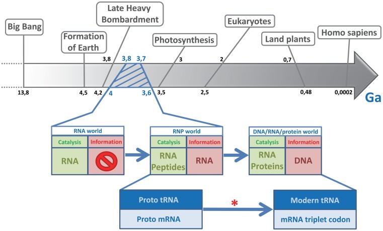
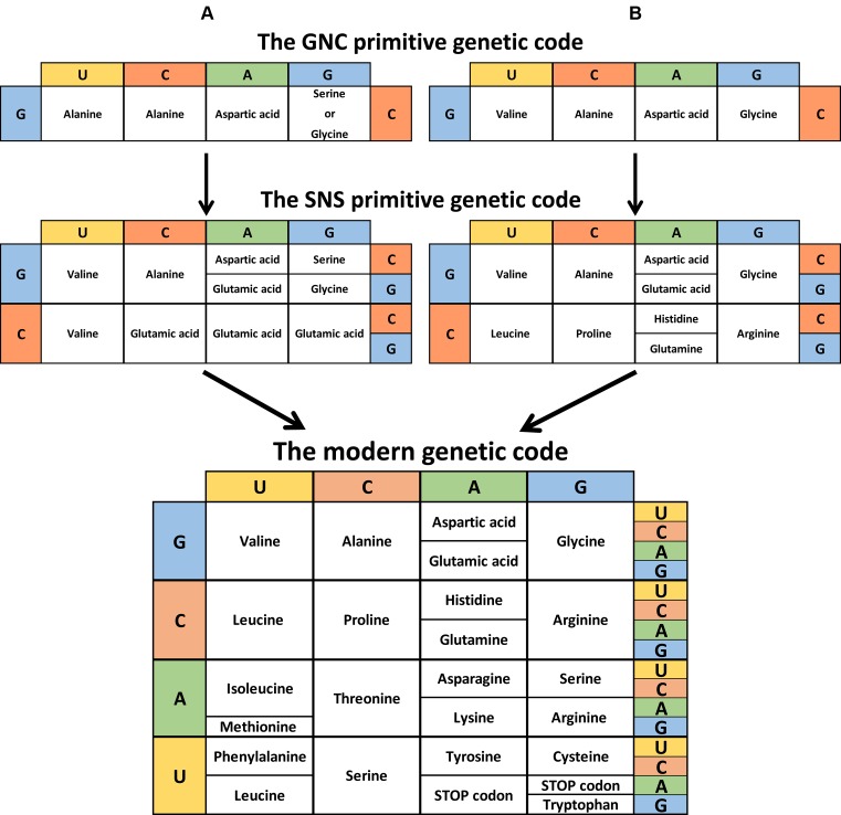
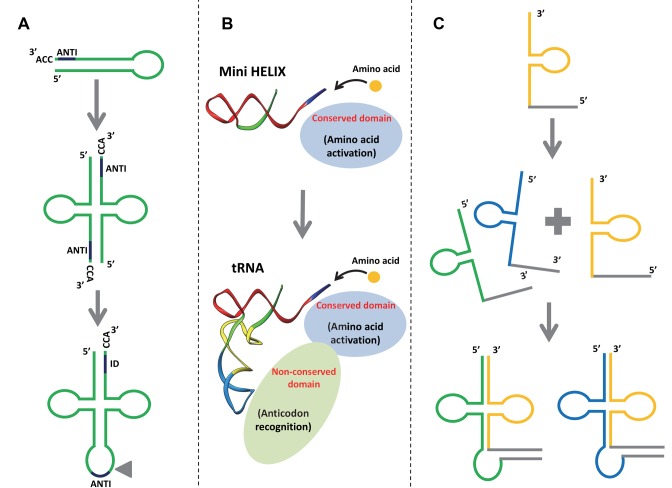
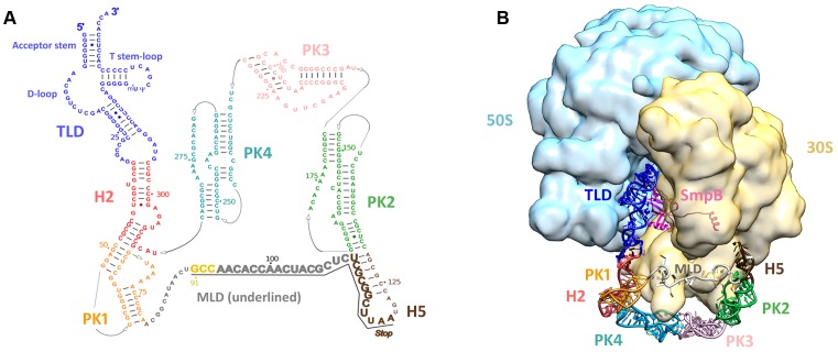
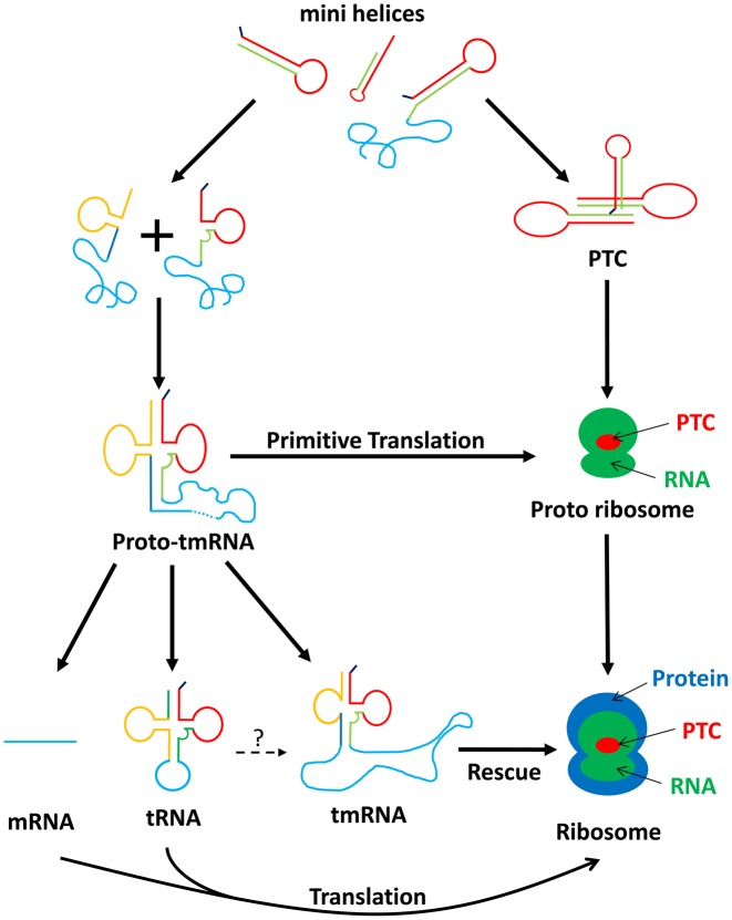
The RNA world hypothesis refers to the early period on earth in which RNA was central in assuring both genetic continuity and catalysis. The end of this era coincided with the development of the genetic code and protein synthesis, symbolized by the apparition of the first non-random messenger RNA (mRNA). Modern transfer-messenger RNA (tmRNA) is a unique hybrid molecule which has the properties of both mRNA and transfer RNA (tRNA). It acts as a key molecule during trans-translation, a major quality control pathway of modern bacterial protein synthesis. tmRNA shares many common characteristics with ancestral RNA. Here, we present a model in which proto-tmRNAs were the first molecules on earth to support non-random protein synthesis, explaining the emergence of early genetic code. In this way, proto-tmRNA could be the missing link between the first mRNA and tRNA molecules and modern ribosome-mediated protein synthesis.


Figures

Figure 1.
Figure 1.
Timeline of early events, emphasizing the transition from an RNA world to modern life. During the history of life on earth, the RNA world lasted from the first appearance of short catalytic RNAs right up to the transition to a modern period in which genetic information carried by DNA and RNA became translated into proteins. The RNP (ribonucleoprotein) world was the intermediate period in which RNA and the first random peptides coexisted as informational and catalytic molecules. The red star indicates when the genetic medium stopped being random. Ga, giga-annum, or 109 (1 000 000 000) years.
Figure 2.
Figure 2.
Theory of the genetic code evolution. This shows the evolutionary pathway going from the GNC code (4 codons) to the SNS code (16 codons) to the universal genetic code (64 codons). (A) Adapted from Massimo Di Giulio (72). (B) Adapted from Kenji Ikehara (10). (C) Instead of the conventional representation, the modern genetic code is shown reflecting the order of codon occurrence (columns G and U inverted).
Figure 3.
Figure 3.
Different models explaining the origins of tRNA. (A) tRNA may originate from the dimerization of two hairpin structures. ANTI, anticodon; ID, the discrimination region for identifying tRNA. The triangle represents the position where the intron is found in tRNA genes (73). (B) tRNA may originate from the late fusion between two RNA minihelices. A new aminoacyl tRNA synthetase (aaRS) domain links the operational RNA code to the trinucleotides of the genetic code (21). (C) tRNA may originate from the fusion of split genes of non-contiguous tRNAs (22).
Figure 4.
Figure 4.
tmRNA secondary and tertiary structures. (A) Secondary structure diagram of Thermus thermophilus tmRNA. Watson–Crick base pairs are connected by lines and GU pairs are represented by dots. Domains are in color: the tRNA-like domain (TLD) is blue; helix 2 (H2) is red; pseudoknot 1 (PK1) is orange; helix 5 (H5) is brown; pseudoknot 2 (PK2) is green; pseudoknot 3 (PK3) is pink; and pseudoknot 4 (PK4) is light blue. The nucleotides within the internal open reading frame (ORF) are underlined and shown in a larger font. The resume codon is yellow and the STOP codon is indicated. (B) Cryo-EM map of the alanyl-tmRNA-SmpB complex bound to a stalled ribosome. 3D molecular model of tmRNA based on the homology modeling of each independent domain followed by flexible fitting into the cryo-EM density map of the accommodated step (74). EMDB entry: EMD-5188. Color code: tmRNA domains are the same as in (A); SmpB is magenta; the small 30S subunit is yellow; the large 50S subunit is light blue.
Figure 5.
Figure 5.
Position and secondary structure similarities between the tRNA intron and tmRNA pseudoknots. (A) The tRNA intron in the three major kingdoms (Bacteria, Archaea and Eukaryota), adapted from Akio Kanai (22). Introns are framboise and mature tRNA is gray. Intron clipping sites are indicated with black arrows. (B) Secondary structure of tmRNA. The tRNA-like (TLD) and mRNA-like (MLD) domains are indicated, and the pseudoknots are framboise. Note the similar positioning of the tRNA introns in the three domains of life and in the other tmRNA domains.
Figure 6.
Figure 6.
Similarity of the positions of G:U mismatches in the tRNA intron and in tmRNA. (A) Secondary structure of the Azoarcus group I intron. Exon sequences are in lower case and blue, while introns are in upper case letters, with red arrows indicating the splice boundaries. The conserved G-U mismatch necessary for self-splicing and the guanine partner are red. (B) Secondary structure of the Escherichia coli tRNA-like domain (TLD) of tmRNA. The conserved G-U mismatches in the TLD are red. A similarity in position between the G-U mismatch of the tRNA intron and the TLD is noticeable. (C) Secondary structure of a tRNAIle (CAU) from Azoarcus. The red arrow indicates the insertion site for the introns shown in (C). The exon sequence common to figures (A) and (C) is blue. Mismatches are signaled by dots.
Figure 7.
Figure 7.
Consensus of amino acids and repartition of first codon of mRNA-like domain in bacteria phyla. (A) Sequence of the consensus sequence and diversity of the amino acids in the MLD. Sequence conservation is measured in bits, going from weakly conserved (0) to highly conserved (4). Alanine is in red and the other amino acids are in black. The alignment was obtained using 708 tag sequences from the UTHSCSA RNP database (http://rnp.uthscsa.edu/rnp/tmRDB/peptide/peptide.html) (66). The consensus logo was created using WebLogo (http://weblogo.berkeley.edu/logo.cgi) (75). (B) Analysis of the first MLD codon in different bacteria phyla. The first codon of the MLD is indexed by the ancestral codons (GNC) and the others. The percentage of each is bold and the sequence number is in parentheses. If the percentage of a codon in a phylum is superior to 50%, it is red. Non-bacterial tmRNA is yellow. Sequence alignments are presented for 940 different tmRNAs. They were taken from the ‘tmRNA website’ (76). Briefly, the first codon is identified thanks to the highly-conserved upstream sequence determinants. Of course, the resume codon sequence itself is another likely determinant. (C) Variations in the first MLD codon in the different bacteria phyla. The green line indicates ancestral codons for the first codon of the MLD, and the blue line indicates other codons. Adapted from the phylogenetic tree of all extant organisms based on 16S rRNA gene sequence data, as originally proposed by Woese.
Figure 8.
Figure 8.
Schematic model representing the different possibilities for the origin of tmRNA. In the beginning of the RNA world, there was a mix of different minihelices. A homodimerisation between two of these would have generated the first proto-ribosome, the Peptidyl Transferase Center (PTC). With aleatory amino acids or aminoacylated minihelices, the PTC created the first random peptide syntheses. A heterodimerization between two minihelices (plus sign) then surely generated a proto-tmRNA. In this step, the proto-tmRNA would have been used as a proto-mRNA. At the same time, the PTC must have evolved into a proto-ribosome by acquiring new RNA that improved its activity. The proto-ribosome would then have continued to evolve via the new synthesized proteins, finally ending up as the ribosomal complex seen today. In contrast to this ribosomal development, the proto-tmRNA took varying evolutionary paths. Through self-splicing, it produced a proto-tRNA that evolved into modern tRNA. It also led to modern tmRNA, which now serves as a rescue system in all bacteria. In addition, proto-tmRNA provided the first non-random genetic medium, which evolved into the RNA genome then into modern mRNA. However, we cannot exclude the theory that a proto-tRNAAla grew into modern tmRNA, through the insertion of nucleotides into its gene (question mark) (77).

References

    1. Crick F.H. The origin of the genetic code. J. Mol. Biol. 1968;38:367–379. - PubMed
    1. Orgel L.E. Evolution of the genetic apparatus. J. Mol. Biol. 1968;38:381–393. - PubMed
    1. Woese C.R. Harper & Row. 1967. The genetic code the molecular basis for genetic expression. ASIN: B000I3M2KK.
    1. Jeffares D.C., Poole A.M., Penny D. Relics from the RNA world. J. Mol. Evol. 1998;46:18–36. - PubMed
    1. Goldberg A.L., Wittes R.E. Genetic code: aspects of organization. Science. 1966;153:420–424. - PubMed

Publication types