

Diffusion tensor imaging and histology of developing hearts
- PMID: 27485033
- PMCID: PMC5160010
- DOI: 10.1002/nbm.3576
Diffusion tensor imaging and histology of developing hearts
Abstract
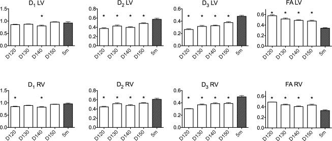
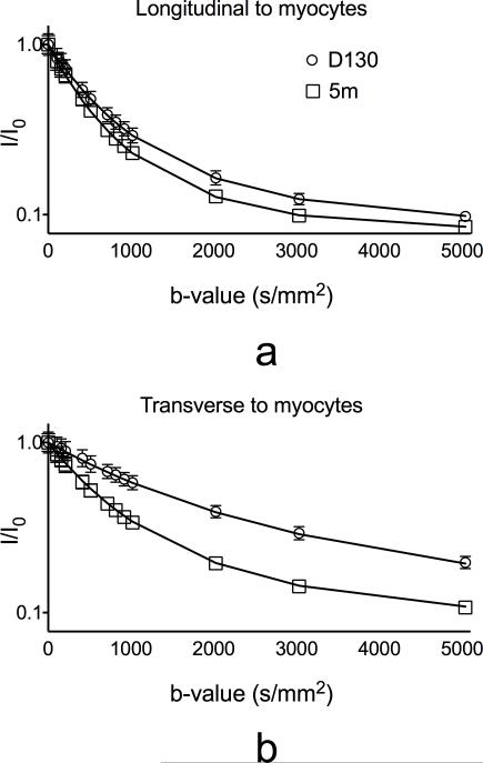
Diffusion tensor imaging (DTI) has emerged as a promising method for noninvasive quantification of myocardial microstructure. However, the origin and behavior of DTI measurements during myocardial normal development and remodeling remain poorly understood. In this work, conventional and bicompartmental DTI in addition to three-dimensional histological correlation were performed in a sheep model of myocardial development from third trimester to postnatal 5 months of age. Comparing the earliest time points in the third trimester with the postnatal 5 month group, the scalar transverse diffusivities preferentially increased in both left ventricle (LV) and right ventricle (RV): secondary eigenvalues D2 increased by 54% (LV) and 36% (RV), whereas tertiary eigenvalues D3 increased by 85% (LV) and 67% (RV). The longitudinal diffusivity D1 changes were small, which led to a decrease in fractional anisotropy by 41% (LV) and 33% (RV) in 5 month versus fetal hearts. Histological analysis suggested that myocardial development is associated with hyperplasia in the early stages of the third trimester followed by myocyte growth in the later stages up to 5 months of age (increased average myocyte width by 198%, myocyte length by 128%, and decreased nucleus density by 70% between preterm and postnatal 5 month hearts.) In a few histological samples (N = 6), correlations were observed between DTI longitudinal diffusivity and myocyte length (r = 0.86, P < 0.05), and transverse diffusivity and myocyte width (r = 0.96, P < 0.01). Linear regression analysis showed that transverse diffusivities are more affected by changes in myocyte size and nucleus density changes than longitudinal diffusivities, which is consistent with predictions of classical models of diffusion in porous media. Furthermore, primary and secondary DTI eigenvectors during development changed significantly. Collectively, the findings demonstrate a role for DTI to monitor and quantify myocardial development, and potentially cardiac disease. Copyright © 2016 John Wiley & Sons, Ltd.
Keywords: DTI; fractional anisotropy; helix angle; mean diffusivity; preterm lamb heart; principal diffusivities; sheet angle.
Copyright © 2016 John Wiley & Sons, Ltd.
Figures


References
-
- Liu L, Oza S, Hogan D, Perin J, Rudan I, Lawn JE, Cousens S, Mathers C, Black RE. Global, regional, and national causes of child mortality in 2000–13, with projections to inform post-2015 priorities: an updated systematic analysis. Lancet. Elsevier. 2014;385(9966):430–40. - PubMed
-
- Albertine K, Jones G, Starcher BC, Bohnsack JF, Davis PL, Cho S-C, Carlton DP, Bland RD. Chronic Lung Injury in Preterm Lambs. Am J Respir Crit Care Med. 1999;159(3):945–58. - PubMed
-
- Reese TG, Weisskoff RM, Smith RN, Rosen BR, Dinsmore RE, Wedeen VJ. Imaging myocardial fiber architecture in vivo with magnetic resonance. Magn Reson Med. 1995;34(6):786–91. - PubMed
-
- Tseng WI, Reese TG, Weisskoff RM, Wedeen VJ. Cardiac Diffusion Tensor MRI In Vivo Without Strain Correction. Magn Reson Med. 1999;42:393–403. - PubMed
Publication types
MeSH terms
Grants and funding
LinkOut - more resources
Full Text Sources
Other Literature Sources
Medical
