Review
. 2016 Oct;17(10):626-42.
doi: 10.1038/nrm.2016.91. Epub 2016 Aug 3.

Structural insights into the catalysis and regulation of E3 ubiquitin ligases

Affiliations
Review

Structural insights into the catalysis and regulation of E3 ubiquitin ligases

Lori Buetow et al. Nat Rev Mol Cell Biol. 2016 Oct.

Abstract

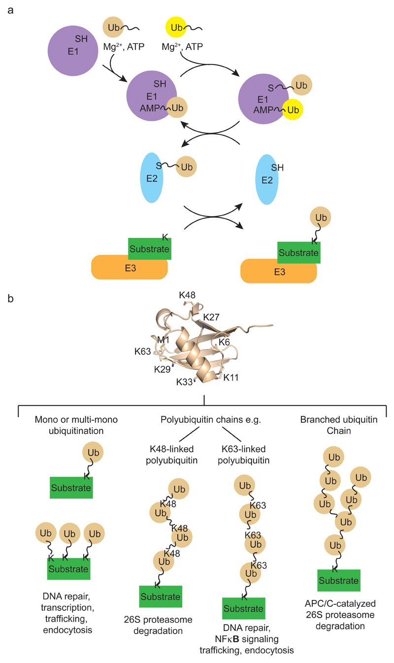
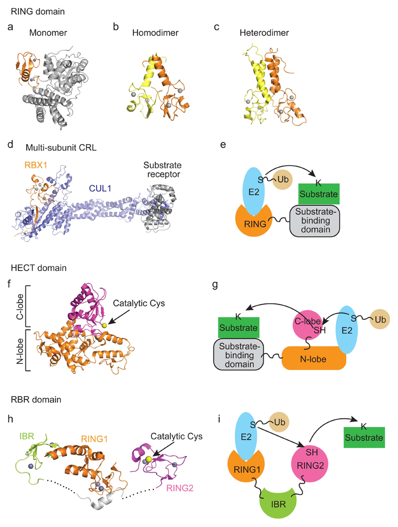
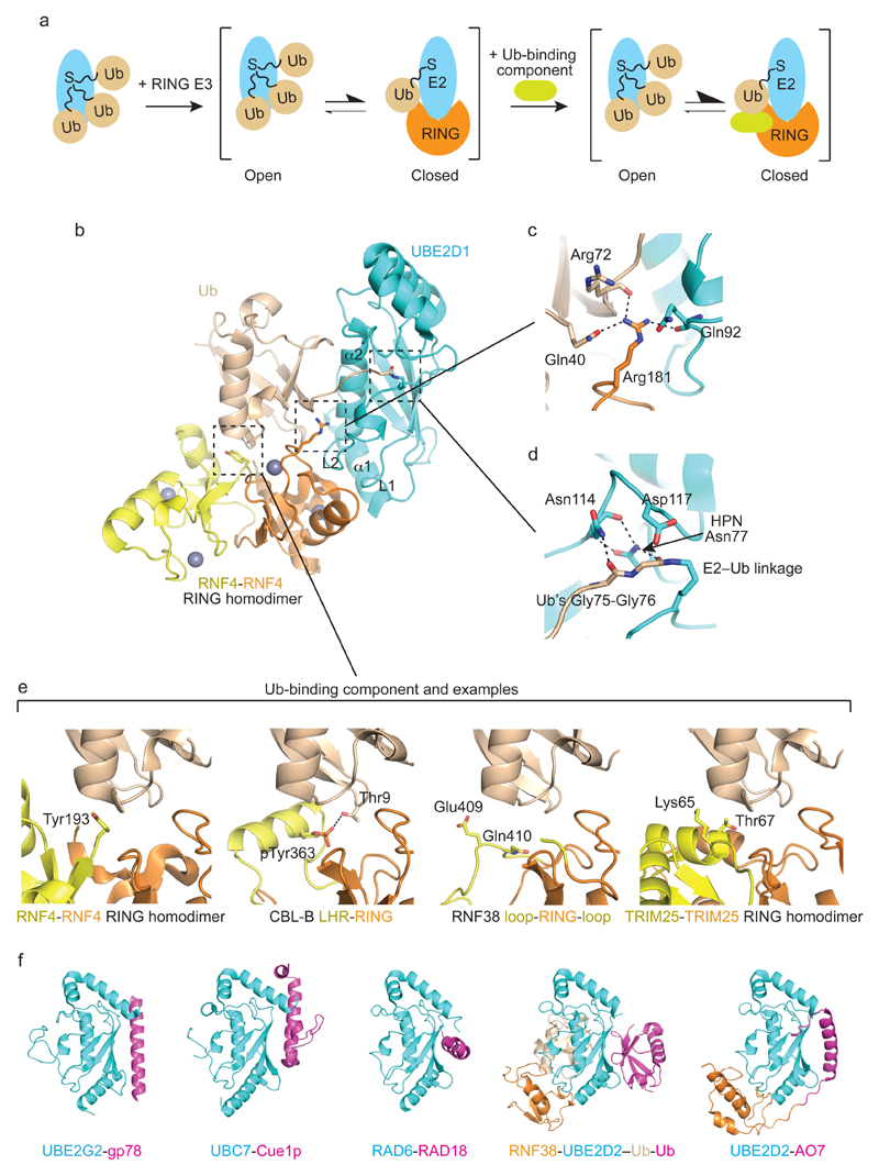
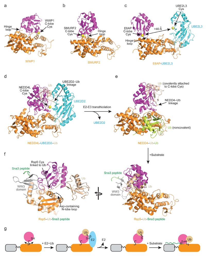
Covalent attachment (conjugation) of one or more ubiquitin molecules to protein substrates governs numerous eukaryotic cellular processes, including apoptosis, cell division and immune responses. Ubiquitylation was originally associated with protein degradation, but it is now clear that ubiquitylation also mediates processes such as protein-protein interactions and cell signalling depending on the type of ubiquitin conjugation. Ubiquitin ligases (E3s) catalyse the final step of ubiquitin conjugation by transferring ubiquitin from ubiquitin-conjugating enzymes (E2s) to substrates. In humans, more than 600 E3s contribute to determining the fates of thousands of substrates; hence, E3s need to be tightly regulated to ensure accurate substrate ubiquitylation. Recent findings illustrate how E3s function on a structural level and how they coordinate with E2s and substrates to meticulously conjugate ubiquitin. Insights regarding the mechanisms of E3 regulation, including structural aspects of their autoinhibition and activation are also emerging.


Figures

Figure 1
Figure 1. Ubiquitin conjugation system.
a | Schematic diagram of ubiquitin conjugation cascade. E1 catalyzes two rounds of adenylation of ubiquitin (labelled Ub in figure) in the presence of Mg2+ and ATP to bind two molecules of ubiquitin: one forms a thioester with E1’s catalytic cysteine and the other is bound non-covalently via its AMP-modified C-terminus to E1’s adenylation domain. E1 then binds E2 and transfers ubiquitin from its catalytic cysteine to E2’s catalytic cysteine to form E2~ubiquitin. E2~ubiquitin dissociates from E1 and E1 re-enters its catalytic cycle. E3 recruits E2~ubiquitin and substrate to catalyze ubiquitin transfer to substrate. Upon transfer, E2 disengages from E3 and E3 binds another E2~ubiquitin to catalyze the next round of substrate ubiquitylation. b | Substrate ubiquitylation outcomes. Ubiquitylated substrates interact with binding partners that harbour ubiquitin-binding domains, thus altering protein-protein interactions and cellular functions of substrates. A single molecule of ubiquitin can be attached to substrate at one (monoubiquitylation) or multiple (multi-monoubiquitylation) lysine sites. Additionally, the seven surface lysine residues (labelled as K and number in the protein structure) and N-terminal methionine (labelled as M1) of ubiquitin can serve as acceptor sites leading to the formation of polyubiquitin chains. These chains can be homogeneous like K48-linked and K63-linked polyubiquitin, where only one site on ubiquitin is modified, or branched, where two molecules of ubiquitin are attached to different acceptor sites on a single ubiquitin molecule. Different ubiquitin linkages vary in structure and therefore recruit discrete interacting partners to produce unique downstream signals, . APC/C synthesizes branched ubiquitin chains on its substrate, resulting in enhanced substrate recognition and degradation by the 26S proteasome.
Figure 2
Figure 2. E3 catalytic mechanisms.
a–e | RING E3-catalyzed ubiquitin transfer. (a) Crystal structure of a monomeric RING E3 from c-CBL (PDB: 1FBV). The RING domain is in orange and the substrate-binding domain is in grey. (b) Crystal structure of a dimeric RING E3 from cIAP2 (PDB: 3EB5). One RING domain is colored orange and the other yellow. (c) NMR structure of BRCA1-BARD1 RING domain heterodimer (PDB: 1JM7). BRCA1 is colored in orange and BARD1 is in yellow. (d) Crystal structure of a multi-subunit RING E3 CRL. The RING domain is in orange, CUL1 is in blue, and the substrate receptor is in grey. (e) A schematic drawing of RING E3-mediated catalysis. The RING domain binds E2~ubiquitin and a substrate-binding domain recruits substrate. Ubiquitin is transferred directly from E2 to a substrate lysine. f,g | HECT E3-catalyzed ubiquitin transfer. (f) Crystal structure of a HECT domain from NEDD4L (PDB: 3JVZ). C- and N-lobes are indicated. C-lobe’s catalytic cysteine is shown as a yellow sphere. (g) A schematic drawing of HECT E3 catalysis. N-lobe of the HECT domain binds E2~ubiquitin and a substrate-binding domain recruits substrate. Ubiquitin is transferred from E2 to the catalytic cysteine of the C-lobe of the HECT domain and subsequently to a substrate lysine. h,i | RBR E3-catalyzed ubiquitin transfer. (h) Crystal structure of an RBR domain from autoinhibited PARKIN (PDB: 5C23). RING1, IBR and RING2 are indicated. RING2’s catalytic cysteine is shown as a yellow sphere. The grey helix is a fragment of the repressor element. Dashed lines indicate disordered regions. This autoinhibited arrangement is specific to PARKIN and may not be representative of all E3s. (i) A schematic drawing of RBR E3 catalysis. RING1 domain binds E2~ubiquitin and ubiquitin is transferred from E2 to RING2’s catalytic cysteine and then to a substrate lysine. How RBR E3s bind substrates remain elusive. PARKIN is recruited to the damaged mitochondria by PINK1 and ubiquitylates proteins bound to the outer mitochondria membrane, . HOIP contains a zinc finger motif in the RING2 domain that binds ubiquitin as described later in Figure 4f. Zinc atoms, which function to stabilize domain folding, are shown as grey spheres.
Figure 3
Figure 3. Mechanism of priming ubiquitin for transfer by RING E3s.
a | RING E3 binding induces the formation of a closed E2~ubiquitin conformation that is primed for catalysis. Left panel, in the absence of RING E3, ubiquitin samples various conformations relative to E2. Middle panel, RING E3 binding shifts the equilibrium toward the closed and primed E2~ubiquitin conformation. Right panel, the presence of an additional ubiquitin-binding component outside the RING domain further stabilizes the primed E2~ubiquitin conformation. Examples of the additional component are shown in e. E2 is colored cyan, ubiquitin wheat, the RING domain orange and the additional component yellow. b | Structure of dimeric RNF4-UBE2D1–ubiquitin complex (PDB 4AP4). Loops L1 and L2 and α1 from UBE2D1, which bind the RING domain, and α2 of UBE2D1, which binds ubiquitin, are indicated. One RNF4 subunit is colored orange, the second yellow, UBE2D1 cyan and ubiquitin wheat. Zinc atoms are shown as grey spheres. c | Close-up view of the linchpin Arg in b with colors as in b. Arg181 forms hydrogen bonds with the sidechain of Gln40 and carbonyl oxygen of Arg72 of ubiquitin and carbonyl oxygen of Gln92 of UBE2D1. This interaction stabilizes ubiquitin in the closed conformation. d | Close-up view of UBE2D1–ubiquitin active site in b. The catalytic cysteine of UBE2D1 was mutated to a lysine to generate a stable amide linkage with the C-terminus of ubiquitin to enable crystallization of the complex. e | Additional ubiquitin-binding components that stabilize E2~ubiquitin in the closed conformation. Left panel, Tyr193 from the C-terminus of the second subunit of RNF4 packs against ubiquitin. Middle left panel, phospho-Tyr363 and the linker helix region (LHR) of CBL-B contact ubiquitin. Middle right panel, loops adjacent to the RING domain in RNF38 bind ubiquitin. Right panel, residues from the C-terminal helix of the second subunit of TRIM25 pack against ubiquitin. RING domains are colored orange, non-RING components yellow and ubiquitin wheat. f | Additional E2-E3 interactions involving the backside of E2. Left to right: UBE2G2-gp78 complex (PDB: 3H8K), UBC7-Cue1p complex (PDB: 4JQU), RAD6-RAD18 complex (PDB: 2YBF), RNF38-UBE2D2–Ub-Ub complex (PDB: 4V3L) and UBE2D2-AO7 complex (PDB: 5D1K). E2 is colored in cyan, E2 backside-binding component is in pink, ubiquitin is in wheat and RING domain is in orange.
Figure 4
Figure 4. Mechanism of ubiquitin transfer by HECT E3s.
a–c | Conformational flexibility of HECT domains. Structural alignment based on the N-lobe of HECT domains from WWP1 (a, PDB: 1ND7), SMURF2 (b, PDB: 1ZVD) and E6AP bound to the E2 UBE2L3 (c, PDB: 1C4Z) reveals different C-lobe conformations. N-lobes are colored orange, C-lobes magenta and UBE2L3 cyan. Arrows indicate the flexible hinge loop. Catalytic cysteines are shown as yellow spheres. The distance between E2 and E3 catalytic cysteines observed in the structure is indicated in c. d | Structure of NEDD4L HECT-UBE2D2–ubiquitin complex (PDB: 3JVZ). UBE2D2–ubiquitin linkage (indicated) and C-lobe’s catalytic cysteine are juxtaposed allowing transthiolation of ubiquitin from the active site of the E2 to the catalytic cysteine of E3. Colors are as in a–c with ubiquitin colored wheat. e | Structure of NEDD4 HECT domain following transthiolation (PDB: 4BBN). In this case NEDD4 is bound to two ubiquitin molecules, one which is a covalently attached to the catalytic cysteine in the C-lobe following transthiolation (shown in wheat) and a second one which is non-covalently bound to the N-lobe that enhances the processivity of substrate ubiquitylation (shown in lime). Colors are as in a–c. f | Structure of Rsp5(E3)–ubiquitin-Sna3 peptide(substrate) complex (PDB: 4LCD). The catalytic Cys777 of Rsp5, Gly75 of ubiquitin and Lys125 of the Sna3 peptide are covalently linked via a three-way chemical cross-linker to mimic substrate ubiquitylation. Colors are as in a–d with Sna3 peptide colored green and the WW3 domain grey. The disordered N-lobe loop harboring Asp495 is depicted as a dashed line. In d, e and f right panel, alignment of the three structures using the N-lobe reveals different C-lobe conformations in E2-E3 transthiolation and substrate ubiquitylation reactions. g | Schematic diagrams showing the HECT E3 catalytic cycle. In the absence of any binding partner, the C-lobe can rotate relative to the N-lobe about the hinge loop. Upon encountering E2~ubiquitin, the N-lobe binds E2 and the C-lobe rotates to bind ubiquitin, thereby juxtaposing the catalytic cysteines from E2 and E3. Upon ubiquitin transfer, E2 is released. E3 binds substrate and the C-lobe undergoes rotation to juxtapose E3’s catalytic cysteine and substrate lysine for ligation. Colors are as in a–f.
Figure 5
Figure 5. Mechanism of ubiquitin transfer by RBR E3s.
a,b | Structures of autoinhibited PARKIN (PDB: 5C23) and HHARI (PBD: 4KBL), respectively. Schematic diagrams above show domain architecture with structural domains colored according to the diagrams. Zinc atoms and RING2’s catalytic cysteines are shown as grey and yellow spheres, respectively. c,d | Structures of PARKIN and HHARI in a and b, respectively, with a modeled E2 in cyan generated by overlaying their RING1 domain with the structure of RNF38 RING domain bound to E2 (PDB: 4V3L). Colors are as in a. Distances between E2 and E3’s catalytic cysteines are indicated. e | Structure of HOIP RBR domain bound to UBE2D2–ubiquitin (PDB: 5EDV). HOIP was crystallized as a domain swapped dimer, but the biological unit of HOIP-UBE2D2-ubiquitin complex is shown here. A schematic diagram shows crystallized HOIP construct’s domain architecture. Structural domains are colored according to this diagram. UBE2D2 is colored cyan, ubiquitin wheat and ubiquitinAllo yellow. E2–ubiquitin linkage and RING2’s catalytic cysteines are shown as yellow spheres. In a, b and e, the helical extension (hE) comprises two segments, hE1 and hE2 (indicated in the structures), which are involved in binding ubiquitin. f | Structure of HOIP together with acceptor ubiquitin (green, serving as substrate) and a second donor ubiquitin molecule (wheat) generated via crystallographic symmetry (PDB: 4LJO). Top panel is in the same orientation as in e. g | HOIP catalytic cycle. Binding of a ubiquitin molecule to the allosteric site (UbAllo) primes HOIP’s RBR conformation for E2~ubiquitin recruitment. The E2~ubiquitin thioester and RING2’s catalytic cysteine are juxtaposed for RING2~ubiquitin formation. Upon formation of the RING2~ubiquitin, E2 is released. Subsequently, RING2 binds an acceptor ubiquitin (UbAcc) and catalyzes linear ubiquitin chain formation. To catalyze additional rounds of ubiquitin transfer, linear ubiquitin must be released to enable E2~ubiquitin recruitment.
Figure 6
Figure 6. Mechanisms of substrate lysine selection.
a | A schematic drawing of Rsp5-catalyzed substrate ubiquitylation. Rsp5~ubiquitin presents its N- and C-lobes and WW3 domain in a conformation where the distance is 25 Å between the Rsp5~ubiquitin thioester and the substrate binding site in the WW3 domain. In this way, lysines at least 10 residues away from the WW3-binding motif PY in the substrate are prioritized for ubiquitylation. The N-lobe is colored orange, the C-lobe magenta, the WW3 domain grey, ubiquitin wheat and substrate in green. b | Structure of RNF2-BMI1-UBE2D3 complex bound to nucleosome core particle (NCP) (PDB: 4R8P). RNF2 is colored orange, BMI1 yellow, UBE2D3 cyan, H2A green, H2B blue, H3 pink, H4 purple and DNA grey. H2A’s Lys119, the target for monoubiquitylation in this reaction, is shown as a black sphere and UBE2D3’s catalytic cysteine is shown as a yellow sphere. The interactions between the RNF2-BMI1-UBE2D3 complex and NCP restrain UBE2D3 and H2A movement such that UBE2D3’s catalytic cysteine can only be juxtaposed with H2A’s Lys119 for site-specific ubiquitylation. c | Schematic drawings of APC/C-catalyzed substrate ubiquitylation. APC11 binds UBE2C~ubiquitin via the canonical RING E3-E2~ubiquitin interaction shown in Figure 3 and APC2’s WHB domain contacts UBE2C’s backside. These bipartite interactions fix the UBE2C~ubiquitin orientation such that the thioester bond is ~40 Å away from the D and KEN box binding sites on the substrate-receptor CDH1, thereby prioritizing substrate lysines located 10 residues away from these motifs for ubiquitylation. Once the substrate is modified with a single ubiquitin, APC/C tracks the tip of the growing ubiquitin chain. APC11 binds the acceptor ubiquitin (colored in lime) using a surface distinct from its E2~ubiquitin-binding site and presents the acceptor ubiquitin to a UBE2S~ubiquitin complex for ubiquitin chain elongation. APC/C is shown as an outline. APC11 is colored orange, UBE2C and UBE2S cyan, ubiquitin wheat, APC2 and WHB light blue and CDH1 grey. Green ribbons depict binding sites on CDH1 for substrates containing a D- or KEN-box.
Figure 7
Figure 7. Mechanisms of autoinhibition and activation of E3s.
a | Schematic showing domain composition of cIAP1 (top) and the mechanism of its autoinhibition and activation (bottom). In solution, cIAP1 adopts a closed conformation where the E2~ubiquitin binding and dimerization surfaces of the RING domain are occluded. SMAC mimetic compound binds the BIR3 domain and shifts cIAP1 to an extended conformation. This enables RING domain dimerization which is critical for binding and priming E2~ubiquitin for catalysis. b | Schematic showing domain composition of SMURF2 (top) and the mechanism of its autoinhibition and activation (bottom). The C2 domain of SMURF2 binds both the N- and C-lobes of the HECT domain, thereby preventing rotation of the two lobes and blocking non-covalent ubiquitin binding. Addition of SMAD7 or other stimuli causes the release of the C2 domain, thereby facilitating E2-E3 transthiolation and non-covalent ubiquitin binding important for processive substrate ubiquitylation. c | Schematic drawings showing domain composition of PARKIN (top) and the mechanism of its autoinhibition and activation. PARKIN adopts an autoinhibited conformation where the E2~ubiquitin binding site on RING1 and catalytic cysteine of RING2 are blocked. Moreover, in this conformation the catalytic cysteine of RING2 is distal from the modeled catalytic cysteine of E2 as shown in Figure 5c. Addition of phospho-Ser65-ubiquitin (shown as pUb in the drawing) and phosphorylation of PARKIN’s Ser65 in the UBL domain (shown as a red ball and stick) synergistically activate PARKIN. The phosphorylated UBL domain is released from the RING1 domain, partially freeing the E2~ubiquitin binding surface. Phospho-Ser65-ubiquitin then binds a pocket created by RING0, RING1, hE and IBR and induces straightening of hE similar to that observed in the HOIP-UBE2D2–ubiquitin structure (Figure 5e,g). Notably, PARKIN’s RING1-hE-IBR-phospho-Ser65-ubiquitin complex adopts a conformation that resembles RING1-hE-IBR-ubiquitinAllo in the HOIP-UBE2D2–ubiquitin structure, suggesting a similar mechanism in E2~ubiquitin recruitment of both enzymes, though this remains to be investigated. For all panels, domain architectures are shown and colored according to the schematic. d | Crystal structure of PARKIN bound to phospho-Ser65-ubiquitin (PDB: 5CAW). Structural domains are colored according to the schematic drawing in c. Phospho-Ser65-ubiquitin is in yellow with pSer65 indicated. RING2’s catalytic cysteine is shown as a yellow sphere.

References

    1. Hershko A, Ciechanover A. The ubiquitin system. Annu Rev Biochem. 1998;67:425–79. - PubMed
    1. Dye BT, Schulman BA. Structural mechanisms underlying posttranslational modification by ubiquitin-like proteins. Annu Rev Biophys Biomol Struct. 2007;36:131–50. - PubMed
    1. Pickart CM, Eddins MJ. Ubiquitin: structures, functions, mechanisms. Biochim Biophys Acta. 2004;1695:55–72. - PubMed
    1. Komander D, Rape M. The ubiquitin code. Annu Rev Biochem. 2012;81:203–29. - PubMed
    1. Chau V, et al. A multiubiquitin chain is confined to specific lysine in a targeted short-lived protein. Science. 1989;243:1576–83. - PubMed