Case Reports
. 2016 Dec 1;22(23):5682-5687.
doi: 10.1158/1078-0432.CCR-16-1031. Epub 2016 Aug 2.

Regression of Chemotherapy-Resistant Polymerase ε (POLE) Ultra-Mutated and MSH6 Hyper-Mutated Endometrial Tumors with Nivolumab

Affiliations
Case Reports

Regression of Chemotherapy-Resistant Polymerase ε (POLE) Ultra-Mutated and MSH6 Hyper-Mutated Endometrial Tumors with Nivolumab

Alessandro D Santin et al. Clin Cancer Res. .

Abstract

Purpose: The management of endometrial carcinoma no longer amenable to treatment with surgery or radiotherapy has not improved significantly with modern chemotherapy. Alternative therapeutic options are desperately needed.

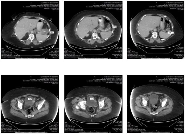
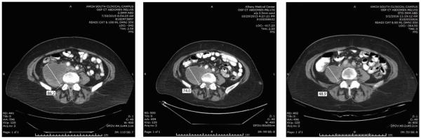
Experimental design: We describe 2 heavily pretreated patients with recurrent disease refractory to surgery, radiotherapy, and chemotherapy who were treated with the anti-PD-1 immune checkpoint inhibitor nivolumab.

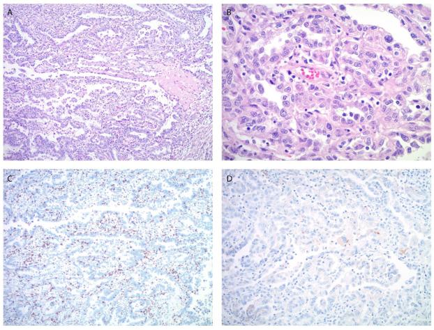
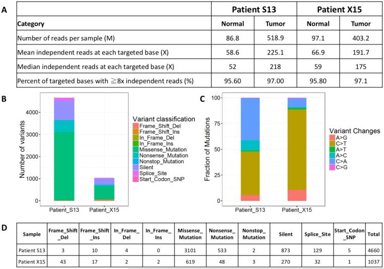
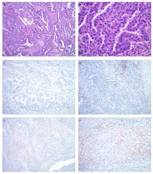
Results: Patient #1 harbored an ultra-mutated tumor (mutation load/MB = 117.3, total mutations = 4,660) driven by mutation in the exonuclease domain of the DNA polymerase ε gene. Patient #2 harbored a hyper-mutated tumor (mutation load/MB = 33.5, total mutations = 1,037) due to a germinal MSH6 gene mutation. Both patients demonstrated a remarkable clinical response to the anti-PD-1 immune checkpoint inhibitor nivolumab. Patients' clinical responses remain unchanged at the time of the writing of this report, with no grade 3 or higher side effects reported to date.

Conclusions: Anti-PD-1 inhibitors represent a novel treatment option for recurrent/metastatic, ultra/hyper-mutated human tumors refractory to salvage treatment. Clin Cancer Res; 22(23); 5682-7. ©2016 AACRSee related commentary by Piulats and Matias-Guiu, p. 5623.


Figures

Figure 1
Figure 1
Microscopic images of the endometrial carcinoma in patient #1. A, B. The tumor shows predominantly a papillary and glandular architecture (A) with high nuclear grade and clear to pale eosinophilic cytoplasm (B), consistent with clear cell carcinoma component. C. Moderate peri- and intratumoral lymphocytic infiltrate is highlighted by CD8 immunostain. D. PD-L1 immunostain shows focal, weak staining predominantly located in the tumor cells. (A. Original magnification 100x, H&E stain, B. Original magnification 400x, H&E stain, C. Original magnification 100x, D. Original magnification 200x).
Figure 2
Figure 2
Representative CAT scans demonstrating activity (ie, partial response) of nivolumab in Patient #1 (ie, POLE-ultra-mutated). Left panels: Pretreatment images with baseline measurements of two representative metastatic tumor deposits [ie, mass abutting the pancreatic tail in the gastro-splenic ligament (Upper Left Panel) and mass involving the bladder dome wall (Lower Left Panel). Middle panels: Regression of the metastatic tumor deposits described above three months after treatment initiation. Right panels: regression (Upper Right) and disappearance (Lower Right) of the metastatic tumor deposits described above seven months after treatment initiation with nivolumab.
Figure 3
Figure 3
Whole Exome Sequencing (WES) results of Patient #1 (ie, POLE-ultra-mutated, S13) and Patient # 2 (i.e., MSH6-hyper-mutated, X15). A) Exome sequencing quality statistics for both patients’ tumor and normal DNA. (B) Exonic mutation burden in Patient S13 and X15 with different variant classifications displayed in the barplot. (C) Distribution of six different mutation conversions in Patient S13 and X15. (D) Number and classification of the distinct variant of mutations detected in Patient S13 and X15.
Figure 4
Figure 4
Microscopic images of the endometrial carcinoma in patient #2. A. The tumor shows a predominant glandular architecture with irregular, slit-like spaces and focal necrosis. The nuclei are large with a high nuclear to cytoplasmic ratio, prominent nucleoli and brisk mitotic activity (B). C. P53 immunostain shows a wild-type staining pattern with weak to moderate nuclear positivity in approximately 10% of tumor cells. D. The lymphocytic infiltrate is predominantly peritumoral; less frequent intratumoral lymphocytes are also highlighted by CD8 immunostain. E, F. PD-L1 immunostain shows diffuse, weak to moderate staining predominantly in peritumoral lymphocytes. (A. Original magnification 100x, H&E stain, B. Original magnification 400x, H&E stain, C. Original magnification 200x, D. Original magnification 100x, E. Original magnification 100x, F. Original magnification 200x).
Figure 5
Figure 5
Representative CAT scans demonstrating activity (ie, partial response) of nivolumab in Patient #2 (ie, MSH6-hyper-mutated). Left panel: Pretreatment image with baseline measurement of a representative metastatic tumor deposit (ie, right common iliac mass invading the psoas muscle and obstructing the right ureter). Middle panel: Regression of the metastatic tumor deposit described above three months after treatment initiation. Right panel: Continue regression (Right Panel) of the metastatic tumor deposit nine months after treatment initiation with nivolumab.

References

    1. Kandoth C, Schultz N, Cherniack AD, Akbani R, Liu Y, Shen H, et al. Integrated genomic characterization of endometrial carcinoma. The Cancer Genome Atlas Research Network. Nature. 2013;497:67–73. - PMC - PubMed
    1. Zhao S, Choi M, Overton JD, Bellone S, Roque DM, Cocco E, et al. Landscape of somatic single-nucleotide and copy-number mutations in uterine serous carcinoma. Proceedings of the National Academy of Sciences of the USA. 2013;110:2916–21. - PMC - PubMed
    1. Howitt BE, Shukla SA, Sholl LM, Ritterhouse SA, Watkins JC, Rodig S, et al. Association of Polymerase e–Mutated and Microsatellite-Instable Endometrial Cancers with Neoantigen Load, Number of Tumor-Infiltrating Lymphocytes, and Expression of PD-1 and PD-L1. JAMA Oncol. 2015;1:1319–23. - PubMed
    1. Le DT, Uram JN, Wang H, Bartlett BR, Kemberling H, Eyring AD, et al. PD-1 Blockade in Tumors with Mismatch-Repair Deficiency. N Engl J Med. 2015;372(26):2509–20. - PMC - PubMed
    1. Rayner E, van Gool IC, Palles C, Kearsey SE, Bosse T, Tomlinson I, et al. A panoply of errors: polymerase proofreading domain mutations in cancer. Nature Reviews Cancer. 2016;16:71–81. - PubMed

Publication types

MeSH terms