. 2016 Aug 2;7(4):e00972-16.
doi: 10.1128/mBio.00972-16.

Lcp1 Is a Phosphotransferase Responsible for Ligating Arabinogalactan to Peptidoglycan in Mycobacterium tuberculosis

Affiliations

Lcp1 Is a Phosphotransferase Responsible for Ligating Arabinogalactan to Peptidoglycan in Mycobacterium tuberculosis

James Harrison et al. mBio. .

Abstract

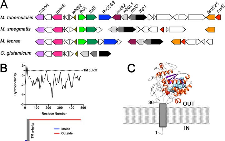
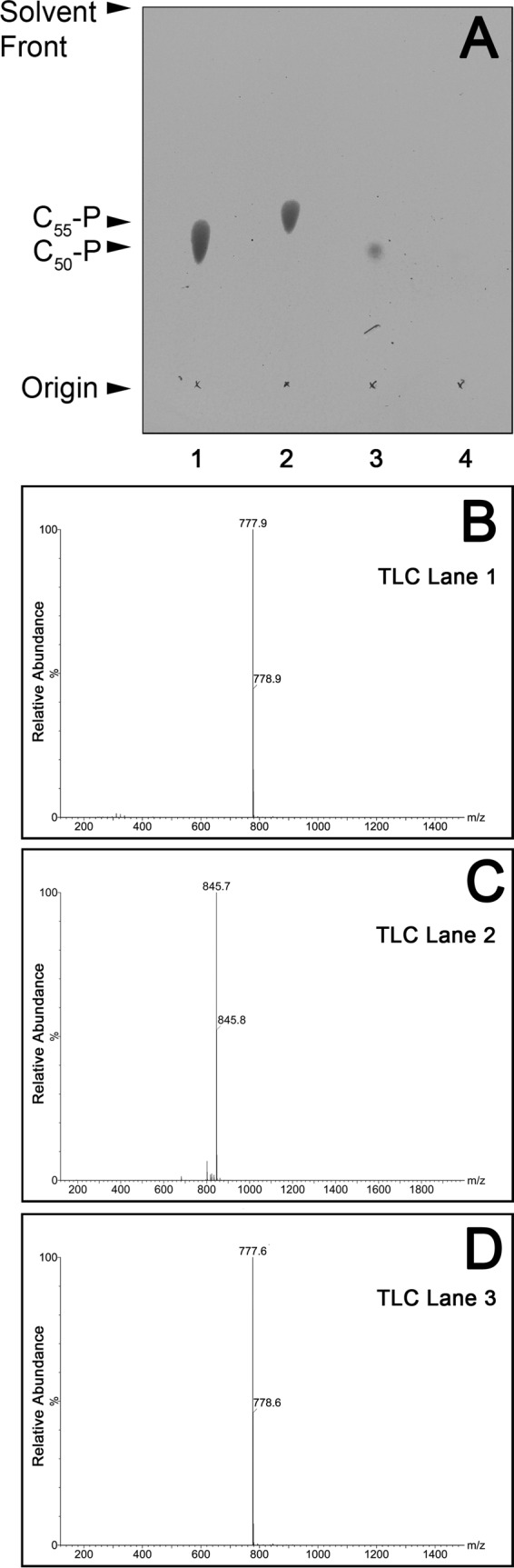
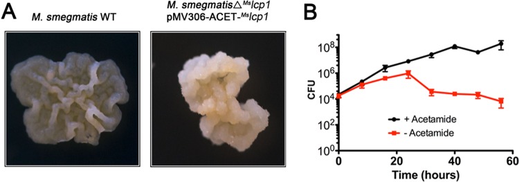
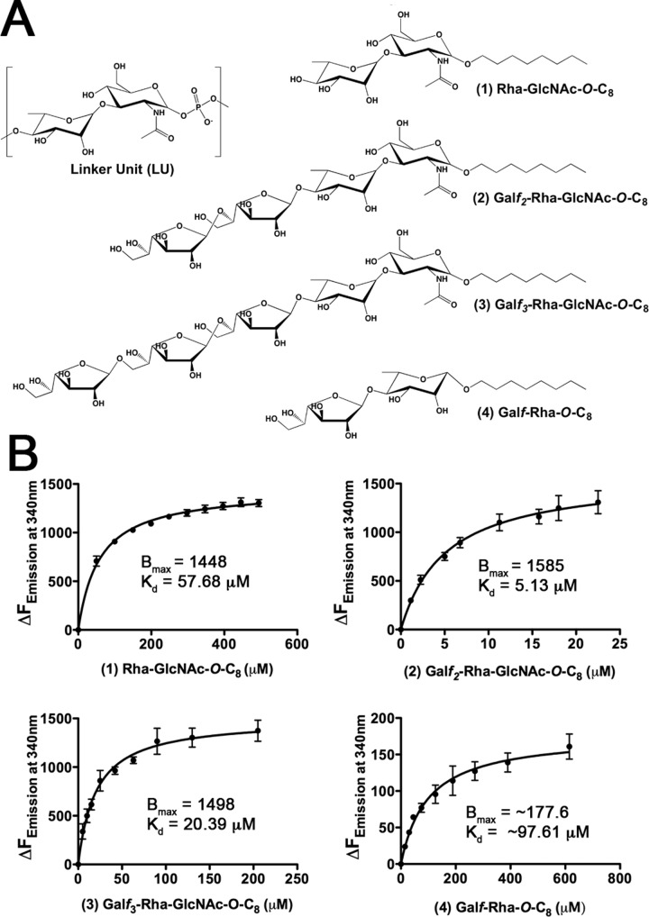
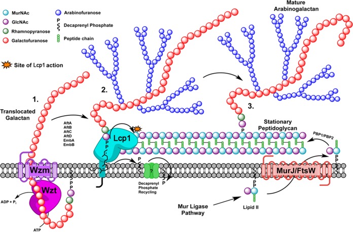
Mycobacterium tuberculosis, the etiological agent of tuberculosis (TB), has a unique cell envelope which accounts for its unusual low permeability and contributes to resistance against common antibiotics. The main structural elements of the cell wall consist of a cross-linked network of peptidoglycan (PG) in which some of the muramic acid residues are covalently attached to a complex polysaccharide, arabinogalactan (AG), via a unique α-l-rhamnopyranose-(1→3)-α-d-GlcNAc-(1→P) linker unit. While the molecular genetics associated with PG and AG biosynthetic pathways have been largely delineated, the mechanism by which these two major pathways converge has remained elusive. In Gram-positive organisms, the LytR-CpsA-Psr (LCP) family of proteins are responsible for ligating cell wall teichoic acids to peptidoglycan, through a linker unit that bears a striking resemblance to that found in mycobacterial arabinogalactan. In this study, we have identified Rv3267 as a mycobacterial LCP homolog gene that encodes a phosphotransferase which we have named Lcp1. We demonstrate that lcp1 is an essential gene required for cell viability and show that recombinant Lcp1 is capable of ligating AG to PG in a cell-free radiolabeling assay.

Importance: Tuberculosis is an infectious disease caused by the bacterial organism Mycobacterium tuberculosis Survival of M. tuberculosis rests critically on the integrity of its unique cell wall; therefore, a better understanding of how the genes and enzymes involved in cell wall assembly work is fundamental for us to develop new drugs to treat this disease. In this study, we have identified Lcp1 as an essential phosphotransferase that ligates together arabinogalactan and peptidoglycan, two crucial cell wall macromolecules found within the mycobacterial cell wall. The discovery of Lcp1 sheds new light on the final stages of mycobacterial cell wall assembly and represents a key biosynthetic step that could be exploited for new anti-TB drug discovery.


Figures

FIG 1
FIG 1
Comparison of the lcp1 locus and in silico analysis of Lcp1 by TMHMM and I-TASSER. (A) The locus in bacteria analyzed consists of lcp1, which in M. tuberculosis has the locus tag Rv3267 and in M. smegmatis has the locus tag MSMEG1824. For M. leprae and C. glutamicum, locus tags are ML0756 and NCgl0708, respectively. (B) The TMHMM server was used to predict the membrane topology of MtbLcp1 (48). (C) I-TASSER homology model of MtbLcp1 positioned in relation to the cytoplasmic membrane and predicted N-terminal TM α-helix (19).
FIG 2
FIG 2
TLC and ES-MS analysis and identification of decaprenyl-1-monophosphate bound to recombinant MtbLcp1. (A) TLC analysis of organic extractable material of recombinant MtbLcp1. Standards of decaprenyl-1-monophosphate and undecaprenyl-1-monophosphate are in lanes 1 and 2, respectively. Lane 3 contains material extracted from MtbLcp1, while lane 4 contains a negative control of material extracted from EmbCCT (21). Lipids were analyzed by TLC using silica gel plates (5735 silica gel 60F254; Merck) and developed in CHCl3-CH3OH-NH4OH-H2O (65:25:0.5:3.6, vol/vol/vol/vol), and plates were sprayed with 5% ethanolic molybdophosphoric acid and charred to visualize the lipids. (B) ES-MS analysis of decaprenyl-1-monophosphate standard (TLC, lane 1). (C) ES-MS analysis of undecaprenyl-1-monophosphate standard (TLC, lane 2). (D) ES-MS organic extracted material from MtbLcp1 (TLC, lane 3).
FIG 3
FIG 3
Essentiality of Mslcp1 in M. smegmatis mc21551. (A) Growth and colony morphology of M. smegmatis and ΔMslcp1 conditional mutant on tryptic soy agar with the inducer acetamide. WT, wild type. (B) CFU counts of M. smegmatis ΔMslcp1 conditional mutant cultured in tryptic soy broth in the presence and absence of the inducer acetamide (data plotted represent the mean ± standard deviation from three independent biological replicate experiments).
FIG 4
FIG 4
Binding of linker unit analogues to MtbLcp1. (A) Chemical structure of the mycobacterial linker unit and novel chemical scaffolds used as ligands to probe the interaction of the linker unit with MtbLcp1. (B) Saturation binding experiments using intrinsic tryptophan fluorescence of MtbLcp1 to study binding of ligands 1 to 4. Data plotted represent the mean ± standard deviation from three independent biological replicate experiments.
FIG 5
FIG 5
AG-PG ligase activity of MtbLcp1 using a radiolabeled cell-free functional assay. (A) Organic solvent-extracted fractions from 14C-labeled assay mixtures were analyzed by TLC and developed in CHCl3-CH2OH-H2O-NH4OH (65:25:3.6:0.5, vol/vol/vol/vol) on aluminum-backed silica gel plates (5735 silica gel 60F254; Merck), and products were visualized by autoradiography, exposing the TLCs to X-ray film (Kodak X-Omat). TLC is representative of three independent biological replicate experiments, and corresponding densitometric data plotted represent the mean ± standard deviation for each band. Associated P values are as follows: *, P ≤ 0.05; **, P ≤ 0.01; ***, P ≤ 0.001. (B) Insoluble material from fraction 2 of the assay mixtures was hydrolyzed in 2 M TFA, reduced, and per-O-acetylated. The resulting 14C-labeled per-O-acetylated alditol acetate derivatives were analyzed by TLC using ethyl acetate-hexane (4:6, vol/vol) on aluminum-backed silica gel plates (5735 silica gel 60F254; Merck), and products were visualized by autoradiography, exposing the TLCs to X-ray film (Kodak X-Omat), and compared to known alditol-acetate standards of d-galactose (Gal), d-glucose (Glc), and d-N-acetylglucosamine (GlcNAc). TLC is representative of three independent biological replicate experiments, and corresponding densitometric data plotted represent the mean ± standard deviation for each band. Associated P values are as follows: *, P ≤ 0.05; **, P ≤ 0.01; ***, P ≤ 0.001.
FIG 6
FIG 6
Schematic diagram of the latter stages of mycobacterial cell wall biosynthesis. (1) Translocation of galactan polymer across plasma membrane. Lcp1 then recognizes and binds the linker unit (i.e., represented by compounds 2 and 3). (2) Phosphotransferase activity links AG to the 6′-OH of muramyl residues, releasing decaprenyl-1-monophosphate for recycling. (3) Newly bound AG is released by Lcp1 to go on to form mature mAGP. Additional synthesis of new PG, requiring AG addition by Lcp1, takes place simultaneously.

References

    1. WHO 2015. Global tuberculosis report 2015. World Health Organization, Geneva, Switzerland.
    1. Loewenberg S. 2012. India reports cases of totally drug-resistant tuberculosis. Lancet 379:205. doi: 10.1016/S0140-6736(12)60085-3. - DOI - PubMed
    1. Lederer E, Adam A, Ciorbaru R, Petit J, Wietzerbin J. 1975. Cell walls of mycobacteria and related organisms; chemistry and immunostimulant properties. Mol Cell Biochem 7:87–104. doi: 10.1007/BF01792076. - DOI - PubMed
    1. Mahapatra S, Crick DC, Brennan PJ. 2000. Comparison of the UDP-N-acetylmuramate:L-alanine ligase enzymes from Mycobacterium tuberculosis and Mycobacterium leprae. J Bacteriol 182:6827–6830. doi: 10.1128/JB.182.23.6827-6830.2000. - DOI - PMC - PubMed
    1. Mahapatra S, Scherman H, Brennan PJ, Crick DC. 2005. N glycolylation of the nucleotide precursors of peptidoglycan biosynthesis of Mycobacterium spp. is altered by drug treatment. J Bacteriol 187:2341–2347. doi: 10.1128/JB.187.7.2341-2347.2005. - DOI - PMC - PubMed

Publication types

MeSH terms

LinkOut - more resources