. 2016 Aug 16;113(33):9304-9.
doi: 10.1073/pnas.1608783113. Epub 2016 Aug 2.

Dishevelled is a NEK2 kinase substrate controlling dynamics of centrosomal linker proteins

Affiliations

Dishevelled is a NEK2 kinase substrate controlling dynamics of centrosomal linker proteins

Igor Cervenka et al. Proc Natl Acad Sci U S A. .

Abstract

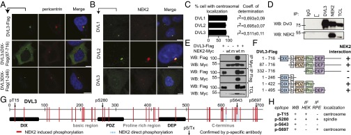
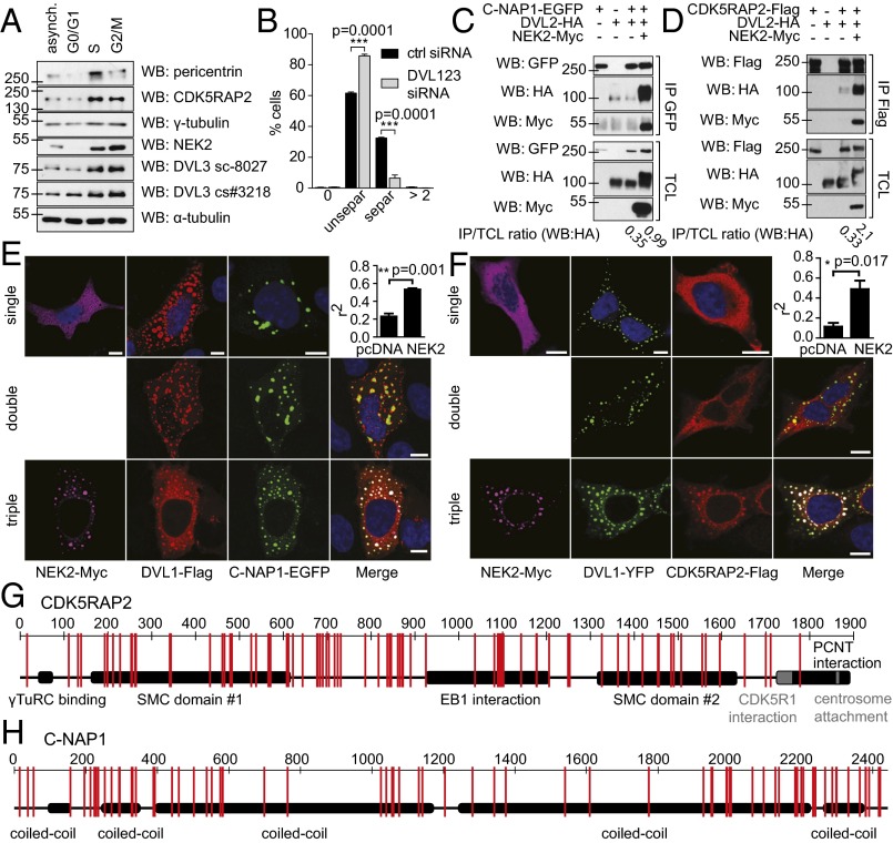
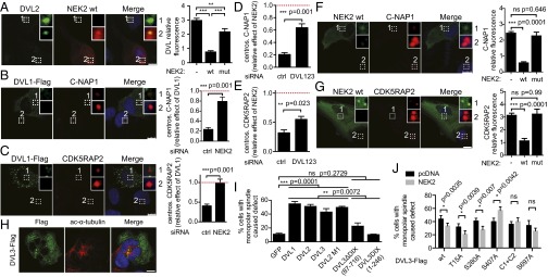
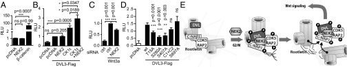
Dishevelled (DVL) is a key scaffolding protein and a branching point in Wnt signaling pathways. Here, we present conclusive evidence that DVL regulates the centrosomal cycle. We demonstrate that DVL dishevelled and axin (DIX) domain, but not DIX domain-mediated multimerization, is essential for DVL's centrosomal localization. DVL accumulates during the cell cycle and associates with NIMA-related kinase 2 (NEK2), which is able to phosphorylate DVL at a multitude of residues, as detected by a set of novel phospho-specific antibodies. This creates interfaces for efficient binding to CDK5 regulatory subunit-associated protein 2 (CDK5RAP2) and centrosomal Nek2-associated protein 1 (C-NAP1), two proteins of the centrosomal linker. Displacement of DVL from the centrosome and its release into the cytoplasm on NEK2 phosphorylation is coupled to the removal of linker proteins, an event necessary for centrosomal separation and proper formation of the mitotic spindle. Lack of DVL prevents NEK2-controlled dissolution of loose centrosomal linker and subsequent centrosomal separation. Increased DVL levels, in contrast, sequester centrosomal NEK2 and mimic monopolar spindle defects induced by a dominant negative version of this kinase. Our study thus uncovers molecular crosstalk between centrosome and Wnt signaling.

Keywords: Dishevelled; NEK2; Wnt signaling; centrosome; linker proteins.


Conflict of interest statement

The authors declare no conflict of interest.

Figures

Fig. 1.
Fig. 1.
DIX domain is required for DVL centrosomal localization, and NEK2 is a DVL kinase. (A) HEK293 cells were transfected with a low amount of DVL3, DVL3ΔDIX, and DVL3 DIX domain (green), and colocalization with pericentrin (red) was assessed. (B) NEK2 (red) colocalizes with DVL1, DVL2, and DVL3 (green) on an endogenous level in RPE cells. (C) Quantification (% of cells where proteins colocalized and coefficient of determination R2). (D) Endogenous DVL3 was co-IPed, using NEK2 as bait; anti-DVL3/IgG isotype was used as a positive and negative control. (E) HEK293 cells were cotransfected with DVL3 and NEK2, as indicated, and subjected to coimmunoprecipitation. DVL3 interacts with and shows an electrophoretic mobility shift, as a result of posttranslational modifications, only after coexpression with the wild-type form of the NEK2 kinase. (F) Summary of interaction of individual DVL3 deletion mutants with NEK2 (raw data, SI Appendix, Fig. S2 A and B). Domain structure is schematized. (b, basic region; Pro, proline-rich region). (G) HEK293 cells were transfected with DVL3, cotransfected with empty vector or wild-type NEK2, coimmunoprecipitated, and subjected to MS analysis. Schematic view of identified phosphorylated residues of DVL3 by MS. Red solid lines represent sites whose phosphorylation was induced by NEK2. Blue dashed lines represent sites directly phosphorylated by NEK2; purified DVL3 domains were incubated with recombinant kinase in presence of ATP and subjected to MS analysis. Arrows, induction of phosphorylation by NEK2 coexpression confirmed by phospho-specific antibody. For a complete list of residues, see SI Appendix, Table S1. (H) Panel indicating the successful use of phospho-specific antibodies using different methods and cell lines. (Scale bars, 10 μm.)
Fig. 2.
Fig. 2.
DVL influences centrosome separation and associates with linker proteins CDK5RAP2 and C-NAP1. (A) HeLa S. Fucci cells were sorted according to the cell cycle phase, subjected to Western blotting, and probed with antibodies against DVL3, centrosomal proteins (pericentrin, CDK5RAP2, γ-tubulin, NEK2), and α-tubulin as loading control. (B) RPE cells were stained for pericentrin and glutamylated tubulin, and distribution of centrosomal signals (SI Appendix, Fig. S4G) was quantified. Cells transfected with DVL1-2-3 siRNA show increased proportion of centrosomes before separation. (C and D) HEK293 cells were transfected, as indicated, and subjected to coimmunoprecipitation and Western blotting. Components of proteinaceous centrosomal linker (C) C-NAP1 and (D) CDK5RAP2 show enhanced interaction with DVL2 after coexpression of NEK2 kinase. (E and F) HEK293 cells were transfected as indicated, and colocalization was inspected by confocal microscopy and quantified. Colocalization of both C-NAP1 (E) and CDK5RAP2 (F) with DVL1 is visibly enhanced after NEK2 coexpression. (G and H) HEK293 cells were transfected with CDK5RAP2 or C-NAP1, cotransfected with empty vector or wild-type NEK2, coimmunoprecipitated, and subjected to analysis by mass-spectrometry. Differentially phosphorylated residues are indicated (schematic view), for a complete list of residues, see SI Appendix, Table S2. Graphs represent mean ± SEM of three independent replicates. *P < 0.05; **P < 0.01; ***P < 0.001; Student t test, E and F. DAPI (blue). (Scale bars, 100 μm.)
Fig. 3.
Fig. 3.
DVL mediates NEK2-triggered displacement of linker proteins from the centrosome. (A) RPE cells with overexpressed NEK2 show significantly reduced fluorescence intensity of endogenous DVL2 at centrosome in a kinase activity-dependent manner. (B and C) Overexpressed DVL1 displaces linker proteins from centrosome manifested by reduced fluorescence of CDK5RAP2 and C-NAP1. This phenotype is rescued by NEK2 siRNA. (D and E) Overexpressed NEK2 wt displaces linker proteins from centrosome manifested by reduced fluorescence of CDK5RAP2 and C-NAP1. This phenotype is rescued by DVL1-2-3 siRNA. Red dashed line shows baseline fluorescence of linker proteins. (F and G) C-NAP1 and CDK5RAP2 are removed from the centrosome after transfection of wt but not mut NEK2. (H) DVL overexpression (green) in HEK293 cells leads to monopolar spindle phenotype; acetylated-α-tubulin (red). (I) Extent of monopolar spindle formation in full-length DVLs, DVL3 with deleted DIX domain (DVLΔDIX87-716), DVL3DIX1-246, and nonpolymerizable mutant of DVL2 (DVL2 M1). (J) Quantification of the monopolar spindle-related phenotypes in S–A mutants at the DVL3 sites phosphorylated by NEK2 in the presence and absence of exogenous NEK2 was quantified. Graphs represent mean ± SEM of three independent replicates. *P < 0.05; **P < 0.01; ***P < 0.001 (ANOVA, Bonferroni's posttest: A, F, G, I; Student t test: B–E; multiple comparison adjusted Student t test: J). DAPI (blue). (Scale bars, 10 μm.)
Fig. 4.
Fig. 4.
DVL phosphorylation by NEK2 affects Wnt signaling. Proposed mechanism of DVL action during centrosome separation. HEK293 cells were transfected as indicated and analyzed for Topflash reporter activity. (A) NEK2 overexpression shows no significant activation of Wnt/β-catenin pathway, unlike β-catenin. (B) NEK2 alone is not able to potentiate Wnt/β-catenin activation induced by DVL3, but synergy is observed in the presence of CK1ε. (C) Knockdown of NEK2 by siRNA decreases Wnt/β-catenin pathway activation by recombinant Wnt-3a. (D) Topflash reporter activity after transfection of Ser to Ala mutants at the DVL3 sites phosphorylated by NEK2. (E) DVL accumulates during cell cycle at the centrosome. In G2/M, when NEK2 activity is peaking, DVL is phosphorylated on several residues, which increases its affinity toward linker proteins. DVL works as a scaffold that facilitates and is required for NEK2-induced release of C-NAP1 and CDK5RAP2 from centrosomes. DVL released from the centrosome by the activity of NEK2 is than available for and increases the Wnt/β-catenin pathway activation. Graphs represent mean ± SEM of at least three independent replicates. *P < 0.05; **P < 0.01 (ANOVA, Bonferroni's posttest, A–D).

References

    1. Hikasa H, Sokol SY. Wnt signaling in vertebrate axis specification. Cold Spring Harb Perspect Biol. 2013;5(1):a007955. - PMC - PubMed
    1. Clevers H, Nusse R. Wnt/β-catenin signaling and disease. Cell. 2012;149(6):1192–1205. - PubMed
    1. Gao C, Chen YG. Dishevelled: The hub of Wnt signaling. Cell Signal. 2010;22(5):717–727. - PubMed
    1. Park TJ, Mitchell BJ, Abitua PB, Kintner C, Wallingford JB. Dishevelled controls apical docking and planar polarization of basal bodies in ciliated epithelial cells. Nat Genet. 2008;40(7):871–879. - PMC - PubMed
    1. Lee KH, et al. Identification of a novel Wnt5a-CK1ε-Dvl2-Plk1-mediated primary cilia disassembly pathway. EMBO J. 2012;31(14):3104–3117. - PMC - PubMed

Publication types

MeSH terms