. 2016 Aug 16;113(33):E4867-76.
doi: 10.1073/pnas.1524915113. Epub 2016 Aug 2.

The stringent response regulates adaptation to darkness in the cyanobacterium Synechococcus elongatus

Affiliations

The stringent response regulates adaptation to darkness in the cyanobacterium Synechococcus elongatus

Rachel D Hood et al. Proc Natl Acad Sci U S A. .

Abstract

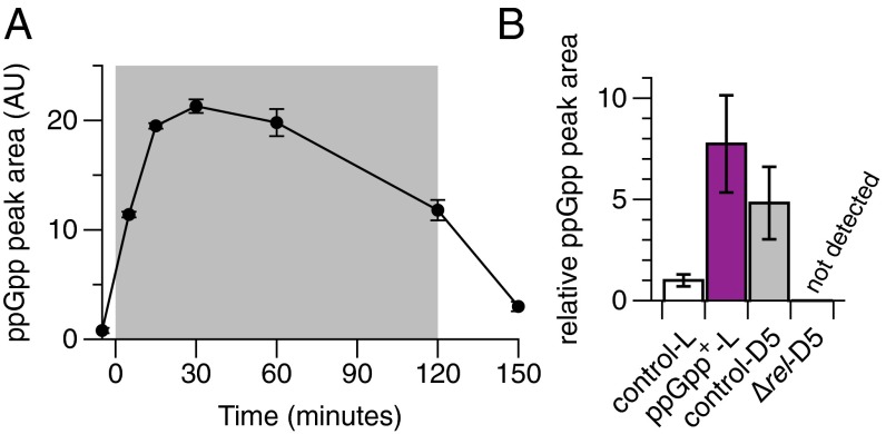
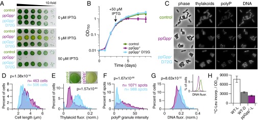
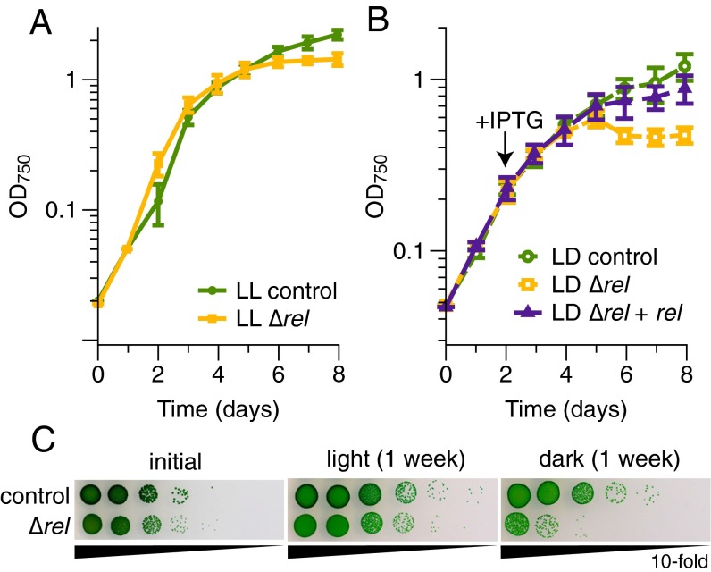
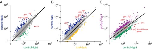
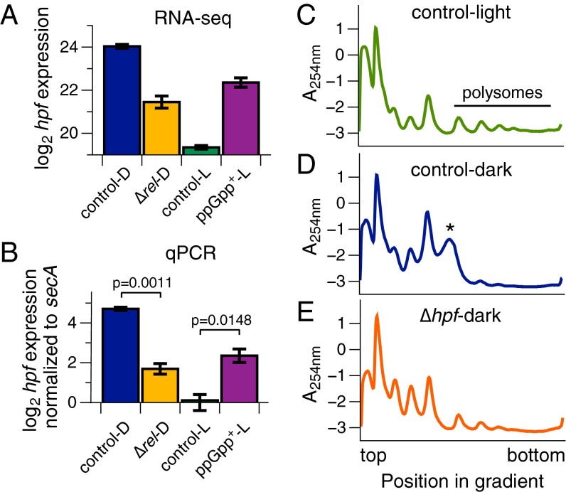
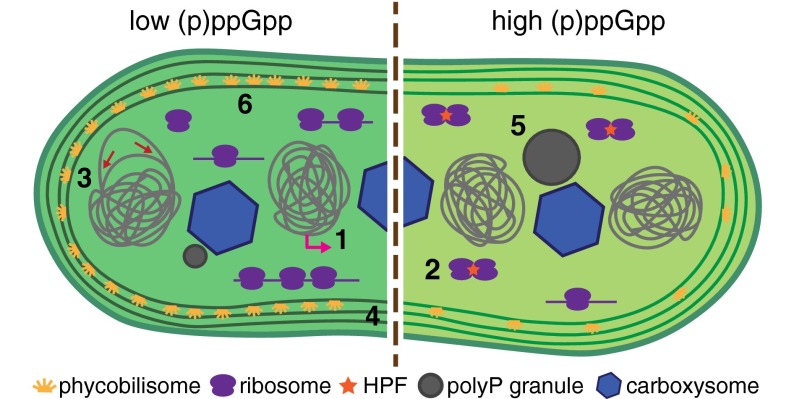
The cyanobacterium Synechococcus elongatus relies upon photosynthesis to drive metabolism and growth. During darkness, Synechococcus stops growing, derives energy from its glycogen stores, and greatly decreases rates of macromolecular synthesis via unknown mechanisms. Here, we show that the stringent response, a stress response pathway whose genes are conserved across bacteria and plant plastids, contributes to this dark adaptation. Levels of the stringent response alarmone guanosine 3'-diphosphate 5'-diphosphate (ppGpp) rise after a shift from light to dark, indicating that darkness triggers the same response in cyanobacteria as starvation in heterotrophic bacteria. High levels of ppGpp are sufficient to stop growth and dramatically alter many aspects of cellular physiology, including levels of photosynthetic pigments and polyphosphate, DNA content, and the rate of translation. Cells unable to synthesize ppGpp display pronounced growth defects after exposure to darkness. The stringent response regulates expression of a number of genes in Synechococcus, including ribosomal hibernation promoting factor (hpf), which causes ribosomes to dimerize in the dark and may contribute to decreased translation. Although the metabolism of Synechococcus differentiates it from other model bacterial systems, the logic of the stringent response remains remarkably conserved, while at the same time having adapted to the unique stresses of the photosynthetic lifestyle.

Keywords: (p)ppGpp; Synechococcus; cyanobacteria; hibernation promoting factor; stringent response.


Conflict of interest statement

The authors declare no conflict of interest.

Figures

Fig. 1.
Fig. 1.
ppGpp levels increase in the dark in Synechococcus and can be genetically manipulated. (A) Synechococcus cultures were shifted from the light (white background) to the dark (gray background) at 0 min and were harvested at the time points shown. Extracts were analyzed by anion exchange HPLC (AU, arbitrary units). Peaks eluting at the same time as a ppGpp standard were integrated to determine relative ppGpp levels. (B) Analysis of ppGpp levels from Synechococcus strains in the light (L) and the dark (D). Cultures harvested in the light were induced with IPTG for 17 h (control, WT-CmR). Cultures in the dark were harvested 5 min after the light-to-dark shift. ppGpp levels in the ∆rel mutant were below the limit of detection (∼1 μM). When analyzed using a one-tailed t test, ppGpp+-L vs. control-L, P = 0.0469; control-D5 vs. control-L, P = 0.0669. Data are presented as mean ± SEM (n = 3).
Fig. 2.
Fig. 2.
High (p)ppGpp levels stop growth and dramatically alter Synechococcus physiology. (A and B) Inducing high (p)ppGpp levels stops growth of Synechococcus. (A) When induced with IPTG, viability of the ppGpp+ strain decreases. Images are representative of three independent experiments. (B) Cultures of the indicated strains (control, WT-KanR) were grown in constant light, induced with IPTG where indicated, and monitored by absorbance at 750 nm (OD750). Data are presented as mean ± SD (n = 3). (C) At 26 h after IPTG induction, cultures were stained with DAPI and imaged by fluorescence microscopy. Representative images from three independent experiments are shown. (Scale bar: 5 μm.) (D–G) Microscopy images from two independent cultures at 26 h after induction were analyzed using MicrobeTracker and SpotFinder. Histogram colors match those in A, B, and C: purple, ppGpp+; light blue, ppGpp+ D72G. The same number of cells (ppGpp+, n = 463 cells; ppGpp+ D72G, n = 506 cells) was used for each analysis shown. Data were analyzed with the Mann–Whitney U test, and P values are indicated on the histograms. (D) ppGpp+ cells are longer than control cells. (E) Levels of light-harvesting pigments are lower in ppGpp+ cells than in control cells. Natural fluorescence from thylakoid membranes was normalized to cell area. (Inset) Pigmentation differences between control and ppGpp+ strains are striking. Cultures were imaged ∼48 h after induction. (F) Intensities of polyphosphate (polyP) granules are higher in ppGpp+ cells than in control cells. (G) DNA content is lower in ppGpp+ cells than in control cells. Histograms of DAPI-DNA fluorescence (AU; normalized to cell area) in ppGpp+ and ppGpp+ D72G cultures. (Inset) Analysis of DNA content by flow cytometry confirms that ppGpp+ cells contain less DNA per cell than control cells. Cultures were stained with Vybrant DyeCycle Green at 26 h after induction with IPTG. A total of 10,000 events were analyzed by flow cytometry for each condition. Histogram colors match those in A, B, and E: green, control; purple, ppGpp+. (H) Translation rates decrease in the dark and in ppGpp+ cells. Incorporation of 14C-Leu into trichloroacetic acid-precipitated proteins was measured by scintillation counting and is plotted as 14C counts per min/OD750. Labeling was performed for 1 h with 0.2 μCi 14C-Leu (for WT D, after a 2-h dark pulse; for ppGpp+, at 12 h after IPTG induction). Data are presented as mean ± SD (n = 6).
Fig. 3.
Fig. 3.
(p)ppGpp is important for maintaining viability during darkness. (A) Growth of the control (WT-CmR) and the ∆rel mutant is similar in constant light (LL) until cells reach stationary phase. (B) Growth of the ∆rel mutant is impaired in 12-h light/12-h dark (LD) cycles, but complementation of the ∆rel mutant restores nearly WT growth (control, WT-CmR/KanR). An IPTG-inducible copy of the rel gene was reintroduced into the ∆rel mutant at a neutral site, and IPTG was added to all cultures when indicated by the arrow. Data are presented as mean ± SD (A, n = 3; B, n = 4). (C) Viability of the ∆rel strain decreases greatly after incubation in constant darkness. Tenfold serial dilutions of cultures were plated at the beginning of the experiment, after a week in constant light, or after a week in constant darkness. Images are representative of two independent experiments.
Fig. 4.
Fig. 4.
(p)ppGpp regulates the expression of many genes in Synechococcus. RNA-seq was performed for four different conditions: control (WT-CmR)-light; ppGpp+-light, ppGpp+ cultures in the light after 18 h of IPTG induction; control-dark, WT-CmR cultures after a 2-h dark pulse; ∆rel-dark, ∆rel cultures after a 2-h dark pulse. RNA-seq data were analyzed and normalized using Rockhopper. For all conditions except ppGpp+, n = 4 biological replicates; for ppGpp+, n = 3 biological replicates. All panels show scatter plots of expression values based on upper-quartile normalization. The regression line used to identify differentially expressed genes is shown (A, R2 = 0.81224; B, R2 = 0.77495; C, R2 = 0.7258). Genes with expression values at least one SD higher or lower than the values predicted by the regression line are colored according to the condition under which they are more highly expressed. Genes not considered differentially expressed are colored in gray. All plots are shown such that the higher (p)ppGpp condition is on the y axis and the lower (p)ppGpp condition is on the x axis: (p)ppGpp–up-regulated genes are above the regression line whereas (p)ppGpp–down-regulated genes are below the regression line. Selected differentially expressed genes discussed in (p)ppGpp Regulates the Expression of Many Genes in Synechococcus are colored in red, with the gene name indicated.
Fig. 5.
Fig. 5.
(p)ppGpp regulates translation through hibernation promoting factor. (A) RNA-seq expression data reveal striking regulation of hpf by (p)ppGpp. Rockhopper-normalized expression values plotted on a log2 scale. Data are presented as mean ± SEM (for all conditions except ppGpp+, n = 4; for ppGpp+, n = 3). (B) Verification of hpf gene regulation by quantitative reverse transcriptase PCR (qPCR). hpf expression was normalized to secA expression and plotted on a log2 scale relative to control-L. A two-tailed t test between the indicated conditions was performed, and P values are shown. Data are presented as mean ± SEM (n = 4 biological replicates). (CE) Polysome profiles from Synechococcus lysates analyzed by sucrose density gradient centrifugation. Cultures were grown to midlog phase and shifted into the dark for 2 h where appropriate. Two minutes before harvesting, all cultures were treated with chloramphenicol to arrest translation elongation. Cell lysates were separated on 10 to 40% sucrose gradients by ultracentrifugation. Abundance of RNA species was monitored by absorbance at 254 nm (A254nm; arbitrary units). All traces are representative of two independent biological replicates. (C) Control (WT-KanR) cells in the light were actively translating, as revealed by their abundant polysomes. (D) After a 2-h dark pulse, control cells exhibited decreased translation and instead contained dimerized ribosomes, as indicated by the asterisk (*). (E) hpf is required for ribosome dimerization and decreased translation in the dark. After a 2-h dark pulse, lysate from a ∆hpf mutant contained abundant polysomes and completely lacked dimerized ribosomes.
Fig. 6.
Fig. 6.
Model of (p)ppGpp regulation in Synechococcus. Our results indicate that many fundamental processes are regulated by (p)ppGpp in Synechococcus. These processes are schematized here and include (site 1) transcription, (site 2) translation, (site 3) DNA replication, (site 4) cell growth and division, (site 5) polyP granule formation, and (site 6) photosynthesis.

Similar articles

Cited by

References

    1. Külheim C, Agren J, Jansson S. Rapid regulation of light harvesting and plant fitness in the field. Science. 2002;297(5578):91–93. - PubMed
    1. Bailey S, Grossman A. Photoprotection in cyanobacteria: Regulation of light harvesting. Photochem Photobiol. 2008;84(6):1410–1420. - PubMed
    1. Ito H, et al. Cyanobacterial daily life with Kai-based circadian and diurnal genome-wide transcriptional control in Synechococcus elongatus. Proc Natl Acad Sci USA. 2009;106(33):14168–14173. - PMC - PubMed
    1. Vijayan V, Zuzow R, O’Shea EK. Oscillations in supercoiling drive circadian gene expression in cyanobacteria. Proc Natl Acad Sci USA. 2009;106(52):22564–22568. - PMC - PubMed
    1. Doolittle WF. The cyanobacterial genome, its expression, and the control of that expression. Adv Microb Physiol. 1979;20:1–102. - PubMed

Publication types

Substances

LinkOut - more resources