. 2016 Jul 19;5(4):6.
doi: 10.1167/tvst.5.4.6. eCollection 2016 Jul.

Efficacy and Safety of Human Retinal Progenitor Cells

Affiliations

Efficacy and Safety of Human Retinal Progenitor Cells

Ma'ayan Semo et al. Transl Vis Sci Technol. .

Erratum in

  • Erratum.
    [No authors listed] [No authors listed] Transl Vis Sci Technol. 2016 Dec 1;5(6):8. doi: 10.1167/tvst.5.6.8. eCollection 2016 Dec. Transl Vis Sci Technol. 2016. PMID: 27933218 Free PMC article.

Abstract

Purpose: We assessed the long-term efficacy and safety of human retinal progenitor cells (hRPC) using established rodent models.

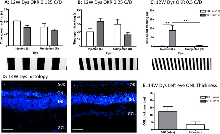
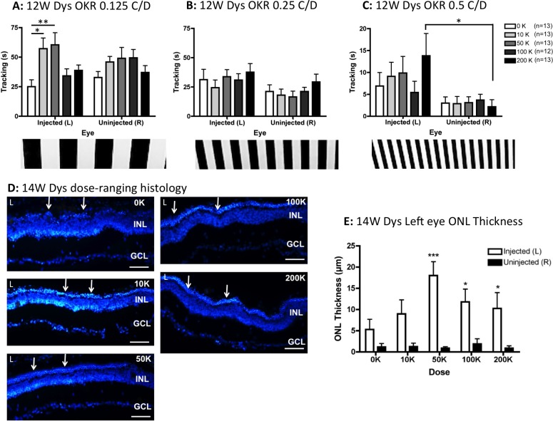
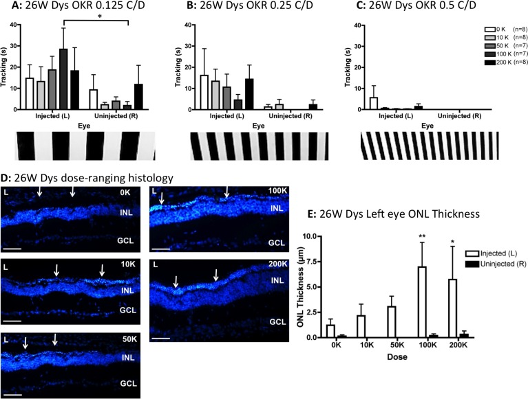
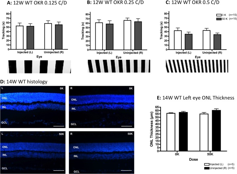
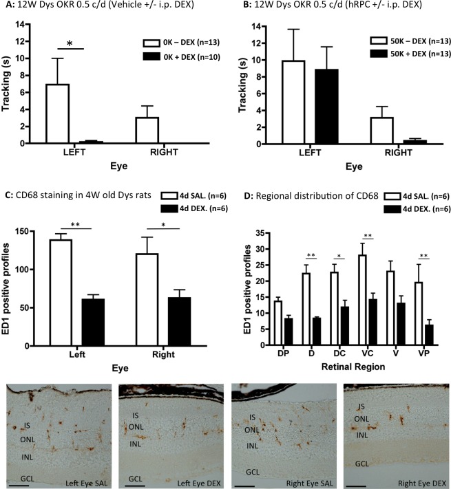
Methods: Efficacy of hRPC was tested initially in Royal College of Surgeons (RCS) dystrophic rats immunosuppressed with cyclosporine/dexamethasone. Due to adverse effects of dexamethasone, this drug was omitted from a subsequent dose-ranging study, where different hRPC doses were tested for their ability to preserve visual function (measured by optokinetic head tracking) and retinal structure in RCS rats at 3 to 6 months after grafting. Safety of hRPC was assessed by subretinal transplantation into wild type (WT) rats and NIH-III nude mice, with analysis at 3 to 6 and 9 months after grafting, respectively.

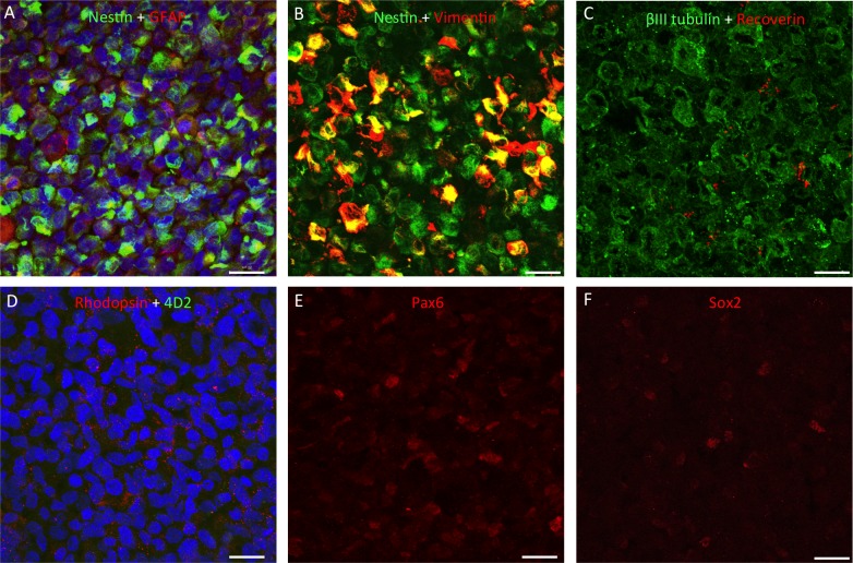
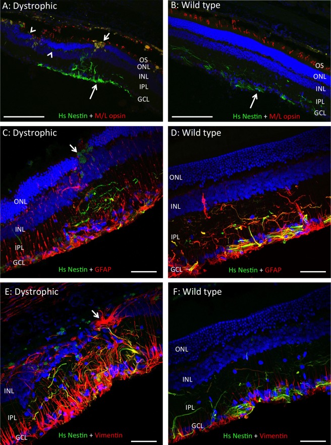
Results: The optimal dose of hRPC for preserving visual function/retinal structure in dystrophic rats was 50,000 to 100,000 cells. Human retinal progenitor cells integrated/survived in dystrophic and WT rat retina up to 6 months after grafting and expressed nestin, vimentin, GFAP, and βIII tubulin. Vision and retinal structure remained normal in WT rats injected with hRPC and there was no evidence of tumors. A comparison between dexamethasone-treated and untreated dystrophic rats at 3 months after grafting revealed an unexpected reduction in the baseline visual acuity of dexamethasone-treated animals.

Conclusions: Human retinal progenitor cells appear safe and efficacious in the preclinical models used here.

Translational relevance: Human retinal progenitor cells could be deployed during early stages of retinal degeneration or in regions of intact retina, without adverse effects on visual function. The ability of dexamethasone to reduce baseline visual acuity in RCS dystrophic rats has important implications for the interpretation of preclinical and clinical cell transplant studies.

Keywords: dexamethasone; nestin; retinal degeneration; retinal progenitor cells, human.


Figures

Figure 1
Figure 1
Subretinal injection of a single dose (50 K) of hRPC into dexamethasone-treated RCS dystrophic rats significantly preserves vision 3 months after grafting, as assessed using the OKR. (A) At 0.125 C/D there is no effect of transplant or eye. (B) At 0.25 C/D there is a significant interaction between transplantation and eye but no significance at the post hoc level. (C) At the highest spatial frequency (0.5 C/D), transplantation of hRPC significantly improves vision, with the 50 K (cell transplanted) left eyes tracking significantly better than either vehicle-injected (0 K) left eyes or uninjected right eyes. (D) Representative histology images from the left eyes (L) of cell-injected (50 K) and vehicle-injected (0 K) animals 14 weeks after grafting. (E) Quantification of ONL thickness 14 weeks after grafting reveals more ONL in cell-grafted eyes. However, this does not reach statistical significance. Graphs show the mean ± SEM with *P < 0.001 by Bonferroni's comparison. DAPI staining in blue. INL, inner nuclear layer; GCL, ganglion cell layer. Scale bars: 50 μm.
Figure 2
Figure 2
In the absence of dexamethasone treatment, subretinal injection of hRPC into RCS dystrophic rats significantly preserves vision and ONL thickness 3 months after grafting. (A) At 0.125 C/D there is a significant effect of dose, with the 10 and 50 K groups tracking more than the 0 K (vehicle) group at the post hoc level. (B) At 0.25 C/D there is a significant effect of eye but not dose. (C) At 0.5 C/D there also was an effect of eye with a dose effect apparent at the post hoc level, where only the eyes injected with 200 K cells tracked significantly better than the uninjected right eyes. (D) Representative histology images from the left eyes (L) of vehicle-injected (0 K) and cell-injected (10–200 K) animals 14 weeks after grafting show clear ONL preservation (arrows) above 50 K. (E) The ONL was significantly preserved in left eyes injected with ≥50K cells (compared to uninjected right eyes). Graphs show the mean ± SEM, with *P < 0.05, **P < 0.01, ***P < 0.001 by Bonferroni's comparison. DAPI staining in blue. Scale bars: 50 μm.
Figure 3
Figure 3
At 6 months after grafting, in the absence of dexamethasone treatment, hRPC injection slows the progressive decline of visual function and ONL thickness in RCS dystrophic rats. (A) Visual function was preserved at the lowest spatial frequency (0.125 C/D), with a significant effect of eye and post hoc significance restricted to eyes injected with 100 K hRPC. (B) At 0.25 C/D, eye remained a significant factor but there was no further significance at the post hoc level. (C) At the finest grating frequency tested (0.5 C/D), dystrophic rats were unable to track with the uninjected right eyes and although some left eye–driven tracking occurred, no significant differences were detectable. (D) Representative histology images from the left eyes (L) of vehicle-injected (0 K) and cell-injected (10–200 K) animals 26 weeks after grafting showing ONL preservation (arrows). (E) The ONL was significantly preserved in left eyes injected with ≥100K cells (compared to uninjected right eyes). Graphs show the mean ± SEM, with *P < 0.05 and **P < 0.01 by Bonferroni's comparison. DAPI staining in blue, animal numbers in € match those used in (A–C). Scale bars: 50 μm.
Figure 4
Figure 4
At 3 months after grafting, in the absence of dexamethasone treatment, subretinal injection of hRPC into WT rats had no significant impact on either visual acuity. The rats tracked well at all spatial frequencies tested by OKR: 0.125 C/D (A), 0.25 C/D (B), and 0.125 C/D (C), with time spent tracking the visual stimulus not significantly affected by either hRPC transplantation (0 vs. 50 K) or eye (left versus right). (D) Representative retinal images from the left (L) and right (R) eyes of vehicle-injected (0 K) and hRPC-injected (50 K) animals. (E) There were no statistically significant differences in ONL thickness between either transplant group (0 vs. 50K) or between left and right eyes. Graphs show the mean ± SEM, DAPI-stained nuclei in blue. Scale bars: 50 μm.
Figure 5
Figure 5
Immunocytochemistry for retinal markers in sections through a pellet of hRPC, fixed immediately upon cessation of surgery. All cells expressed nestin and βIII tubulin, with some positive staining for GFAP and variable levels of vimentin expression (A–C). Human retinal progenitor cells were found to be largely negative for recoverin (red in [C]), negative for rhodopsin using the 4D2 antibody (green in [D]) but positive for this opsin using a rabbit polyclonal antibody (red in [D]). As shown in adjacent sections, subpopulations of hRPC expressed the retinal progenitor markers Pax6 and Sox2 ([E] and [F], respectively). Proteins of interest are color coded and colocalization appears yellow. Cell nuclei stained blue with DAPI. Scale bars: 20 μm.
Figure 6
Figure 6
In the absence of dexamethasone, hRPC survive for 6 months following subretinal injection into RCS dystrophic and wild type rats. Representative images from left eyes showing hRPC stained green with an antibody to human-specific nestin (Hs Nestin) 6 months after grafting into the RCS dystrophic (A, C, E) and WT (B, D, F) retina. Human cells were located in the inner retina where they extend processes into IPL, with human material also seen at the vitreoretinal interface (large arrows in [A] and [B]). Human processes were negative for M/L opsin but positive for GFAP and vimentin to varying degrees (yellow colocalization in [C–F]). Small arrows in (A) and (C) indicate macrophages in the vicinity of the injection site, while arrowheads highlight hRPC processes extending along the outer plexiform layer in (A). Images in (A) and (C) are from rats injected with 200 K hRPC, while all others are from rats injected with 50 K hRPC. Arrow in (E) indicates GFAP-positive subretinal scarring. IPL, inner plexiform layer, outer segments (OS). DAPI staining in blue and proteins of interest color-coded. Scale bars: (A–B) 200 μm, (C–F) 50 μm.
Figure 7
Figure 7
Dexamethasone treatment reduces baseline visual acuity in RCS dystrophic rats. At the highest spatial frequency tested (0.5 C/D), dexamethasone was a significant factor, reducing the amount of time spent head tracking by vehicle (0 K)–injected left eyes (P < 0.05) and abolishing the head tracking response from uninjected right eyes (A). In contrast, the level of high spatial frequency head tracking was maintained in the presence of dexamethasone following 50 K hRPC injections into the left eye (B). A brief period of dexamethasone exposure (4 days of IP injection DEX into 23-day-old RCS dystrophic rats) significantly reduced the number of CD68 (ED1)–positive profiles compared to systemic saline injected (SAL) controls in left (vehicle-injected) and right (uninjected) eyes (C). This effect was significant across several regions of dorsal and ventral retina (D). Representative ED1-stained images from left (vehicle-injected) and right (uninjected) eyes are shown below the graphs (all from dorsal retina). ED1-positive staining is brown. Graphs show the mean ± SEM with post hoc significance as indicated: *P < 0.05; **P < 0.01. Scale bars: 50 μm.

Similar articles

Cited by

References

    1. Marc RE,, Jones BW,, Watt CB,, Strettoi E. Neural remodeling in retinal degeneration. Prog Retin Eye Res. 2003; 22: 607–655. - PubMed
    1. Gal A,, Li Y,, Thompson DA,, et al. Mutations in MERTK, the human orthologue of the RCS rat retinal dystrophy gene, cause retinitis pigmentosa. Nat Genet. 2000; 26: 270–271. - PubMed
    1. Vugler AA. Progress toward the maintenance and repair of degenerating retinal circuitry. Retina. 2010; 30: 983–1001. - PubMed
    1. Hamel C. Retinitis pigmentosa. Orphanet J of Rare Diseases. 2006; 1: 40. - PMC - PubMed
    1. Singh MS,, Charbel Issa P,, Butler R,, et al. Reversal of end-stage retinal degeneration and restoration of visual function by photoreceptor transplantation. Proc Natl Acad Sci U S A. 2013; 110: 1101–1106. - PMC - PubMed