Macro-to-micro interfacing to microfluidic channels using 3D-printed templates: application to time-resolved secretion sampling of endocrine tissue
- PMID: 27486597
- PMCID: PMC5048548
- DOI: 10.1039/c6an01055e
Macro-to-micro interfacing to microfluidic channels using 3D-printed templates: application to time-resolved secretion sampling of endocrine tissue
Abstract
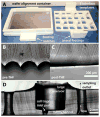
Employing 3D-printed templates for macro-to-micro interfacing, a passively operated polydimethysiloxane (PDMS) microfluidic device was designed for time-resolved secretion sampling from primary murine islets and epidiymal white adipose tissue explants. Interfacing in similar devices is typically accomplished through manually punched or drilled fluidic reservoirs. We previously introduced the concept of using hand fabricated polymer inserts to template cell culture and sampling reservoirs into PDMS devices, allowing rapid stimulation and sampling of endocrine tissue. However, fabrication of the fluidic reservoirs was time consuming, tedious, and was prone to errors during device curing. Here, we have implemented computer-aided design and 3D printing to circumvent these fabrication obstacles. In addition to rapid prototyping and design iteration advantages, the ability to match these 3D-printed interface templates with channel patterns is highly beneficial. By digitizing the template fabrication process, more robust components can be produced with reduced fabrication variability. Herein, 3D-printed templates were used for sculpting millimetre-scale reservoirs into the above-channel, bulk PDMS in passively-operated, eight-channel devices designed for time-resolved secretion sampling of murine tissue. Devices were proven functional by temporally assaying glucose-stimulated insulin secretion from <10 pancreatic islets and glycerol secretion from 2 mm adipose tissue explants, suggesting that 3D-printed interface templates could be applicable to a variety of cells and tissue types. More generally, this work validates desktop 3D printers as versatile interfacing tools in microfluidic laboratories.
Figures


Similar articles
-
3D-templated, fully automated microfluidic input/output multiplexer for endocrine tissue culture and secretion sampling.Lab Chip. 2017 Jan 17;17(2):341-349. doi: 10.1039/c6lc01201a. Lab Chip. 2017. PMID: 27990542 Free PMC article.
-
Culture and Sampling of Primary Adipose Tissue in Practical Microfluidic Systems.Methods Mol Biol. 2017;1566:185-201. doi: 10.1007/978-1-4939-6820-6_18. Methods Mol Biol. 2017. PMID: 28244052 Free PMC article.
-
3D printed mold leachates in PDMS microfluidic devices.Sci Rep. 2020 Jan 22;10(1):994. doi: 10.1038/s41598-020-57816-y. Sci Rep. 2020. PMID: 31969661 Free PMC article.
-
3D Printed Microfluidics.Annu Rev Anal Chem (Palo Alto Calif). 2020 Jun 12;13(1):45-65. doi: 10.1146/annurev-anchem-091619-102649. Epub 2019 Dec 10. Annu Rev Anal Chem (Palo Alto Calif). 2020. PMID: 31821017 Free PMC article. Review.
-
3D-printed microfluidic devices.Biofabrication. 2016 Jun 20;8(2):022001. doi: 10.1088/1758-5090/8/2/022001. Biofabrication. 2016. PMID: 27321137 Review.
Cited by
-
Micro-Macro: Selective Integration of Microfeatures Inside Low-Cost Macromolds for PDMS Microfluidics Fabrication.Micromachines (Basel). 2019 Aug 30;10(9):576. doi: 10.3390/mi10090576. Micromachines (Basel). 2019. PMID: 31480301 Free PMC article.
-
Automated microfluidic droplet sampling with integrated, mix-and-read immunoassays to resolve endocrine tissue secretion dynamics.Lab Chip. 2018 Sep 26;18(19):2926-2935. doi: 10.1039/c8lc00616d. Lab Chip. 2018. PMID: 30112543 Free PMC article.
-
Understanding Signal and Background in a Thermally Resolved, Single-Branched DNA Assay Using Square Wave Voltammetry.Anal Chem. 2018 Mar 6;90(5):3584-3591. doi: 10.1021/acs.analchem.8b00036. Epub 2018 Feb 9. Anal Chem. 2018. PMID: 29385341 Free PMC article.
-
A Microfluidic Hanging-Drop-Based Islet Perifusion System for Studying Glucose-Stimulated Insulin Secretion From Multiple Individual Pancreatic Islets.Front Bioeng Biotechnol. 2021 May 12;9:674431. doi: 10.3389/fbioe.2021.674431. eCollection 2021. Front Bioeng Biotechnol. 2021. PMID: 34055765 Free PMC article.
-
Rapid lipolytic oscillations in ex vivo adipose tissue explants revealed through microfluidic droplet sampling at high temporal resolution.Lab Chip. 2020 Apr 21;20(8):1503-1512. doi: 10.1039/d0lc00103a. Epub 2020 Apr 2. Lab Chip. 2020. PMID: 32239045 Free PMC article.
References
-
- McDonald JC, Duffy DC, Anderson JR, Chiu DT, Wu H, Schueller OJ, Whitesides GM. Electrophoresis. 2000;21:27–40. - PubMed
-
- Sia SK, Whitesides GM. Electrophoresis. 2003;24:3563–76. - PubMed
-
- Bhatia SN, Ingber DE. Nat Biotechnol. 2014;32:760–772. - PubMed
-
- Byun CK, Abi-Samra K, Choi MY, Takayama S. Electrophoresis. 2013;35:245–257. - PubMed
MeSH terms
Substances
Grants and funding
LinkOut - more resources
Full Text Sources
Other Literature Sources

