Case Reports
. 2016 Aug 4;99(2):470-80.
doi: 10.1016/j.ajhg.2016.06.017.

Isolated and Syndromic Retinal Dystrophy Caused by Biallelic Mutations in RCBTB1, a Gene Implicated in Ubiquitination

Affiliations
Case Reports

Isolated and Syndromic Retinal Dystrophy Caused by Biallelic Mutations in RCBTB1, a Gene Implicated in Ubiquitination

Frauke Coppieters et al. Am J Hum Genet. .

Abstract

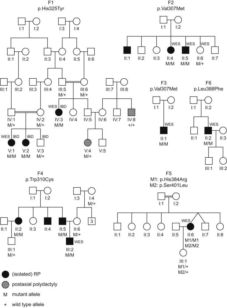
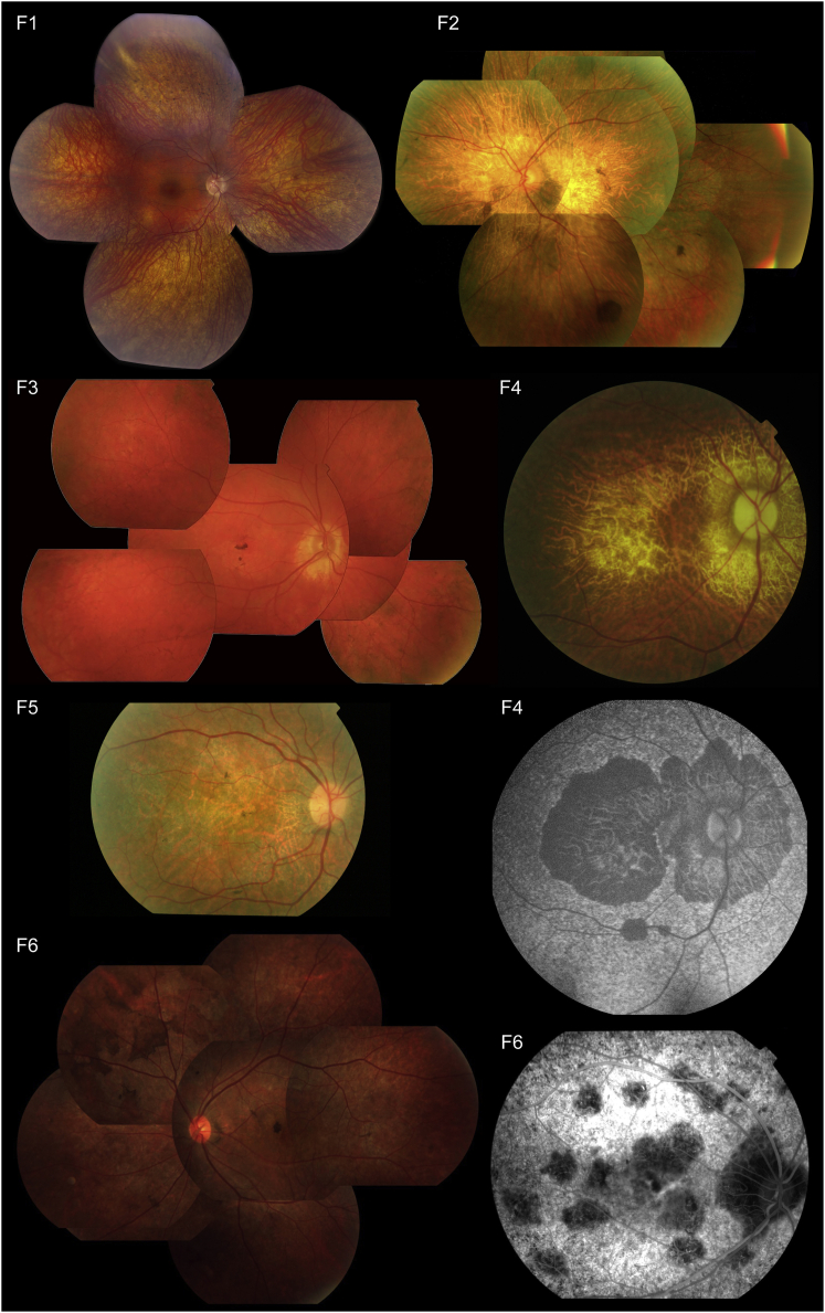
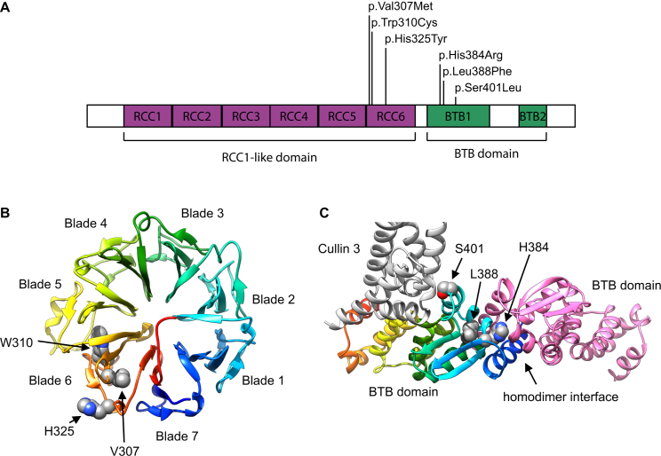
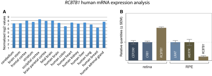
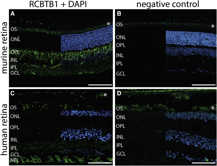
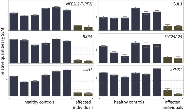
Inherited retinal dystrophies (iRDs) are a group of genetically and clinically heterogeneous conditions resulting from mutations in over 250 genes. Here, homozygosity mapping and whole-exome sequencing (WES) in a consanguineous family revealed a homozygous missense mutation, c.973C>T (p.His325Tyr), in RCBTB1. In affected individuals, it was found to segregate with retinitis pigmentosa (RP), goiter, primary ovarian insufficiency, and mild intellectual disability. Subsequent analysis of WES data in different cohorts uncovered four additional homozygous missense mutations in five unrelated families in whom iRD segregates with or without syndromic features. Ocular phenotypes ranged from typical RP starting in the second decade to chorioretinal dystrophy with a later age of onset. The five missense mutations affect highly conserved residues either in the sixth repeat of the RCC1 domain or in the BTB1 domain. A founder haplotype was identified for mutation c.919G>A (p.Val307Met), occurring in two families of Mediterranean origin. We showed ubiquitous mRNA expression of RCBTB1 and demonstrated predominant RCBTB1 localization in human inner retina. RCBTB1 was very recently shown to be involved in ubiquitination, more specifically as a CUL3 substrate adaptor. Therefore, the effect on different components of the CUL3 and NFE2L2 (NRF2) pathway was assessed in affected individuals' lymphocytes, revealing decreased mRNA expression of NFE2L2 and several NFE2L2 target genes. In conclusion, our study puts forward mutations in RCBTB1 as a cause of autosomal-recessive non-syndromic and syndromic iRD. Finally, our data support a role for impaired ubiquitination in the pathogenetic mechanism of RCBTB1 mutations.


Figures

Figure 1
Figure 1
RCBTB1 Mutations Identified in Six Families Affected by Syndromic and Non-syndromic iRD Filled symbols represent affected individuals, whereas clear symbols represent unaffected individuals. A double line represents reported consanguinity. Genotypes of different family members are indicated below them. Individuals who underwent identity-by-descent mapping and/or whole-exome sequencing are indicated by IBD and WES, respectively.
Figure 2
Figure 2
Representative Retinal Pictures of Index Individuals from the Six Families Affected by RCBTB1-Associated iRD (F1) Composite fundus picture of the retinal epithelium of individual V:2 shows outer retinal atrophy, which is more pronounced in the retinal periphery with predominantly spicular intraretinal pigmentation, and a better preserved macula. Overall, this is compatible with a diagnosis of RP. (F2–F6) Fundus pictures show progressive pattern-like reticular dystrophy in the retinal periphery, fine heterogeneity of pigment epithelium alterations, and rounded spots of chorioretinal macular atrophy (which enlarge with age). (F2) Fundus picture of the left eye of II:4 at age 68 years shows features similar to those of II:2 (F6), i.e., some pigment deposits in the form of large brown spots, as well as retinal atrophy. (F3) Fundus picture of the right eye of II:1 shows central coalescent areas with chorioretinal atrophy and peripheral reticular dystrophy. (F4) Second and third panels, right column: fundus picture and autofluorescence of the right eye of II:5 (67 years) show central and peripapillary chorioretinal atrophy. (F5) Fundus picture of the right eye of II:6 at 55 years. A detailed macular view shows a discolored retina, which reflects the retinal atrophy and a few fine pigment deposits. (F6) Left: fundus picture of LE of II:2 displays features similar to those of II:4 (F2). Right: fluorescence angiography of the right eye of II:2 displays irregular hypofluorescent areas in the posterior pole.
Figure 3
Figure 3
Location and Structural Modeling of Identified RCBTB1 Missense Variants (A) Schematic diagram of RCBTB1 shows the location of the missense variants within two distinct domains: three (F1–F4) in the sixth repeat of the RCC1-like domain (RLD), i.e., RCC6, and two (F5 and F6) in the first BTB domain (BTB1). (B) A homology model for the β-propeller structure of the RLD is shown in rainbow colors, evolving from blue (N-terminal) to red (C-terminal). The RCBTB1 RLD contains seven repeats that form a seven-bladed β-propeller, in which each blade consists of a four-stranded antiparallel β sheet. The p.Val307Met (c.919G>A), p.Trp310Cys (c.930G>T), and p.His325Tyr (c.973C>T) variants are all found in the sixth blade. Val307 and Trp310 are part of the third strand of blade 6. Trp310 is a conserved aromatic. The bulky indole group of Trp310 is buried in the hydrophobic core between blades five and six and makes extensive Van der Waals contacts with three aliphatic sidechains of blade five. p.Trp310Cys therefore introduces a big void between both blades and is probably highly destabilizing. Val307 is part of a hydrophobic core between blades six and seven. The more bulky methionine side chain introduced by p.Val307Met is clashing with residues of blade seven. His325 cannot be modeled accurately because alignments with different methods and against different templates give diverging outcomes for the exact position of this residue. Most likely, His325 is surface exposed at the end of the fourth strand of blade six. (C) A homology model for the RCBTB1 BTB domain is shown in rainbow colors, evolving from blue (N-terminal) to red (C-terminal). Another interacting RCBTB1 BTB domain is shown in pink. Part of an interacting CUL3 molecule is shown in gray. His384 is found at the BTB homodimerization interface. A histidine is present at this position in nine of ten BTB structures aligned with the RCBTB1 BTB domain. His384 forms an extensive hydrogen-bond network at the interfacial area and makes a direct Van der Waals contact with the homodimerization partner. p.His384Arg (c.1151A>G) disrupts the hydrogen-bonding network, and accommodation of two bulky arginine residues in the homodimer interface is impossible. Leu388 is an extremely conserved residue of the hydrophobic BTB core and is identical in all ten crystal structures. p.Leu388Phe (c.1164G>T) introduces drastic steric clashes of the phenyl ring with surrounding hydrophobic residues. Ser401 is a surface-exposed residue that is either close to or at the edge of the BTB-CUL3 interface, depending on the template that was used. It is unclear whether p.Ser401Leu (c.1202C>T) can disrupt the interaction with CUL3. Homology models were built on the basis of different structure templates with YASARA Structure., Additional models were built in MODELER and YASARA Structure with alignments based on HHPRED and Phyre2., , , , On the basis of the initial models, the alignments were edited for model improvement, as judged by the DOPE score in MODELER, visual inspection, and Verify_3D 3D profile analysis of the models., The models are based on template structures of RLD (PDB: 4O2W) and the BTB domain complex (PDB: 4J8Z and 4AP2). The effects of the variants were analyzed in YASARA Structure., Figures were generated with UCSF Chimera.
Figure 4
Figure 4
Expression Analysis of RCBTB1 mRNA (A) Expression analysis was performed according to the manufacturer’s instructions with an in-house-designed custom array (SurePrint G3 Human Gene Expression array version 2, AMADID 041648, Agilent Technologies) covering all protein-coding genes and 22,980 long non-coding RNA transcripts (LNCipedia version 2.1). Data normalization was performed with the VSN package in R. All values were log2 transformed. Samples included total RNA from whole brain, colon, heart, kidney, liver, lung, breast, and adrenal gland (Stratagene Europe; all adult tissues); cerebellum, brain stem, striatum, frontal cortex, occipital cortex, and parietal cortex (Agilent; adult tissues); and fetal whole brain (Agilent). (B) qPCR-based expression analysis of mRNA from RCBTB1 and two positive control genes strongly expressed in the retina and retinal pigment epithelium (RPE) was performed as previously described on commercial human cDNA from retina (BioChain) and RPE (3H Biomedical). High retinal and limited RPE expression was observed. Error bars represent the SE of the relative quantities.
Figure 5
Figure 5
RCBTB1 Staining on Human and Murine Retinal Sections (A and B) Representative fluorescence images of murine cryosections stained with RCBTB1 antibody (1:100, Abcam) (A) or negative control (B). RCBTB1 immunoreactivity in the murine retina mainly localized to the inner retina. (C and D) Representative fluorescence images of human paraffin-embedded sections stained with RCBTB1 antibody (1:100, Abcam) (C) or negative control (D). Human RCBTB1 also localized to the inner retina; the strongest signals were detected in the nerve fiber layer. Sections were counterstained with DAPI (blue) and are displayed as split images. Immunohistochemistry was performed as previously described. Asterisks mark autofluorescence of photoreceptor outer segments. Scale bars represent 100 μm. Abbreviations are as follows: OS, outer segment; IS, inner segment; ONL, outer nuclear layer; OPL, outer plexiform layer; INL, inner nuclear layer; IPL, inner plexiform layer; GCL, ganglion cell layer; and NFL, nerve fiber layer.
Figure 6
Figure 6
Expression Analysis of NFE2L2, CUL3, and Four NRF2 Target Genes The expression of NFE2L2, CUL3, RXRA, SLC25A25, and IDH1 was significantly lower in two affected individuals (V:1 and V:2 from F1) than in six healthy control individuals (respective p values are 0.001, 0.005, 0.001, 0.026, and 0.002). For EPHX1, the observed decrease was not significant (p value of 0.076). qPCR expression analysis was performed as previously described. Error bars represent the SE of the relative quantities.

References

    1. Berger W., Kloeckener-Gruissem B., Neidhardt J. The molecular basis of human retinal and vitreoretinal diseases. Prog. Retin. Eye Res. 2010;29:335–375. - PubMed
    1. Lee K., Garg S. Navigating the current landscape of clinical genetic testing for inherited retinal dystrophies. Genet. Med. 2015;17:245–252. - PubMed
    1. Lenassi E., Vincent A., Li Z., Saihan Z., Coffey A.J., Steele-Stallard H.B., Moore A.T., Steel K.P., Luxon L.M., Héon E. A detailed clinical and molecular survey of subjects with nonsyndromic USH2A retinopathy reveals an allelic hierarchy of disease-causing variants. Eur. J. Hum. Genet. 2015;23:1318–1327. - PMC - PubMed
    1. Roosing S., van den Born L.I., Sangermano R., Banfi S., Koenekoop R.K., Zonneveld-Vrieling M.N., Klaver C.C., van Lith-Verhoeven J.J., Cremers F.P., den Hollander A.I., Hoyng C.B. Mutations in MFSD8, encoding a lysosomal membrane protein, are associated with nonsyndromic autosomal recessive macular dystrophy. Ophthalmology. 2015;122:170–179. - PubMed
    1. Xu M., Yamada T., Sun Z., Eblimit A., Lopez I., Wang F., Manya H., Xu S., Zhao L., Li Y. Mutations in POMGNT1 cause non-syndromic retinitis pigmentosa. Hum. Mol. Genet. 2016;25:1479–1488. - PMC - PubMed

Publication types

MeSH terms

LinkOut - more resources