. 2016 Aug 11;536(7615):229-33.
doi: 10.1038/nature19079. Epub 2016 Aug 3.

Mechanism of arginine sensing by CASTOR1 upstream of mTORC1

Mechanism of arginine sensing by CASTOR1 upstream of mTORC1

Robert A Saxton et al. Nature. .

Abstract

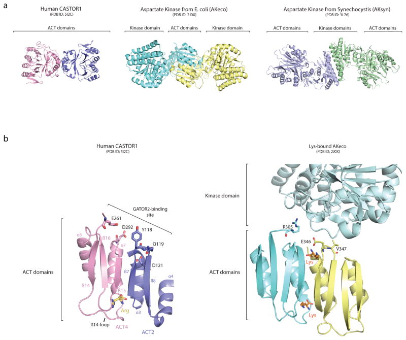
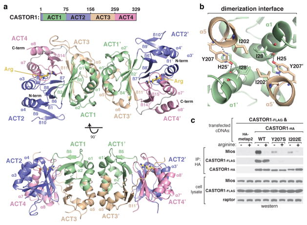
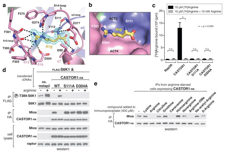
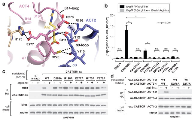
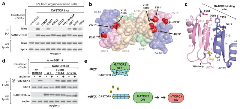
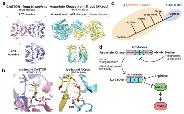
The mechanistic Target of Rapamycin Complex 1 (mTORC1) is a major regulator of eukaryotic growth that coordinates anabolic and catabolic cellular processes with inputs such as growth factors and nutrients, including amino acids. In mammals arginine is particularly important, promoting diverse physiological effects such as immune cell activation, insulin secretion, and muscle growth, largely mediated through activation of mTORC1 (refs 4, 5, 6, 7). Arginine activates mTORC1 upstream of the Rag family of GTPases, through either the lysosomal amino acid transporter SLC38A9 or the GATOR2-interacting Cellular Arginine Sensor for mTORC1 (CASTOR1). However, the mechanism by which the mTORC1 pathway detects and transmits this arginine signal has been elusive. Here, we present the 1.8 Å crystal structure of arginine-bound CASTOR1. Homodimeric CASTOR1 binds arginine at the interface of two Aspartate kinase, Chorismate mutase, TyrA (ACT) domains, enabling allosteric control of the adjacent GATOR2-binding site to trigger dissociation from GATOR2 and downstream activation of mTORC1. Our data reveal that CASTOR1 shares substantial structural homology with the lysine-binding regulatory domain of prokaryotic aspartate kinases, suggesting that the mTORC1 pathway exploited an ancient, amino-acid-dependent allosteric mechanism to acquire arginine sensitivity. Together, these results establish a structural basis for arginine sensing by the mTORC1 pathway and provide insights into the evolution of a mammalian nutrient sensor.


Figures

Extended Data Figure 1
Extended Data Figure 1. Multiple sequence alignment of CASTOR1 homologues
a) Expanded Multiple Sequence Alignment of CASTOR1 homologues from various organisms. Positions are colored white to blue according to increasing sequence identity. Secondary structure features are labeled and colored by ACT domain as in 1A.
Extended Data Figure 2
Extended Data Figure 2. Dimerization deficient CASTOR1 mutants bind arginine but fail to inhibit mTORC1 in cells
a) The dimerization deficient CASTOR1 Y207S and I202E mutants bind arginine in vitro. FLAG-immunoprecipitates prepared from HEK-293T cells transiently expressing indicated FLAG-tagged proteins were used in binding assays with [3H]Arginine as described in the methods. Unlabeled arginine was included as a competitor where indicated. Values are Mean ± SD for 3 technical replicates from one representative experiment. b) Dimerization deficient CASTOR1 Y207S and I202E mutants fail to inhibit mTORC1. HEK-293T cells transiently expressing FLAG-S6K1 and HA-tagged CASTOR1 WT, Y207S, or I202E were starved of arginine for 50 min and, where indicated, restimulated for 10 min. FLAG- immunoprecipitates were prepared from lysates and analyzed as in 1C. Phospho-S6K1 was used as an indicator of mTORC1 activity.
Extended Data Figure 3
Extended Data Figure 3. Model of lysine-binding in CASTOR1
a) Comparison of the arginine-bound pocket of human CASTOR1 with a model of the pocket with lysine in place of arginine. Arginine and lysine stick representations are shown in yellow and orange, respectively. The distances in the lysine-bound model, 3.8 Å and 5.0 Å, are beyond the range of standard hydrogen-bonds and salt-bridges, respectively. ACT domains are labeled as in 1A. b) Chemical structures of arginine analogues used in Fig. 2E. Differences relative to L-Arginine are highlighted in oranges boxes.
Extended Data Figure 4
Extended Data Figure 4. Differences in the arginine-binding capacities of CASTOR1 and CASTOR2
a) Multiple sequence alignment of human CASTOR1 and CASTOR2, highlighting differences in amino acid sequence that are in close proximity to arginine binding residues in CASTOR1. b) The CASTOR1 HHV108–110QNI mutant constitutively binds GATOR2 in cells. HEK-293T cells transiently expressing HA-metap2 or the indicated HA-tagged CASTOR1 constructs were starved of arginine for 50 min and, where indicated, restimulated for 10 min. HA-immunoprecipitates prepared and analyzed as in 1C. c) The CASTOR1 HHV108–110QNI mutant displays reduced arginine-binding capacity in vitro. Binding assays were performed with the indicated CASTOR1 or CASTOR2 constructs and immunoprecipitates analyzed as in 2C. Values are Mean ± SD for 3 technical replicates from one representative experiment. d) Comparison of the CASTOR1 HHV108–110QNI mutant and WT CASTOR2. HEK-293T cells transiently expressing HA-metap2 or the indicated HA-tagged CASTOR1 or CASTOR2 constructs were starved of arginine for 50 min and, where indicated, restimulated for 10 min. HA-immunoprecipitates prepared and analyzed as in 1C.
Extended Data Figure 5
Extended Data Figure 5. GATOR2-binding deficient CASTOR1 mutants still bind arginine and homodimerize
a) The CASTOR1 YQ118–119AA, D121A, E261A and D292A mutants bind arginine in vitro. FLAG-immunoprecipitates prepared from HEK-293T cells transiently expressing indicated FLAG-tagged proteins were used in binding assays with [3H]Arginine as described in the methods. Unlabeled arginine was included as a competitor where indicated. Values are Mean ± SD for 3 technical replicates from one representative experiment. b) The CASTOR1 YQ118–119AA, D121A, E261A and D292A mutants dimerize in cells. HA-immunoprecipitates prepared from HEK293T-cells transiently expressing CASTOR1-FLAG and HA-metap2 or the indicated HA-tagged CASTOR1 constructs were analyzed as in 1C.
Extended Data Figure 6
Extended Data Figure 6. Similarities between human CASTOR1 and prokaryotic Aspartate Kinases
a) Ribbon diagram views of human CASTOR1 (this paper), AKeco (PDB ID: 2J0x) and AKsyn (PDB ID: 3L76), highlighting the different modes of dimerization. AKs can dimerize through an interlocked-ACT domain conformation (as in AKeco) or through their kinase domains (AKsyn), both of which are distinct from the side-by-side ACT-domain dimerization in CASTOR1. b) View of AKeco depicting positions of residues R305, E346, and V347, which correspond to the positions of GATOR2-interacting residues of CASTOR1.
Figure 1
Figure 1. Architecture of human CASTOR1
a, Two orthogonal views of the CASTOR1 homodimer (ribbon diagram), with ACT-domains 1–4 colored in green, purple, wheat, and pink, respectively. The bound arginine is shown in yellow. Disordered regions not observed in the crystal structure are omitted. b, View of the CASTOR1 dimerization interface, with side chains of key residues represented in stick form. c, Dimerization deficient CASTOR1 Y207S and I202E mutants display weaker interactions with endogenous GATOR2. HEK-293T cells transiently expressing FLAG-tagged CASTOR1 wild type (WT) and the indicated HA-tagged constructs were starved of arginine for 50 min and, where indicated, restimulated for 10 min. HA-immunoprecipitates were generated from cell lysates and analyzed by immunoblotting for the indicated proteins. Mios was used as a representative GATOR2 component.
Figure 2
Figure 2. The arginine-binding pocket of CASTOR1
a, View of the arginine-binding pocket in CASTOR1, together with its Fo-Fc electron density map calculated and contoured at 4σ from an omit map lacking arginine. The bound arginine is shown in yellow. Hydrogen bonds or salt-bridges are shown as black dashed lines. Residues 269–273 are omitted for clarity. b, Steric view of the arginine-binding pocket, depicting the surface representation of CASTOR1 and stick model of arginine (yellow). The β14-loop (residues 269–280) is omitted for clarity. c, CASTOR1 S111A and D304A mutants do not bind arginine in vitro. FLAG-immunoprecipitates prepared from HEK-293T cells transiently expressing indicated FLAG-tagged proteins were used in binding assays with [3H]Arginine as described in the methods. Values are Mean ± SD for 3 technical replicates from one representative experiment. d, The CASTOR1 S111A and D304A mutants constitutively bind GATOR2 and inhibit mTORC1 signaling in cells. HEK-293T cells transiently expressing FLAG-S6K1 and the indicated HA-tagged constructs were starved of arginine for 50 min and, where indicated, restimulated for 10 min. Both FLAG- and HA-immunoprecipitates were prepared from lysates and analyzed as in 1c. e, Effects of various arginine analogues on the CASTOR1-GATOR2 interaction in vitro. HEK-293T cells transiently expressing HA-CASTOR1 WT were starved of arginine for 50 min. HA-immunoprecipitates were prepared from cell lysates then incubated with 400 μM of the indicated compounds for 20 min and analyzed as in 1c.
Figure 3
Figure 3. Arginine facilitates the intramolecular association of the ACT2 and ACT4 domains of CASTOR1
a, Top-down view of the arginine- and β14-loop-mediated contacts between ACT2 and ACT4. Hydrogen bonds and salt-bridges are shown as black dashed lines. b, CASTOR1 D276A, R126A, E277A, H175A, and C278A mutants display reduced arginine-binding capacity in vitro. Binding assays were performed and immunoprecipitates analyzed as in 2c. Values are Mean ± SD for 3 technical replicates from one representative experiment. c, The CASTOR1 D276A, R126A, E277A, H175A, and C278A mutants constitutively bind GATOR2 in cells. HEK-293T cells transiently expressing the indicated HA-tagged constructs were starved of arginine for 50 min and, where indicated, restimulated for 10 min. HA-immunoprecipitates prepared and analyzed as in 1c. d, CASTOR1 ACT1–2 (1–169) and CASTOR1 ACT3–4 (169–329) associate in an arginine- and β14-loop dependent manner. HEK-293T cells transiently expressing the indicated HA-tagged constructs were starved of arginine for 60 min and, where indicated, restimulated for 60 min. HA-immunoprecipitates were prepared and analyzed as in 1c.
Figure 4
Figure 4. The GATOR2 binding site of CASTOR1 is at the ACT2-ACT4 interface and is required for signalling arginine deprivation to mTORC1
a, The CASTOR1 D292A, E261A, D121A, and YQ118–119AA mutants are deficient in GATOR2 binding. HA-immunoprecipitates prepared from HEK293T-cells transiently expressing the indicated HA-tagged constructs were analyzed as in 1c. b, Solvent-exposed surface view of the CASTOR1 homodimer highlighting the GATOR2-binding sites (red). Residue E261 is in a partially disordered loop and not visible in one monomer (left). c, Cross-sectional view of the ACT2-ACT4 interface showing the positions of the critical GATOR2-binding residues relative to the bound arginine (yellow) and the β14-loop. d, The GATOR2-binding-deficient YQ118–119AA and D121A mutants of CASTOR1 fail to inhibit the mTORC1 pathway and render cells insensitive to arginine starvation. HEK-293T cells were transiently transfected with FLAG-S6K1 and the indicated HA-tagged constructs. FLAG-immunoprecipitates were prepared and analyzed as in 1d. e, Model of how arginine releases CASTOR1 from GATOR2 to activate mTORC1.
Figure 5
Figure 5. Insights into the evolution of arginine sensing by CASTOR1
a, (Top) Ribbon view of human CASTOR1 dimer (pink and purple) and AKeco dimer (blue and yellow, PDB ID 2J0X). (Bottom) Ribbon view of the human CASTOR1 monomer (left) and regulatory domain from AKeco (right). b, Comparison of the arginine-binding pocket in human CASTOR1 with the lysine-binding pocket in AKeco. Arginine and lysine are shown in yellow and orange, respectively. Hydrogen bonds and salt-bridges are shown as black dashed lines. c, Phylogenetic distribution of Aspartate Kinase (orange) and CASTOR1 homologues (purple). d, Model of the evolution of CASTOR1 from the regulatory domain of an ancestral Aspartate Kinase.

References

    1. Laplante M, Sabatini DM. mTOR signaling in growth control and disease. Cell. 2012;149:274. - PMC - PubMed
    1. Dibble CC, Manning BD. Signal integration by mTORC1 coordinates nutrient input with biosynthetic output. Nature Cell Biology. 2013;15:555–564. - PMC - PubMed
    1. Jewell JL, Russell RC, Guan KL. Amino acid signaling upstream of mTOR. Nature Reviews Molecular Cell Biology. 2013;14:133. - PMC - PubMed
    1. Ban H, et al. Arginine and Leucine regulate p70 S6 kinase and 4E-BP1 in intestinal epithelial cells. International journal of molecular medicine. 2004;13:537–543. - PubMed
    1. Bronte V, Zanovello P. Regulation of immune responses by L-arginine metabolism. Nat Rev Immunol. 2005;5:641–654. - PubMed

Publication types

MeSH terms